NAV2 / Neuron navigator 2 · IHC design guide

Design Immunohistochemistry for NAV2

Plan NAV2 chromogenic IHC in paraffin sections using cytoplasmic staining in glandular cells as a reference (HPA tissue IHC). This guide covers controls, fixation, detection and scoring, with an IHC antibody dilution range of 1:100–1:300 (datasheet A05540).

Evidence assembled Oct 2026 · For research use; verify linked source records and product datasheet before use
Immunohistochemistry protocol sheet for NAV2 (IHC for NAV2): expected localisation Variable cytoplasmic staining in tissues (HPA tissue IHC), antibody A05540, validated IHC image, and IHC protocol steps
Printable NAV2 IHC protocol sheet — expected localisation Variable cytoplasmic staining in tissues (HPA tissue IHC), antibody A05540, controls and protocol steps. Open the full NAV2 IHC guide →

NAV2 Immunohistochemistry Experimental Design Guide

Expected localisation, validated protocols, controls and antibodies — the at-a-glance facts below, then the full design guide.

Must know before staining
Expected localisation Variable cytoplasmic staining in tissues (HPA tissue IHC)
Staining pattern Variable cytoplasmic staining in glandular cells (HPA tissue IHC)
Antigen retrieval Tris-EDTA pH 9.0 HIER, heat-mediated (datasheet A05540)
Positive control ⓘ Appendix+4 more · see all
Negative control ⓘ Adipose tissue+3 more · see all
Important caveats
Reasons your staining may differ from the expected pattern.
Fixation Matched tissue-IHC evidence does not establish the fixation claim. Validate the specimen-specific method before use. (selected-SKU IHC image A05540)
Caveat Tissue levels vary, so weak staining needs controls (HPA tissue IHC)
Regulation Expression varies by tissue (UniProt)
Isoform / epitope 13 isoforms; check epitope coverage across variants (UniProt)
Section 1

Recommended NAV2 IHC & IF Protocols

The catalog antibody’s IHC-P protocol (datasheet A05540) appears alongside published NAV2 protocols for colorectal cancer tissue (PMC4600204) and rat spleen and ankle joints (PMC13615836).

Recommended immunohistochemistry (IHC-P) protocol parameters
SampleParaffin-embedded human spleen tissue; fixative not specified (datasheet A05540)
FixationImage fixative and duration unreported (datasheet A05540); verify before use.
Sectioning4–5 µm sections on charged slides (standard)
DeparaffinisationXylene, graded ethanol series to water (standard)
Antigen retrievalHeat retrieval: Tris-EDTA pH 9.0 (datasheet A05540); 20 min, 95–100 °C (standard)
Peroxidase block3% H2O2, 10 min, room temperature (standard)
Blocking10% normal serum of the secondary host, 30 min, room temperature (standard)
Primary antibodyRabbit anti-NAV2, 1:100-1:300 (datasheet A05540)
Primary incubationOvernight at 4 °C (standard)
DetectionHRP-polymer secondary, DAB chromogen 5–10 min (standard)
CounterstainHematoxylin, blue, dehydrate and mount (standard)
Expected resultNAV2-positive staining in glandular cells of appendix (HPA tissue IHC: High). HPA tissue profile: Cytoplasmic expression at variable levels in most tissues. No signal in the no-primary control.
💡Decision noteStart with Tris-EDTA pH 9.0 heat retrieval for the catalog antibody (datasheet A05540); the published protocols use different retrieval buffers (PMC4600204; PMC13615836).
Section 2

What Is the Expected NAV2 Staining Pattern?

In paraffin-section IHC, expect predominantly cytoplasmic NAV2 staining at variable levels across tissues (HPA tissue IHC: Approved; medium consistency with RNA; pending external verification). Glandular cells in several organs and kidney tubule cells are reported as strongly positive (HPA tissue IHC: High). NAV2 is also annotated in the nucleus, cytoskeleton, axons and perikaryon, with no transmembrane segment (UniProt Q8IVL1: subcellular location and topology).

What am I looking at on my slide?
Cytoplasmic staining in appendix, breast, colon or duodenal glandular cells, or kidney tubule cells.This matches reported High staining in those cell populations (HPA tissue IHC). Judge the labelled cells and compartment together; staining throughout a section is less informative than the reported cell distribution (general IHC interpretation).
Only crisp plasma-membrane rims or extracellular deposits are visible, without a cytoplasmic pattern.That compartment is discordant with the reported cytoplasmic IHC profile and NAV2's lack of a transmembrane segment (HPA tissue IHC; UniProt Q8IVL1: topology). Check the detection controls and antibody conditions before assigning the signal to NAV2 (general IHC practice).
Adipocytes or soft-tissue fibroblasts stain as strongly as nearby expected-positive cells.Those cell types are reported as Not detected (HPA tissue IHC). Unexpected staining warrants evaluation for cross-reactivity or endogenous detection activity; it does not, by itself, establish either cause (general IHC interpretation).
Broad haze covers tissue, empty areas or many cell types, obscuring cell boundaries.A diffuse deposit cannot be scored reliably against the reported cell-specific cytoplasmic pattern (HPA tissue IHC; general IHC interpretation). Review background controls and washing before interpreting intensity (general IHC practice).
A section containing reported High-positive cells shows no detectable signal.Absence of staining in, for example, kidney tubule cells or colon glandular cells conflicts with the HPA reference pattern (HPA tissue IHC: High). First check whether the section and detection controls worked; a single negative slide cannot establish NAV2 absence (general IHC practice).
💡Expected NAV2 appearanceCall a positive result when identifiable glandular or kidney tubule cells show predominantly cytoplasmic chromogen, potentially High in the listed tissues (HPA tissue IHC); uniform extracellular or membrane-only colour is discordant (HPA tissue IHC; UniProt Q8IVL1: topology).
How each factor affects the staining
Tissue and cell identityHPA reports High staining in several glandular populations, esophageal squamous epithelium and kidney tubules, but Not detected in adipocytes and soft-tissue fibroblasts (HPA tissue IHC). Score the named cell population rather than treating an entire organ as uniformly positive (general IHC interpretation).
Compartment and assayCytoplasmic staining is the tissue-IHC profile, whereas supported ICC/IF images place NAV2 in the nucleoplasm (HPA tissue IHC; HPA subcellular ICC/IF). UniProt also annotates nuclear and cytoskeletal locations (UniProt Q8IVL1: subcellular location). Keep assay-specific reference patterns distinct when reviewing a nuclear signal.
Antibody validationTwo listed antibodies have IHC status Approved; HPA011755 has ICC status Supported (HPA antibodies). The tissue profile has medium consistency with RNA and awaits external verification (HPA tissue IHC). These labels support cautious pattern comparison, not a guarantee for every specimen or staining run.
NAV2 formsUniProt lists 13 isoforms and a single annotated chain spanning residues 1–2488 (UniProt Q8IVL1: isoforms and processing). Without an antibody epitope or isoform-specific staining result in the supplied evidence, do not infer which forms contribute to a slide's signal.
Why is my staining missing, weak or wrong?
SituationLikely causeNext action
No staining in a reported positive control tissue.A failed staining run or unsuitable assay conditions are possibilities; the slide alone cannot distinguish them (general IHC practice).Confirm that the control contains identifiable High-positive glandular or kidney tubule cells (HPA tissue IHC). Check section quality, retrieval settings, antibody dilution and detection controls using the established IHC workflow (general IHC practice).
Strong colour appears in adipocytes or soft-tissue fibroblasts.The result conflicts with their Not detected HPA profile; cross-reactivity or endogenous detection activity is possible (HPA tissue IHC; general IHC interpretation).Review a no-primary control and the detection-system controls, then compare staining in reported positive cells on the same run (general IHC practice; HPA tissue IHC).
Staining forms a diffuse brown haze.Background from the staining workflow may obscure the expected cytoplasmic distribution (general IHC practice; HPA tissue IHC).Inspect no-primary controls, blocking, washes and detection development before scoring cell intensity; assess whether identifiable cells still show the expected compartment (general IHC practice; HPA tissue IHC).
Only a membrane-like rim or extracellular deposit is present.That distribution disagrees with the cytoplasmic tissue profile and the absence of a transmembrane segment (HPA tissue IHC; UniProt Q8IVL1: topology).Check morphology and background controls, and confirm that staining is intracellular in reported positive cells before calling NAV2 positive (general IHC practice; HPA tissue IHC).
A predominantly nuclear IHC signal appears.Nuclear NAV2 is annotated, but HPA describes the tissue-IHC profile as cytoplasmic; nuclear predominance therefore needs cautious interpretation (UniProt Q8IVL1: subcellular location; HPA tissue IHC).Compare the same run with reported cytoplasmic positive cells and detection controls before interpreting the nuclear pattern (HPA tissue IHC; general IHC practice).
Why does an ICC/IF image look nuclear when this IHC guide expects cytoplasm?HPA's supported ICC/IF localisation is nucleoplasmic, while its tissue-IHC profile is cytoplasmic (HPA subcellular ICC/IF; HPA tissue IHC).Interpret each result against its own assay reference and record the observed compartment; use the separate ICC/IF guide for that assay's workflow (HPA subcellular ICC/IF; HPA tissue IHC).

Sample controls for NAV2 IHC & IF

🧪Run colon first: glandular cells should stain (HPA: High in colon glandular cells). Use adipose tissue as a negative comparator, focusing on adipocytes (HPA: Not detected in adipocytes); on the colon slide, assess any unstained adjacent cells as an internal background reference without assuming their NAV2 status.
Positive control tissue: Appendix (Glandular cells, HPA High)
Negative control tissue: Adipose tissue (HPA Not detected)
ICC-IF cell lines (HPA subcellular resource): HPA ICC-IF images show NAV2 in A-431, U-251MG, U2OS, with annotated localisation: Nucleoplasm (supported) (HPA subcellular).
Technical controls: Include no-primary (secondary-only) and isotype controls; match the isotype reagent to the primary antibody’s host species and class, using host-matched nonimmune IgG for a polyclonal primary (standard IHC practice). A NAV2 knockout specimen is the preferred biological negative; quench endogenous peroxidase and inspect blood or inflammatory cells in colon for misleading chromogen signal (standard IHC practice).
⚠️Feasibility: A target-specific fixation window or fixation effect is unreported, and the fixative is unreported in the selected A05540 paraffin-section caption (selected-SKU caption). That caption uses Tris-EDTA at pH 9.0 for retrieval, but does not establish that NAV2 staining depends on that condition (selected-SKU caption). The supplied evidence does not establish that frozen sections or IF are easier; for colon IHC, interpret staining alongside the peroxidase controls because blood or inflammatory cells can produce misleading chromogen signal (standard IHC practice).

HPA tissue IHC evidence for NAV2

Comprehensive Human Protein Atlas IHC scoring per tissue (reliability: Approved — Medium consistency between antibody staining and RNA expression data. Pending external verification.). Rows are taken directly from the HPA tissue chart — click any row's HPA link to view the source.

Positive expression · recommended positive controls

TissueCell typeLevelEvidenceSource
Appendix Glandular cells High Protein (IHC) HPA →
Breast Glandular cells High Protein (IHC) HPA →
Colon Glandular cells High Protein (IHC) HPA →
Duodenum Glandular cells High Protein (IHC) HPA →
Esophagus Squamous epithelial cells High Protein (IHC) HPA →

Undetected expression · recommended negative controls

TissueCell typeLevelEvidenceSource
Adipose tissue Adipocytes Not detected Protein (IHC) HPA →
Caudate Glial cells Not detected Protein (IHC) HPA →
Hippocampus Glial cells Not detected Protein (IHC) HPA →
Soft tissue Fibroblasts Not detected Protein (IHC) HPA →
Section 3

Advanced NAV2 IHC Tips

Troubleshoot NAV2 staining in paraffin sections by checking retrieval, tissue controls and compartment patterns before assigning biological meaning to chromogenic signal.

Which retrieval condition should I start with for NAV2 in paraffin sections?
Start with heat-mediated Tris-EDTA at pH 9.0 for NAV2 antigen retrieval (datasheet A05540). The selected paraffin-section spleen image used that buffer, followed by primary antibody at 1:200 overnight at 4°C (datasheet A05540). If staining is weak, compare a shorter and longer heating interval on matched sections while keeping dilution and detection constant (standard IHC practice). Check whether glandular cells in colon or breast stain in the expected cytoplasmic compartment (HPA: high NAV2 staining in these cells; HPA: variable cytoplasmic expression). Excessive retrieval can damage section morphology, so assess tissue preservation alongside signal (standard IHC practice).
How should I troubleshoot fixation when NAV2 staining is weak?
The selected NAV2 image identifies paraffin-embedded spleen but does not report its fixative, so target-specific fixation sensitivity remains unknown (datasheet A05540). Record the fixative, fixation duration and processing history for each specimen before comparing staining across cases (standard IHC practice). If sections differ in staining, process a matched control section through the same Tris-EDTA pH 9.0 retrieval and 1:200 primary dilution (datasheet A05540; standard IHC practice). Inspect morphology and compare cell-specific signal with adjacent controls before changing fixation or retrieval variables (standard IHC practice). Neither NAV2 topology nor its phosphorylation annotations establish a preferred fixative (UniProt Q8IVL1).
Where should convincing NAV2 chromogenic staining appear?
Evaluate cytoplasmic staining first: tissue IHC shows variable cytoplasmic expression across most tissues (HPA: tissue profile). NAV2 is also annotated in the nucleus, cytoskeleton, axon and perikaryon, with reported punctate microtubule and neurofilament association in neuronal cells (UniProt Q8IVL1). Nucleoplasmic localisation is supported by cell imaging, but that observation alone does not validate an isolated nuclear chromogenic signal in paraffin sections (HPA: subcellular; standard IHC interpretation). Compare the stained compartment with the relevant cell type and a negative reagent control before scoring (standard IHC practice). Record nuclear and cytoplasmic patterns separately when both appear (standard IHC practice).
Could NAV2 isoforms or epitope location explain discordant staining?
NAV2 has 13 annotated isoforms, so antibody recognition may depend on whether its mapped epitope is retained in the isoform present (UniProt Q8IVL1; standard immunostaining principle). Check the catalog antibody's immunogen or epitope information before interpreting a tissue difference as altered NAV2 abundance (standard IHC practice). NAV2 has no annotated transmembrane segment and contains a calponin-homology domain at residues 85–192 (UniProt Q8IVL1). Phosphoserines are annotated at residues 1480, 1484, 1488 and 1977; their effect on this antibody's staining is unestablished (UniProt Q8IVL1). Compare matched sections under identical retrieval and detection conditions when assessing epitope-related discrepancies (standard IHC practice).
How can IF help assess a NAV2 IHC localisation pattern?
Use IF as a separate localisation check, pairing NAV2 with a marker for the expected cell type, such as a glandular epithelial marker in colon (HPA: high NAV2 staining in colon glandular cells; standard IF practice). Choose fluorophores after inspecting unstained tissue for autofluorescence, and separate their emission channels during acquisition (standard IF practice). NAV2 has no transmembrane segment and is annotated in intracellular compartments, so permeabilise fixed cells to expose intracellular epitopes while checking whether extraction changes the pattern (UniProt Q8IVL1; standard IF practice). Compare nuclear and cytoplasmic channels separately because tissue IHC and cell imaging report different predominant patterns (HPA: tissue profile; HPA: nucleoplasm supported).
What should I check when NAV2 staining appears diffuse or patchy?
Start with a no-primary control and inspect whether color persists around vessels, damaged areas or section edges (standard chromogenic IHC practice). Block endogenous peroxidase before enzyme-based detection, and check that the detection reagent alone does not produce color (standard chromogenic IHC practice). The selected spleen image used primary antibody at 1:200 overnight at 4°C and secondary antibody at 1:200 for 45 minutes at room temperature (datasheet A05540). If background remains high, compare primary dilutions and washing conditions on adjacent sections while retaining the same retrieval method (standard IHC practice). Score only cell-associated staining with preserved morphology (standard IHC practice).
How should I quantify NAV2 staining across paraffin sections? ⚠ ANSWER MARKED FOR VERIFICATION
Define the compartment and cell population before scoring; HPA reports variable cytoplasmic tissue staining, including high staining in colon glandular cells (HPA: tissue profile; HPA: colon). For those cells, record the percentage positive and staining intensity, then calculate an H-score from 0–300 if intensity categories are reproducible (standard IHC practice). Alternatively, report positive cells per mm² when cell density, rather than staining intensity, is the study endpoint (standard IHC practice). Normalise counts to the annotated viable tissue area or eligible cell population, and keep exposure, counterstain and scoring thresholds consistent across sections (standard IHC practice). Exclude folds and necrotic regions during annotation (standard IHC practice).
How can I distinguish genuine NAV2 signal from staining artefact?
Look for reproducible, cell-associated staining in an expected compartment: tissue IHC reports variable cytoplasmic NAV2, including high staining in kidney tubule cells (HPA: tissue profile; HPA: kidney). Treat isolated edge staining, necrotic deposits and color retained in a no-primary control as potential artefacts (standard chromogenic IHC practice). Check endogenous peroxidase blocking if color persists without primary antibody, and compare the pattern with an adjacent section (standard chromogenic IHC practice). A nuclear signal needs separate scrutiny because nucleoplasmic localisation is supported in cell imaging while the reported tissue IHC profile is cytoplasmic (HPA: subcellular; HPA: tissue profile). HPA tissue reliability is Approved with medium staining-to-RNA consistency and pending external verification (HPA: tissue reliability).
Boster reagents

Best NAV2 / Neuron navigator 2 IHC Antibodies

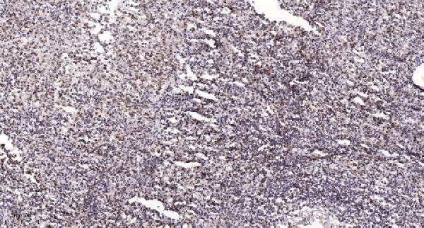
A05540 is a human-reactive anti-NAV2 antibody listed for IHC and IF (catalog: applications, reactivity). Its IHC figure shows paraffin-embedded human spleen (A05540 IHC image caption).

Real IHC data Immunohistochemical analysis of paraffin-embedded human spleen. 1, Antibody was diluted at 1:200 (4° overnight). 2, Tris-EDTA, pH9.0 was used for antigen retrieval. 3, Secondary antibody was diluted at 1:200 (room temperature, 45min).
Anti-Neuron Navigator 2 NAV2 Antibody
Cat # A05540

A05540 will render with an IHC figure of paraffin-embedded human spleen (A05540 IHC image caption). It is also listed for human IF, but no IF figure is supplied (catalog: applications, reactivity, IF image alts).

Which to pick: For tissue IHC, choose A05540, a rabbit polyclonal antibody listed for human IHC at 1:100–1:300 (catalog: host, clonality, reactivity, applications, IHC dilution); its paraffin-section figure used 1:200 and Tris-EDTA retrieval at pH 9.0, but did not report the fixative (A05540 IHC image caption). For IF, A05540 is listed at 1:50 for human samples; ICC validation and an IF figure are not documented (catalog: applications, reactivity, IF dilution, IF image alts). No cross-species option is documented because A05540 lists only Human reactivity (catalog: reactivity).

Each figure is that product's own IHC / IF validation image from its datasheet.

References

  1. UniProt Consortium. UniProt entry Q8IVL1 (NAV2_HUMAN, Neuron navigator 2).
  2. Human Protein Atlas. NAV2 tissue IHC expression (reliability: Approved).
  3. Human Protein Atlas. NAV2 subcellular location (ICC-IF): Localized to the nucleoplasm..
  4. Human Protein Atlas. NAV2 antibody validation summary (2 antibodies).
  5. Neuron navigator 2 overexpression indicates poor prognosis of colorectal cancer and promotes invasion through the SSH1L/cofilin-1 pathway. Journal of experimental & clinical cancer research : CR 2015 — PMC4600204.
  6. Astragaloside IV Modulates the Immune Response in Rheumatoid Arthritis and is Associated with Inhibition of the NAV2-Wnt/β-Catenin Signaling Cascade. Journal of inflammation research 2026 — PMC13615836.
  7. Gene expression signatures differentiate uterine endometrial stromal sarcoma from leiomyosarcoma. Gynecologic oncology 2013 — PMC4051229.
  8. Exploration of potential biomarkers in salivary duct carcinoma based on bioinformatics analysis. Scientific reports 2026 — PMC12886760.
  9. PubMed PMID:11904404 — UniProt-cited evidence.
  10. PubMed PMID:12214280 — UniProt-cited evidence.
  11. PubMed PMID:15158073 — UniProt-cited evidence.