NBN / Nibrin · IHC design guide

Design Immunohistochemistry for NBN

Plan chromogenic paraffin IHC for NBN using widespread nuclear tissue staining as the reference pattern (HPA tissue IHC). This guide covers fixation consistency, antibody titration and nuclear scoring, including the focal recruitment seen after DNA damage (UniProt).

Evidence assembled Oct 2026 · For research use; verify linked source records and product datasheet before use
Immunohistochemistry protocol sheet for NBN (IHC for NBN): expected localisation Nuclear staining in tissue sections (HPA tissue IHC), antibody PB9615, validated IHC image, and IHC protocol steps
Printable NBN IHC protocol sheet — expected localisation Nuclear staining in tissue sections (HPA tissue IHC), antibody PB9615, controls and protocol steps. Open the full NBN IHC guide →

NBN Immunohistochemistry Experimental Design Guide

Expected localisation, validated protocols, controls and antibodies — the at-a-glance facts below, then the full design guide.

Must know before staining
Expected localisation Nuclear staining in tissue sections (HPA tissue IHC)
Staining pattern Widespread nuclear staining across cell types (HPA tissue IHC)
Antigen retrieval EDTA pH 8.0 HIER, heat-mediated (datasheet PB9615)
Positive control ⓘ Lung+4 more · see all
Negative control ⓘ Skeletal muscle+2 more · see all
Important caveats
Reasons your staining may differ from the expected pattern.
Fixation Keep fixation consistent across paraffin sections. (standard IHC practice; not target-specific)
Caveat Genotoxic stress can produce discrete nuclear foci (UniProt)
Regulation DNA damage promotes focal recruitment (UniProt)
Isoform / epitope 0 listed isoforms; one full-length chain (UniProt)
Section 1

Recommended NBN IHC & IF Protocols

The catalog antibody protocol is paired with one published NBN IHC workflow (PMC5029606).

Recommended immunohistochemistry (IHC-P) protocol parameters
SampleParaffin-embedded human breast cancer tissue; fixative not specified (datasheet PB9615)
FixationImage fixative and duration unreported (datasheet PB9615); verify before use.
Sectioning4–5 µm sections on charged slides (standard)
DeparaffinisationXylene, graded ethanol series to water (standard)
Antigen retrievalHeat retrieval: EDTA pH 8.0 (datasheet PB9615); 20 min, 95–100 °C (standard)
Peroxidase block3% H2O2, 10 min, room temperature (standard)
Blocking10% goat serum (datasheet PB9615)
Primary antibodyRabbit anti-NBN, 0.5-1μg/ml recommended; image 2 μg/ml (datasheet PB9615)
Primary incubationOvernight at 4 °C (datasheet PB9615)
DetectionHRP-conjugated secondary, DAB chromogen (datasheet PB9615)
CounterstainHematoxylin, blue, dehydrate and mount (standard)
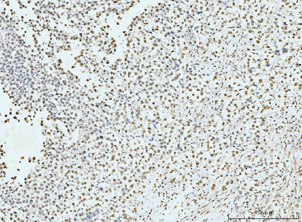
Expected resultNBN-positive staining in alveolar cells type I of lung (HPA tissue IHC: High). HPA tissue profile: Ubiquitous nuclear expression. No signal in the no-primary control.
💡Decision noteStart with heat-mediated EDTA retrieval at pH 8.0 (datasheet: PB9615); the published workflow does not specify retrieval conditions (PMC5029606).
Section 2

What Is the Expected NBN Staining Pattern?

NBN is a nuclear, chromosome-associated protein with no transmembrane segment (UniProt O60934). In paraffin-section IHC, expect predominantly nuclear staining across many cell types; HPA describes a ubiquitous nuclear pattern with supported tissue-IHC reliability (HPA tissue IHC). Alveolar type I cells show high staining, while several other listed cell types show medium staining (HPA tissue IHC). Interpret each cell population against its own HPA observation rather than expecting equal intensity throughout a section.

What am I looking at on my slide?
Predominantly nuclear signal in alveolar type I cells, with weaker or variable nuclear signal elsewhere.This fits the high level reported for alveolar type I cells and the broader ubiquitous nuclear profile (HPA tissue IHC). Compare nuclei within identified cell populations; a uniformly high signal across all cells is not the HPA intensity pattern (HPA tissue IHC).
Discrete nuclear foci, especially in a DNA-damage experiment.NBN can accumulate in nuclear foci at DNA double-strand breaks after genotoxic treatment (UniProt O60934). Foci are a context-dependent finding, so their absence in an untreated tissue section does not by itself contradict the expected nuclear pattern (UniProt O60934).
Predominantly cytoplasmic or extracellular chromogen with little nuclear signal.This conflicts with the nuclear tissue-IHC pattern and UniProt localization (HPA tissue IHC; UniProt O60934). Treat it as suspect staining; inspect the negative control and detection background before assigning it to NBN (general IHC practice).
Strong signal in skeletal-muscle myocytes, soft-tissue fibroblasts, or vaginal squamous cells.These particular cell populations were not detected in the supplied HPA tissue observations (HPA tissue IHC). Check cell identity and controls: cross-reactivity or endogenous detection activity may explain unexpected signal (general IHC practice). Their HPA result does not imply that every other cell in those tissues is negative.
No nuclear signal in an otherwise suitable lung section containing alveolar type I cells.HPA reports high staining in alveolar type I cells (HPA tissue IHC). Confirm that those cells are present and that the run controls worked; absent signal can reflect an assay failure, so a single negative section is insufficient to infer absent NBN (general IHC practice).
💡Expected NBN appearanceCall the stain positive when identified cells show predominantly nuclear chromogen, with strong signal plausible in alveolar type I cells (HPA tissue IHC); broad cytoplasmic color or signal in HPA-undetected cell populations warrants a control check (HPA tissue IHC; general IHC practice).
How each factor affects the staining
Tissue and cell identityHPA reports high staining in lung alveolar type I cells, medium staining in bone-marrow hematopoietic cells, and no detection in skeletal-muscle myocytes (HPA tissue IHC). Score the named cell population, not the tissue as a whole.
Cellular compartmentNBN is nuclear and chromosome-associated, with no transmembrane segment or signal peptide (UniProt O60934). A membrane or secreted pattern has no support in this record; investigate compartment-specific background before interpreting it as target signal (general IHC practice).
Damage stateAfter genotoxic treatment, NBN can form discrete nuclear foci at DNA damage sites (UniProt O60934). Record treatment status when comparing focal patterns; routine untreated IHC need not display conspicuous foci.
Antibody evidenceThree listed HPA antibodies have supported IHC status: HPA001429, CAB003836, and CAB080233 (HPA antibodies). This supports the general tissue pattern, but does not establish the performance of an unlisted antibody or a particular retrieval setting.
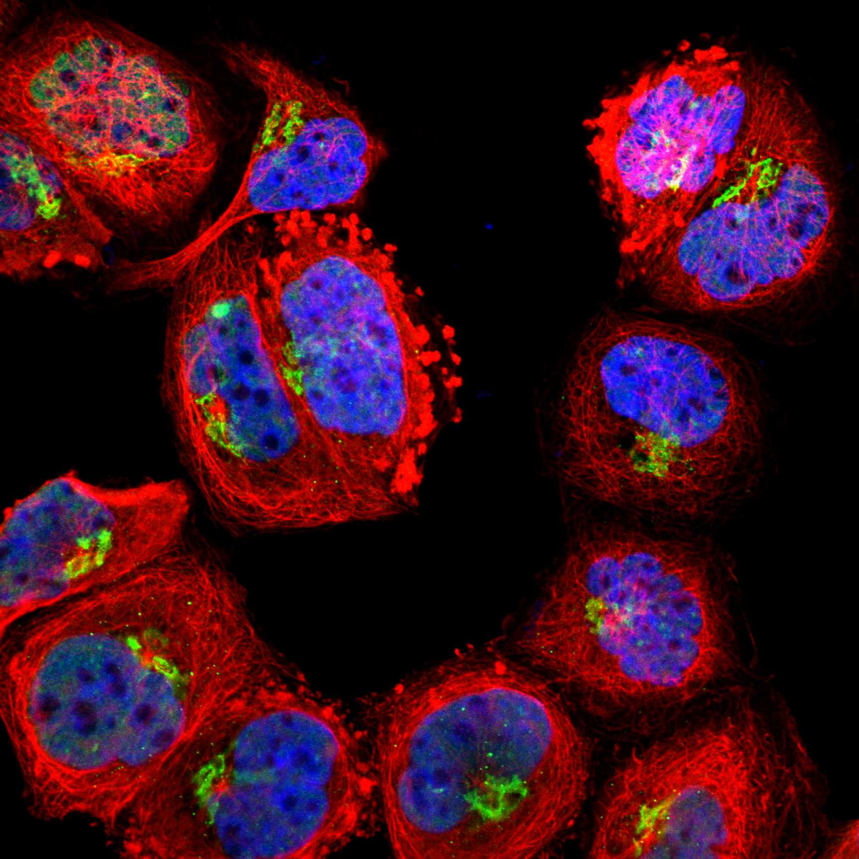
IF/ICC cross-check — Q: What localization should I expect?A: Mainly nucleoplasmic signal; mitotic-chromosome localization is approved, while the reported Golgi localization is uncertain (HPA ICC-IF). This is a localization cross-check, not an IF protocol or a reason to score Golgi signal as definitive.
Why is my staining missing, weak or wrong?
SituationLikely causeNext action
Known-positive alveolar type I cells lack nuclear stain.The run may have failed, or the intended cells may be absent from the evaluated field (general IHC practice).Verify cell identity, a suitable positive control, and the detection run; then optimize the IHC-validated antibody workflow as needed (general IHC practice; HPA tissue IHC: high in alveolar type I cells).
Color is widespread outside nuclei.Diffuse chromogen may reflect background or nonspecific detection (general IHC practice); the expected tissue pattern is nuclear (HPA tissue IHC).Review negative-control staining, blocking, washes, and detection conditions before scoring NBN (general IHC practice).
Unexpectedly strong stain appears in HPA-undetected cells.Cross-reactivity, endogenous detection activity, or mistaken cell identification is possible (general IHC practice; HPA tissue IHC: specified cells not detected).Identify the stained cells, compare a matched negative control, and confirm the pattern with an independently supported antibody if available (general IHC practice; HPA antibodies).
Only a few nuclei contain puncta.Focal accumulation can follow genotoxic treatment (UniProt O60934); sparse puncta alone cannot establish treatment or damage status.Record exposure and compare treated and untreated samples under the same scoring rules (general IHC practice; UniProt O60934).
Nuclear intensity varies between cell populations.The supplied HPA observations span high, medium, low, and undetected levels in named cell types (HPA tissue IHC).Score each identified population separately and compare it with its corresponding HPA observation; do not impose one intensity threshold across the section.
IF/ICC shows apparent Golgi staining alongside nuclear signal.HPA labels the Golgi location uncertain, while nucleoplasm is supported (HPA ICC-IF).Interpret the nuclear pattern first and require an independent control before attributing Golgi signal to NBN (HPA ICC-IF; general IF practice).

Sample controls for NBN IHC & IF

🧪Run lung first and require nuclear staining in alveolar type I cells (HPA: High in lung alveolar cells type I; UniProt O60934: nuclear localization). Use skeletal muscle myocytes as the negative tissue (HPA: Not detected in skeletal muscle myocytes); on the lung slide, assess background in adjacent cells without nuclear staining, without assigning an undocumented cell type as an internal negative (HPA: lung row identifies alveolar cells type I only).
Positive control tissue: Lung (Alveolar cells type I, HPA High)
Negative control tissue: Skeletal muscle (HPA Not detected)
ICC-IF cell lines (HPA subcellular resource): HPA ICC-IF images show NBN in A-431, U-251MG, THP-1, U2OS, with annotated localisation: Nucleoplasm (supported) (HPA subcellular).
Technical controls: Include a no-primary, secondary-only control, a concentration-matched rabbit IgG isotype control with clonality matched where known, and an NBN-knockout specimen as a biological specificity control (PB9615 tissue-IHC caption: rabbit primary; standard IHC practice). For lung HRP/DAB staining, quench endogenous peroxidase and assess pigment background; for IF, check tissue autofluorescence on the no-primary slide (PB9615 tissue-IHC caption: HRP/DAB; standard IHC/IF practice).
⚠️Feasibility: A target-specific fixation window or fixation effect is unreported, and the selected PB9615 paraffin-section caption does not state a fixative (PB9615 tissue-IHC caption). Start IHC-P optimization with heat-mediated EDTA retrieval at pH 8.0; whether retrieval is required in lung is unreported (PB9615 tissue-IHC caption: breast cancer section). An advantage for frozen sections is unreported; IF/ICC may make nuclear foci easier to resolve after DNA damage, while lung autofluorescence needs a background check (UniProt O60934: damage-induced nuclear foci; standard IF practice).

HPA tissue IHC evidence for NBN

Comprehensive Human Protein Atlas IHC scoring per tissue (reliability: Supported — High consistency between antibody staining and RNA expression data.). Rows are taken directly from the HPA tissue chart — click any row's HPA link to view the source.

Positive expression · recommended positive controls

TissueCell typeLevelEvidenceSource
Lung Alveolar cells type I High Protein (IHC) HPA →
Adrenal gland Glandular cells Medium Protein (IHC) HPA →
Appendix Glandular cells Medium Protein (IHC) HPA →
Bone marrow Hematopoietic cells Medium Protein (IHC) HPA →
Breast Adipocytes Medium Protein (IHC) HPA →

Undetected expression · recommended negative controls

TissueCell typeLevelEvidenceSource
Skeletal muscle Myocytes Not detected Protein (IHC) HPA →
Soft tissue Fibroblasts Not detected Protein (IHC) HPA →
Vagina Squamous epithelial cells Not detected Protein (IHC) HPA →
Section 3

Advanced NBN IHC Tips

Troubleshoot NBN staining in paraffin sections by checking nuclear localisation, the validated retrieval condition, detection controls and cell specific scoring.

What retrieval should I try when NBN staining is weak in paraffin sections?
Start with heat mediated retrieval in EDTA pH 8.0 for the catalog antibody (datasheet PB9615). The published product image used that condition in a paraffin section of human breast cancer; its caption does not report the fixative (caption PB9615). If nuclear staining remains weak, vary heating duration in small increments while keeping the buffer and detection steps constant, and check section integrity after each run (standard IHC practice). Compare runs using the same control tissue and score nuclear staining separately from background, because NBN is predominantly nuclear and may form discrete foci after DNA damage (HPA subcellular; UniProt O60934).
Could fixation explain weak or uneven NBN staining?
Target specific fixation sensitivity is unknown from the supplied evidence; the product caption identifies a paraffin section but does not state its fixative (caption PB9615). Record the actual fixative, fixation duration and processing history before comparing sections, because these variables can change antigen accessibility in routine IHC (standard IHC practice). Process a control section alongside the sample, then change retrieval or antibody incubation one variable at a time if nuclear staining is weak (standard IHC practice; HPA tissue IHC). Avoid attributing a fixation effect to NBN phosphorylation or topology without a direct comparison under the relevant fixation conditions (UniProt O60934; standard IHC practice).
Where should convincing NBN staining appear in chromogenic IHC?
Expect staining primarily within nuclei: tissue IHC reports ubiquitous nuclear expression, and subcellular imaging places NBN mainly in the nucleoplasm (HPA tissue IHC; HPA subcellular). Discrete nuclear foci can occur after genotoxic treatment and at DNA double strand breaks, so record treatment history before interpreting a punctate pattern (UniProt O60934). Use a nuclear counterstain to distinguish chromogen inside nuclei from pigment or precipitate lying over them (standard IHC practice). Review any strong cytoplasmic pattern against a matched control and the no primary control; Golgi localisation is listed as uncertain, so it should not override the supported nuclear pattern (HPA subcellular; standard IHC practice).
How should I assess an unexpected NBN staining pattern when the epitope is unspecified?
The supplied record lists one NBN chain, residues 1–754, and zero isoforms; it does not establish the catalog antibody’s epitope (UniProt O60934; caption PB9615). NBN has an FHA domain at residues 24–83 and BRCT domains at 105–181 and 224–315, but those locations do not identify what this antibody binds (UniProt O60934). Phosphorylation is documented at several residues, including 278 and 343; do not infer that it alters staining without epitope and comparative evidence (UniProt O60934). Check the antibody’s documented antigen information, then compare nuclear localisation and control sections before assigning an unexpected pattern to an isoform or modification (standard IHC practice; HPA tissue IHC).
How can I check an NBN pattern by multiplex IF?
For the separate IF/ICC application, pair NBN with an established marker for the cell type being examined and a nuclear counterstain, then assess whether NBN signal lies within those nuclei (HPA subcellular; standard IF practice). Choose fluorophores after inspecting the tissue’s autofluorescence and reserve a clearly separated channel for the weaker signal (standard IF practice). NBN is mainly nucleoplasmic and has no transmembrane segment, so use a validated permeabilisation step that permits antibody access to nuclear epitopes (HPA subcellular; UniProt O60934; standard IF practice). Include single stain and no primary controls when assessing channel bleed through or background; the paraffin IHC caption supplies no IF fixation or dilution condition (standard IF practice; caption PB9615).
What should I change if DAB obscures nuclear NBN staining?
Check a no primary control and examine whether brown signal follows nuclei or instead tracks tissue edges, damaged areas or deposits (standard IHC practice; HPA tissue IHC). The product image used 10% goat serum blocking, 2 μg/ml primary antibody overnight at 4°C, and an HRP secondary followed by DAB (caption PB9615). If background persists, optimise blocking, washing, primary concentration and chromogen development one variable at a time while retaining a control section (standard IHC practice). Include an endogenous peroxidase block and inspect the no primary slide for residual enzyme associated signal; these are general chromogenic IHC safeguards, not demonstrated NBN specific requirements (standard IHC practice).
How should I quantify NBN staining across tissue sections? ⚠ ANSWER MARKED FOR VERIFICATION
Define the cell population and analysis region before scoring, because HPA reports different staining levels across cell types despite a broadly nuclear profile (HPA tissue IHC). For chromogenic sections, record the percentage of nuclei in preset intensity categories and calculate an H score, or report positive nuclei per mm² when cell density differs (standard IHC practice). Normalise positive counts to the number of evaluable nuclei in the same cell population, and keep exposure, threshold and exclusion rules consistent across sections (standard IHC practice). Report diffuse nuclear staining separately from discrete foci if DNA damage is under study, since NBN can accumulate at damage sites (UniProt O60934).
How can I distinguish true NBN staining from artefact?
Give the most weight to signal inside intact nuclei in an identified cell population, consistent with supported nucleoplasmic and tissue nuclear localisation (HPA subcellular; HPA tissue IHC). A control containing a documented positive cell population, such as lung alveolar type I cells, can help assess staining performance; skeletal muscle myocytes are listed as not detected (HPA tissue IHC). Treat staining confined to cut edges, necrotic regions or the no primary control as suspect and check for precipitate or endogenous peroxidase activity (standard IHC practice). Nuclear foci can reflect DNA damage recruitment, but a brown punctum alone cannot establish a double strand break without appropriate biological and staining controls (UniProt O60934; standard IHC practice).
Boster reagents

Best NBN / Nibrin IHC Antibodies

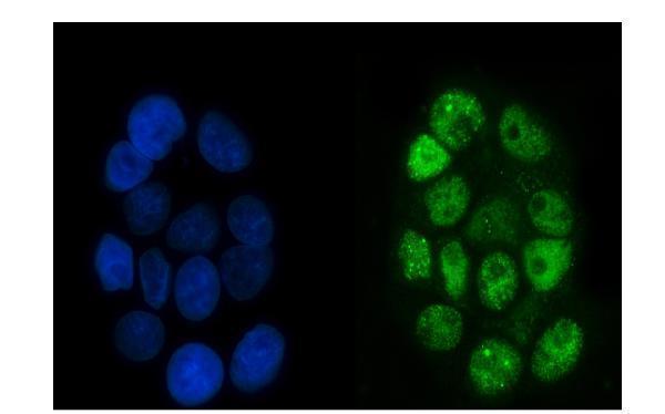
Anti-NBN antibodies have IHC images from human cancer, mouse intestine, and rat kidney paraffin sections, plus IF/ICC images from A431 and U20S cells (catalog image captions).

Real IHC data IHC analysis of p95 NBS1 using anti-p95 NBS1 antibody (PB9615). p95 NBS1 was detected in a paraffin-embedded section of human breast cancer tissue. Heat mediated antigen retrieval was performed in EDTA buffer (pH 8.0, epitope retrieval solution). The tissue section was blocked with 10% goat serum. The tissue section was then incubated with 2 μg/ml rabbit anti-p95 NBS1 Antibody (PB9615) overnight at 4°C. Peroxidase Conjugated Goat Anti-rabbit IgG was used as secondary antibody and incubated for 30 minutes at 37°C. The tissue section was developed using HRP Conjugated Rabbit IgG Super Vision Assay Kit (Catalog # SV0002) with DAB as the chromogen.
Anti-p95 NBS1/NBN Antibody ®
Cat # PB9615
Real IHC data IHC analysis of p95 NBS1 using anti-p95 NBS1 antibody (M00732-1). p95 NBS1 was detected in a paraffin-embedded section of human testis cancer tissue. Heat mediated antigen retrieval was performed in EDTA buffer (pH 8.0, epitope retrieval solution). The tissue section was blocked with 10% goat serum. The tissue section was then incubated with 2 μg/ml mouse anti-p95 NBS1 Antibody (M00732-1) overnight at 4°C. Biotinylated goat anti-mouse IgG was used as secondary antibody and incubated for 30 minutes at 37°C. The tissue section was developed using Strepavidin-Biotin-Complex (SABC) (Catalog # SA1021) with DAB as the chromogen.
Anti-p95 NBS1 Antibody ® (monoclonal, 5D7A1)
Cat # M00732-1
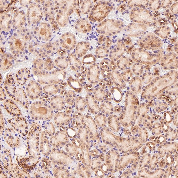
Real IHC data Immunohistochemical analysis of paraffin-embedded Rat kidney, using the Antibody at 1:100 dilution.
Anti-p95/NBS1 NBN Rabbit Monoclonal Antibody
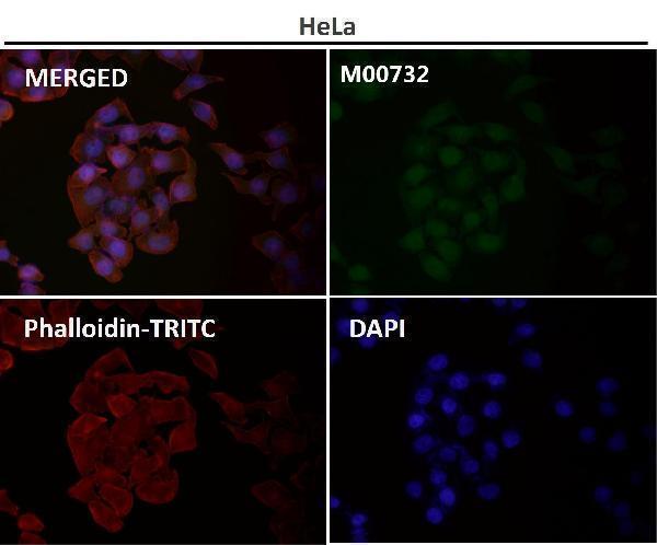
Cat # M00732

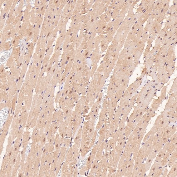
PB9615 has IHC images from human breast and intestinal cancer and mouse intestine, plus an IF/ICC image from U20S cells (PB9615 image captions). M00732-1 has human testis cancer IHC and A431 IF/ICC images; M00732 has rat kidney and heart and human lung cancer and glioblastoma IHC images, plus an IF image with no sample identified (respective image captions).

Which to pick: For tissue IHC, PB9615 offers illustrated human and mouse paraffin sections, while M00732-1 offers a mouse monoclonal option illustrated in human testis cancer paraffin sections (respective IHC image captions; M00732-1 catalog: clone 5D7A1). For IF/ICC, choose between PB9615 and M00732-1 using their illustrated U20S and A431 cell examples, respectively (respective IF image captions). For cross-species IHC planning, all three list human, mouse, and rat reactivity, but the illustrated species differ: PB9615 shows human and mouse, M00732 shows rat and human, and M00732-1 shows human; the paraffin captions do not report the fixative (catalog reactivity; respective IHC image captions).

Each figure is that product's own IHC / IF validation image from its datasheet.

References

  1. UniProt Consortium. UniProt entry O60934 (NBN_HUMAN, Nibrin).
  2. Human Protein Atlas. NBN tissue IHC expression (reliability: Supported).
  3. Human Protein Atlas. NBN subcellular location (ICC-IF): Mainly localized to the nucleoplasm. In addition localized to the mitotic chromosome and the Golgi apparatus..
  4. Human Protein Atlas. NBN antibody validation summary (3 antibodies).
  5. Successful pregnancy after complete resection of leiomyomatosis peritonealis disseminate without recurrence:a case report with next-generation sequencing analysis and literature review. World journal of surgical oncology 2020 — PMC7196235.
  6. Epidermal Nbn deletion causes premature hair loss and a phenotype resembling psoriasiform dermatitis. Oncotarget 2016 — PMC5029606.
  7. Copy Number Amplification of DNA Damage Repair Pathways Potentiates Therapeutic Resistance in Cancer. Theranostics 2020 — PMC7086350.
  8. A novel DNA damage repair-related signature for predicting prognositc and treatment response in non-small lung cancer. Frontiers in oncology 2022 — PMC9673481.
  9. PubMed PMID:9590180 — UniProt-cited evidence.
  10. PubMed PMID:9590181 — UniProt-cited evidence.
  11. PubMed PMID:9620777 — UniProt-cited evidence.