NECTIN1 / Nectin-1 · IHC design guide

Design Immunohistochemistry for NECTIN1

Plan NECTIN1 staining in paraffin sections with the catalog antibody at 0.5–1 μg/ml (datasheet A04988-1). Use colon glandular cells as a high staining reference and assess membranous and cytoplasmic signal in light of the reported secreted variant (HPA tissue IHC).

Evidence assembled Oct 2026 · For research use; verify linked source records and product datasheet before use
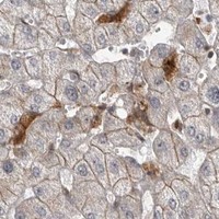
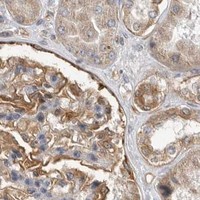
Immunohistochemistry protocol sheet for NECTIN1 (IHC for NECTIN1): expected localisation Membranous and cytoplasmic tissue staining (HPA tissue IHC), antibody A04988-1, validated IHC image, and IHC protocol steps
Printable NECTIN1 IHC protocol sheet — expected localisation Membranous and cytoplasmic tissue staining (HPA tissue IHC), antibody A04988-1, controls and protocol steps. Open the full NECTIN1 IHC guide →

NECTIN1 Immunohistochemistry Experimental Design Guide

Expected localisation, validated protocols, controls and antibodies — the at-a-glance facts below, then the full design guide.

Must know before staining
Expected localisation Membranous and cytoplasmic tissue staining (HPA tissue IHC)
Staining pattern Variable cytoplasmic and membranous staining across tissues (HPA tissue IHC)
Antigen retrieval EDTA pH 8.0 HIER, heat-mediated (datasheet A04988-1)
Positive control ⓘ Appendix+4 more · see all
Negative control ⓘ Adipose tissue+4 more · see all
Important caveats
Reasons your staining may differ from the expected pattern.
Fixation Keep fixation conditions consistent across sections. (standard IHC practice; not target-specific)
Caveat A secreted variant may complicate tissue staining (HPA tissue IHC)
Regulation No expression regulator annotated (UniProt)
Isoform / epitope 3 isoforms; verify extracellular vs cytoplasmic epitope (UniProt)
Section 1

Recommended NECTIN1 IHC & IF Protocols

Compare the catalog antibody’s IHC-P protocol (datasheet A04988-1) with published NECTIN1 protocols for human melanoma sections (PMC9729115) and placental tissue microarrays (PMC10661505).

Recommended immunohistochemistry (IHC-P) protocol parameters
SampleParaffin-embedded human rectal cancer tissue; fixative not specified (datasheet A04988-1)
FixationImage fixative and duration unreported (datasheet A04988-1); verify before use.
Sectioning4–5 µm sections on charged slides (standard)
DeparaffinisationXylene, graded ethanol series to water (standard)
Antigen retrievalHeat retrieval: EDTA pH 8.0 (datasheet A04988-1); 20 min, 95–100 °C (standard)
Peroxidase block3% H2O2, 10 min, room temperature (standard)
Blocking10% goat serum (datasheet A04988-1)
Primary antibodyRabbit anti-NECTIN1, 0.5-1μg/ml (datasheet A04988-1)
Primary incubationOvernight at 4 °C (datasheet A04988-1)
DetectionStreptavidin-biotin complex (SABC), DAB chromogen (datasheet A04988-1)
CounterstainHematoxylin, blue, dehydrate and mount (standard)
Expected resultNECTIN1-positive staining in glandular cells of appendix (HPA tissue IHC: High). HPA tissue profile: Cytoplasmic and membranous expression at variable levels in several different tissue types. No signal in the no-primary control.
💡Decision noteStart with heat-mediated EDTA retrieval at pH 8.0 for the catalog antibody (datasheet A04988-1). The melanoma study used a different retrieval solution (PMC9729115).
Section 2

What Is the Expected NECTIN1 Staining Pattern?

NECTIN1 is a cell-membrane protein found at adherens junctions, with an extracellular region spanning residues 31–355 and a cytoplasmic tail spanning 377–517 (UniProt Q15223 topology; UniProt Q15223 subcellular). In paraffin-section IHC, expect membranous and sometimes cytoplasmic staining in glandular and other HPA-positive cells (HPA tissue IHC). Interpret intensity cautiously: HPA rates the tissue staining Approved but reports low agreement with RNA expression (HPA tissue IHC reliability).

What am I looking at on my slide?
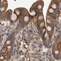
Distinct cell borders, with possible cytoplasmic staining, in colon or duodenal glandular cells (HPA tissue IHC).This fits the HPA tissue profile; both glandular populations are reported High (HPA tissue IHC). Border staining also fits NECTIN1 membrane and adherens-junction localisation (UniProt Q15223 subcellular).
Signal appears predominantly nuclear, with little corresponding cell-border or cytoplasmic staining.A predominantly nuclear pattern is unsupported by the listed UniProt locations and HPA tissue profile (UniProt Q15223 subcellular; HPA tissue IHC). Treat it as suspect and compare controls before scoring.
Strong signal appears in adipocytes or bone-marrow hematopoietic cells (HPA tissue IHC: Not detected).Those cell populations are useful negative comparators (HPA tissue IHC). Unexpected staining warrants checks for antibody cross-reactivity or endogenous chromogen-generating activity (general IHC practice).
Color is widespread across cells and surrounding tissue, obscuring cell borders.This cannot establish the reported cell-specific pattern (HPA tissue IHC). Review background controls, blocking, antibody concentration and detection conditions before interpreting it (general IHC practice).
No signal is visible in appendix glandular cells or liver cholangiocytes (HPA tissue IHC: High).A failed positive control is possible; check detection reagents, retrieval conditions and the antibody's IHC validation (general IHC practice). HPA's Approved rating does not guarantee every section will stain (HPA tissue IHC reliability).
💡Expected NECTIN1 appearanceCall positive when defined membranous, sometimes cytoplasmic, staining marks HPA-positive glandular cells at the expected High level; isolated nuclear or indiscriminate tissue-wide color is suspect (HPA tissue IHC; UniProt Q15223 subcellular).
How each factor affects the staining
Cell and tissue choice (HPA tissue IHC).Appendix, colon, duodenum, endometrium and gallbladder glandular cells are High; salivary glandular cells are Not detected (HPA tissue IHC). Compare named cell populations rather than whole-section color.
Membrane topology and unknown antibody epitope (UniProt Q15223 topology).NECTIN1 has extracellular residues 31–355 and cytoplasmic residues 377–517 (UniProt Q15223 topology). Neither source specifies the catalog antibody epitope; topology alone cannot predict its retrieval requirements.
Secreted protein and variant complexity (UniProt Q15223 subcellular; HPA tissue IHC reliability).UniProt lists a secreted location, and HPA notes at least one secreted variant (UniProt Q15223 subcellular; HPA tissue IHC reliability). Cytoplasmic or extracellular color needs cell-context checks before it is assigned to junctional NECTIN1.
Antibody validation strength (HPA antibodies; HPA tissue IHC reliability).HPA026846 and CAB016135 are each IHC Approved, while HPA reports low staining–RNA consistency (HPA antibodies; HPA tissue IHC reliability). Approved supports use of the reported pattern, with that limitation.
IF/ICC Q&A: is a cell-line fluorescence pattern established? (HPA subcellular).HPA summarizes NECTIN1 as Membrane, Secreted but lists no ICC-IF image cell lines or main location (HPA subcellular). The supplied HPA antibody records give no ICC status (HPA antibodies).
Why is my staining missing, weak or wrong?
SituationLikely causeNext action
A High reference cell population stains weakly or not at all (HPA tissue IHC).The staining run may have failed; HPA's reported High level is an observation, not a guarantee for this section (HPA tissue IHC; general IHC practice).Confirm section integrity and positive-control staining, then review the IHC-validated antibody's documented retrieval, dilution and detection steps (general IHC practice).
Adipocytes stain strongly although HPA reports Not detected (HPA tissue IHC).Off-target antibody binding or endogenous detection activity may contribute (general IHC practice).Compare a no-primary control and a tissue positive control; address endogenous activity for the chosen chromogen system (general IHC practice).
Diffuse chromogen masks glandular cell borders (HPA tissue IHC).Excess antibody, inadequate blocking or detection background can obscure localisation (general IHC practice).Compare background controls and optimize antibody dilution, blocking and detection time using the antibody's IHC instructions (general IHC practice).
Signal is nuclear instead of membranous or cytoplasmic (UniProt Q15223 subcellular; HPA tissue IHC).The observed compartment does not match the supplied localisation evidence; its cause is unresolved (UniProt Q15223 subcellular; HPA tissue IHC).Check the no-primary control and examine a second HPA-positive tissue before assigning that color to NECTIN1 (general IHC practice; HPA tissue IHC).
An HPA-negative comparator and a High reference both stain (HPA tissue IHC).Broad background or cross-reactivity may erase the expected cell-population contrast (general IHC practice; HPA tissue IHC).Reassess blocking and antibody concentration, then score named cell populations against controls rather than total tissue color (general IHC practice; HPA tissue IHC).
Cytoplasmic color appears without a clear cell-border signal (HPA tissue IHC).HPA reports both compartments, and a secreted variant complicates a membrane-only interpretation (HPA tissue IHC reliability; UniProt Q15223 subcellular).Record the compartment separately, compare a reported High population with a Not detected comparator, and avoid calling junctional staining without visible borders (HPA tissue IHC; general IHC practice).

Sample controls for NECTIN1 IHC & IF

🧪Run colon first: glandular cells should show NECTIN1 staining (HPA: High in colon glandular cells). Use salivary gland as the tissue negative (HPA: Not detected in salivary gland glandular cells); on the colon slide, nonglandular areas should lack the distinct cell-border signal expected for a junctional membrane protein, but HPA does not establish those cells as target-negative (UniProt Q15223: cell membrane and adherens junction localization).
Positive control tissue: Appendix (Glandular cells, HPA High)
Negative control tissue: Adipose tissue (HPA Not detected)
ICC-IF cell lines (HPA subcellular resource): HPA carries no ICC-IF cell line for NECTIN1; derive a cell-line control from the positive tissue's cell type (Glandular cells) and confirm it by RNA or western blot first.
Technical controls: Include a no-primary, secondary-only control and a matched rabbit IgG isotype control (caption: rabbit primary antibody and goat anti-rabbit secondary). A matched NECTIN1 knockout specimen can test specificity; with the caption’s SABC–DAB detection, block endogenous peroxidase and check for endogenous biotin background (caption: SABC–DAB).
⚠️Feasibility: No target-specific fixation window or fixation effect is reported, and the selected A04988-1 caption does not state a fixative (A04988-1 caption: fixative not stated). The paraffin-section example uses heat retrieval in EDTA at pH 8.0, which provides a starting condition but does not establish retrieval dependence (A04988-1 caption: heat retrieval in EDTA, pH 8.0). Frozen sections or IF cannot be judged easier from these data; the SABC–DAB method makes endogenous biotin background a practical concern in the chosen tissue (HPA: High in colon glandular cells; A04988-1 caption: SABC–DAB).

HPA tissue IHC evidence for NECTIN1

Comprehensive Human Protein Atlas IHC scoring per tissue (reliability: Approved — Low consistency between antibody staining and RNA expression data. At least one protein variant secreted, tissue location of RNA and protein might differ and correlation is complex.). Rows are taken directly from the HPA tissue chart — click any row's HPA link to view the source.

Positive expression · recommended positive controls

TissueCell typeLevelEvidenceSource
Appendix Glandular cells High Protein (IHC) HPA →
Colon Glandular cells High Protein (IHC) HPA →
Duodenum Glandular cells High Protein (IHC) HPA →
Endometrium Glandular cells High Protein (IHC) HPA →
Gallbladder Glandular cells High Protein (IHC) HPA →

Undetected expression · recommended negative controls

TissueCell typeLevelEvidenceSource
Adipose tissue Adipocytes Not detected Protein (IHC) HPA →
Bone marrow Hematopoietic cells Not detected Protein (IHC) HPA →
Salivary gland Glandular cells Not detected Protein (IHC) HPA →
Skeletal muscle Myocytes Not detected Protein (IHC) HPA →
Spleen Cells in red pulp Not detected Protein (IHC) HPA →
Section 3

Advanced NECTIN1 IHC Tips

Troubleshoot NECTIN1 staining in paraffin sections by checking retrieval, compartment, cell type and controls before interpreting chromogenic signal.

What retrieval condition should I start with for NECTIN1 in paraffin sections?
Start with heat-mediated retrieval in EDTA at pH 8.0 (datasheet A04988-1). This condition accompanies NECTIN1 staining in a paraffin section of human rectal cancer using 1 μg/mL catalog antibody (caption A04988-1). If staining is weak, adjust heating duration on matched sections while holding antibody concentration and detection conditions constant (standard IHC practice). Assess membrane-associated staining separately from diffuse signal, since NECTIN1 occupies cell membranes and adherens junctions (UniProt Q15223 localisation). Compare a high-staining reference such as colon glandular cells with a low-background control on the same run (HPA: High in colon glandular cells; standard IHC practice).
How should I troubleshoot fixation-related loss of NECTIN1 staining?
The selected paraffin-section caption does not state a fixative, so NECTIN1-specific sensitivity to fixation is unknown (caption A04988-1). Record each specimen’s fixative, fixation duration and processing history before comparing staining intensities (standard IHC practice). If a batch stains weakly, compare similarly processed reference sections and verify retrieval, antibody incubation and detection before attributing the result to fixation (standard IHC practice). The documented incubation is 1 μg/mL catalog antibody overnight at 4°C, following EDTA retrieval at pH 8.0 (caption A04988-1). Avoid treating differences between specimens as biological until their processing and staining conditions are comparable (standard IHC practice).
Which staining compartments support a NECTIN1 assignment?
Prioritise staining at cell borders and contacts because NECTIN1 is assigned to the cell membrane and adherens junctions (UniProt Q15223 localisation). Its extracellular region spans residues 31–355, its transmembrane segment 356–376, and its cytoplasmic region 377–517 (UniProt Q15223 topology). Cytoplasmic staining can also occur in tissue IHC, so assess it alongside morphology and controls instead of rejecting it automatically (HPA tissue IHC profile; standard IHC practice). In colon, examine glandular cells as a reference compartment with reported high staining (HPA: High in colon glandular cells). Record border and cytoplasmic patterns separately so a change in distribution is not mistaken for a change in total staining (standard IHC practice).
Could isoforms or epitope position explain discordant NECTIN1 staining?
NECTIN1 has three listed isoforms—Delta, Alpha and Gamma—so establish which sequence region the antibody recognises before comparing results (UniProt Q15223 isoforms; standard IHC practice). The record places Ig-like domains extracellularly at residues 31–334 and the cytoplasmic region at 377–517 (UniProt Q15223 domains and topology). It also lists eight extracellular glycosylation sites and several cytoplasmic phosphorylation sites, making epitope location relevant to interpretation (UniProt Q15223 modifications; standard IHC practice). An antibody’s epitope is not specified in the supplied caption, so do not assign its staining to a particular isoform or modification (caption A04988-1). Compare staining patterns only after confirming epitope coverage and consistent section processing (standard IHC practice).
How can I assess NECTIN1 by IF alongside a cell-type marker?
Treat IF as a separate validation task: the selected catalog evidence documents chromogenic paraffin-section IHC, with no IF result supplied (caption A04988-1). Pair NECTIN1 with a marker identifying the expected glandular cells, such as the reported high-staining colon population, and inspect whether signals occupy the same cells (HPA: High in colon glandular cells; standard IF practice). Choose spectrally separated fluorophores and a detection channel with low tissue autofluorescence, using single-stain controls to check bleed-through (standard IF practice). If the antibody recognises residues 377–517, test permeabilisation; an extracellular epitope at 31–355 may be assessed without it (UniProt Q15223 topology; standard IF practice). Confirm any apparent border signal against morphology and appropriate background controls (standard IF practice).
What controls help distinguish NECTIN1 signal from chromogenic background?
Run a no-primary control and inspect tissue edges, folds and damaged regions before calling diffuse DAB signal positive (standard IHC practice). Include a peroxidase block in the chromogenic workflow because endogenous enzyme activity can produce signal unrelated to antibody binding (standard IHC practice). The selected example used 10% goat serum, a biotinylated secondary antibody, streptavidin-biotin detection and DAB (caption A04988-1). With that detection scheme, check for background from secondary reagents and endogenous biotin using suitable reagent controls (caption A04988-1; standard IHC practice). Judge residual signal against expected cell borders and the reported variable cytoplasmic and membranous pattern (UniProt Q15223 localisation; HPA tissue IHC profile).
How should I score heterogeneous NECTIN1 staining across sections? ⚠ ANSWER MARKED FOR VERIFICATION
Define the cell population and compartment before scoring, because HPA reports variable cytoplasmic and membranous tissue staining (HPA tissue IHC profile). For glandular regions, record the percentage of positive cells and an H-score from intensity categories; report membrane and cytoplasmic scores separately (HPA tissue IHC profile; standard IHC practice). If counting positive cells, express density per mm² of viable tissue and exclude folds, necrosis and blank space (standard IHC practice). Normalise comparisons to the same eligible cell population or viable area, with matched section processing and staining runs (standard IHC practice). State the positivity threshold and assess a high-staining reference such as colon glandular cells across runs (HPA: High in colon glandular cells; standard IHC practice).
When should I question an apparent NECTIN1-positive result?
Question staining confined to an implausible compartment or cell population, since NECTIN1 is associated with membranes and junctions, while tissue IHC can also show cytoplasmic signal (UniProt Q15223 localisation; HPA tissue IHC profile). Compare the candidate result with glandular colon cells reported as high and adipocytes reported as not detected (HPA: High in colon glandular cells; HPA: Not detected in adipocytes). Disregard edge effects, necrotic regions and signal present in no-primary or enzyme controls when assigning positive cells (standard IHC practice). The HPA tissue assay is Approved but has low staining-to-RNA consistency, partly because at least one protein variant is secreted (HPA reliability description). Interpret unexpected distribution with matched controls and morphology rather than RNA abundance alone (HPA reliability description; standard IHC practice).
Boster reagents

Best NECTIN1 / Nectin-1 IHC Antibodies

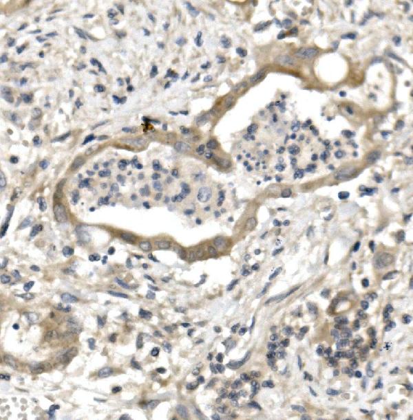
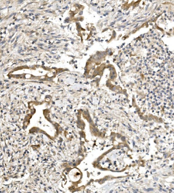
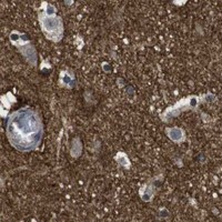
One anti-NECTIN1 antibody has IHC images from paraffin-embedded human rectal cancer and rat brain sections (A04988-1 IHC captions); no IF/ICC validation is reported (catalog applications).

Real IHC data IHC analysis of PVRL1/NECTIN1 using anti-PVRL1/NECTIN1 antibody (A04988-1). PVRL1/NECTIN1 was detected in paraffin-embedded section of human rectal cancer tissue. Heat mediated antigen retrieval was performed in EDTA buffer (pH8.0, epitope retrieval solution). The tissue section was blocked with 10% goat serum. The tissue section was then incubated with 1μg/ml rabbit anti-PVRL1/NECTIN1 Antibody (A04988-1) overnight at 4°C. Biotinylated goat anti-rabbit IgG was used as secondary antibody and incubated for 30 minutes at 37°C. The tissue section was developed using Strepavidin-Biotin-Complex (SABC) (Catalog # SA1022) with DAB as the chromogen.
Anti-PVRL1/NECTIN1 Antibody ®
Cat # A04988-1

A04988-1 is shown in IHC on paraffin-embedded human rectal cancer and rat brain sections (A04988-1 IHC captions). The catalog lists IHC and human, mouse, and rat reactivity, but provides no IF/ICC image or application listing (A04988-1 catalog).

Which to pick: Choose A04988-1 for tissue IHC: its own captions document paraffin-embedded human and rat sections; the fixative is unreported (A04988-1 IHC captions). No listed SKU is validated for IF/ICC (catalog applications). For cross-species IHC, A04988-1 has human and rat image evidence; mouse reactivity is listed, but mouse IHC evidence is unreported (A04988-1 IHC captions; catalog reactivity).

Each figure is that product's own IHC / IF validation image from its datasheet.

References

  1. UniProt Consortium. UniProt entry Q15223 (NECT1_HUMAN, Nectin-1).
  2. Human Protein Atlas. NECTIN1 tissue IHC expression (reliability: Approved).
  3. Human Protein Atlas. NECTIN1 subcellular location (ICC-IF): Membrane, Secreted.
  4. Human Protein Atlas. NECTIN1 antibody validation summary (2 antibodies).
  5. FAM83H and Nectin1 expression are related with survival and relapse of bladder urothelial carcinoma patients. BMC urology 2021 — PMC8501681.
  6. Loss of NECTIN1 triggers melanoma dissemination upon local IGF1 depletion. Nature genetics 2022 — PMC9729115.
  7. Secreted proteins in plasma and placenta as novel non-invasive biomarkers for intrahepatic cholestasis of pregnancy: A case-control study. Heliyon 2023 — PMC10661505.
  8. Disrupted presynaptic nectin1-based neuronal adhesion in the entorhinal-hippocampal circuit contributes to early-life stress-induced memory deficits. Translational psychiatry 2022 — PMC8980071.
  9. PubMed PMID:7721102 — UniProt-cited evidence.
  10. PubMed PMID:9616127 — UniProt-cited evidence.
  11. PubMed PMID:11356977 — UniProt-cited evidence.