This website uses cookies to ensure you get the best experience on our website.


- Table of Contents
Plan paraffin section NINJ1 IHC using the catalog antibody's 5 μg/mL starting concentration (datasheet). Liver hepatocytes show medium staining, but tissue IHC reliability is uncertain (HPA tissue IHC).

Expected localisation, validated protocols, controls and antibodies — the at-a-glance facts below, then the full design guide.
| Expected localisation | Cell membrane expected; tissue compartment unverified (UniProt) | |
| Staining pattern | Endothelial cells and hepatocytes; compartment unspecified (HPA tissue IHC) | |
| Antigen retrieval | Citrate pH 6.0 HIER, 95–98 °C, 20 min (rule: cytoplasmic / membrane antigen) | |
| Positive control | Liver | |
| Negative control | Adipose tissue+4 more · see all |
| Fixation | Keep fixation consistent across sections. (standard IHC practice; not target-specific) | |
| Caveat | Tissue staining reliability is uncertain (HPA tissue IHC) | |
| Regulation | Expression regulation is not established (UniProt) | |
| Isoform / epitope | No isoforms; the secreted 1–56 chain lacks the cytoplasmic region (UniProt) |
The catalog antibody’s IHC-P protocol is accompanied by one published NINJ1 IHC protocol for neonatal intestinal tissue (PMC13237964).
| Sample | Tissue sections; selected-image fixative not specified (standard IHC workflow) |
| Fixation | Image fixative and duration unreported (datasheet A08377); verify before use. |
| Sectioning | 4–5 µm sections on charged slides (standard) |
| Deparaffinisation | Xylene, graded ethanol series to water (standard) |
| Antigen retrieval | Heat-induced epitope retrieval in citrate buffer, pH 6.0, 20 min at 95–98 °C (standard rule: cytoplasmic / membrane antigen) |
| Peroxidase block | 3% H2O2, 10 min, room temperature (standard) |
| Blocking | 10% normal serum of the secondary host, 30 min, room temperature (standard) |
| Primary antibody | Rabbit anti-NINJ1, 5 μg/mL (datasheet A08377) |
| Primary incubation | Overnight at 4 °C (standard) |
| Detection | HRP-polymer secondary, DAB chromogen 5–10 min (standard) |
| Counterstain | Hematoxylin, blue, dehydrate and mount (standard) |
| Expected result | NINJ1-positive staining in hepatocytes of liver (HPA tissue IHC: Medium). HPA tissue profile: Expressed in endothelial cells and liver. No signal in the no-primary control. |
NINJ1 is a membrane protein with two transmembrane segments and extracellular regions; a secreted form is also annotated (UniProt Q92982 topology and processing). In paraffin-section IHC, look for staining in liver hepatocytes, reported at medium intensity, and assess endothelial cells identified in the tissue profile (HPA tissue IHC). Treat this as a provisional pattern: HPA rates tissue staining and the listed antibody’s IHC validation as Uncertain (HPA tissue IHC; HPA antibody HPA045063).
| Hepatocytes show a discernible cell-edge signal of moderate strength. | This fits the reported medium hepatocyte staining and the predicted membrane compartment (HPA tissue IHC: liver hepatocytes Medium; UniProt Q92982 topology). HPA does not assign a subcellular pattern to those hepatocytes, so judge the edge pattern against controls rather than treating it as independently validated. |
| Signal outlines endothelial cells in a vessel-associated pattern. | Endothelial expression is part of HPA’s tissue profile, but the supplied profile gives no endothelial intensity or antibody-specific confirmation (HPA tissue IHC: expressed in endothelial cells and liver; reliability Uncertain). Confirm the cell identity from section morphology before scoring it as NINJ1. |
| Strong staining is confined to nuclei, with no plausible cell-edge signal. | An exclusively nuclear pattern conflicts with the annotated membrane and secreted locations (UniProt Q92982 subcellular location). Consider it an artefact or off-target signal and compare it with the no-primary control; UniProt topology alone cannot validate the antibody’s staining pattern. |
| Prominent signal appears in cells reported as unstained by HPA, or blankets unrelated cell types. | For example, HPA reports adipocytes and bone-marrow hematopoietic cells as Not detected by tissue IHC (HPA tissue IHC). Check cross-reactivity and endogenous detection activity, while allowing for HPA’s Uncertain reliability and UniProt’s broad tissue-expression annotation (HPA tissue IHC; UniProt Q92982 tissue specificity). |
| The slide is diffusely colored, or the liver control has no discernible hepatocyte signal. | Diffuse color reduces confidence that staining marks a cell compartment; absent liver staining misses the supplied positive reference (HPA tissue IHC: hepatocytes Medium). Use control sections to separate background, detection failure, and a possible discrepancy with HPA’s uncertain tissue result (HPA tissue IHC: reliability Uncertain). |
| Antibody evidence | The listed rabbit polyclonal antibody HPA045063 has an Uncertain IHC assessment, and the tissue profile is also Uncertain (HPA antibody HPA045063; HPA tissue IHC). Use the pattern as a working expectation and interpret isolated strong staining cautiously. |
| Membrane topology and epitope position | NINJ1 has extracellular residues 1–78 and 139–152, separated by two transmembrane segments and a short cytoplasmic region (UniProt Q92982 topology). The supplied record does not locate the antibody epitope, so it cannot predict which region the IHC antibody detects. |
| Processing and extracellular signal | UniProt annotates a secreted NINJ1 chain spanning residues 1–56 alongside the full-length 1–152 chain (UniProt Q92982 processing). Extracellular staining is therefore biologically possible, but these annotations cannot establish that a particular extracellular deposit on a section is specific. |
| Tissue reference limits | HPA reports medium staining in liver hepatocytes and Not detected staining in several listed cell populations (HPA tissue IHC). Its Uncertain reliability and UniProt’s broad tissue-specificity statement limit how firmly any single negative population can exclude NINJ1 (HPA tissue IHC; UniProt Q92982 tissue specificity). |
| IF/ICC expectation? | A membrane pattern is plausible from UniProt topology and HPA’s subcellular summary (UniProt Q92982 topology; HPA subcellular: Membrane). The supplied HPA entry has no ICC-IF image cell lines and no ICC validation for HPA045063; it cannot provide an IF/ICC reference pattern or protocol (HPA subcellular; HPA antibody HPA045063). |
| Situation | Likely cause | Next action |
|---|---|---|
| Liver control shows no hepatocyte signal. | The run may lack detectable staining, or this specimen may differ from HPA’s medium hepatocyte result (HPA tissue IHC: hepatocytes Medium; reliability Uncertain). | Check that tissue and detection controls worked, then optimize general IHC conditions such as retrieval and antibody concentration. No NINJ1-specific retrieval condition or fixation sensitivity is supplied. |
| Color covers the section without clear cell boundaries. | Diffuse staining cannot be assigned confidently to the annotated membrane compartment (UniProt Q92982 subcellular location); general IHC background is another possibility. | Compare a no-primary control, inspect blocking and wash performance, and score only signal distinguishable from background. Do not interpret a uniform haze as the HPA hepatocyte pattern (HPA tissue IHC). |
| Signal appears mainly inside nuclei. | Nuclear confinement disagrees with UniProt’s membrane and secreted annotations (UniProt Q92982 subcellular location). | Review morphology and the no-primary control, then reassess antibody specificity before reporting nuclear NINJ1. The supplied HPA IHC assessment is Uncertain (HPA antibody HPA045063). |
| Adipocytes or bone-marrow hematopoietic cells stain strongly. | Those populations are reported as Not detected in the supplied HPA tissue IHC entries; cross-reactivity or endogenous detection activity merits investigation (HPA tissue IHC). | Inspect no-primary and detection controls and compare staining with cell morphology. Treat the discrepancy as unresolved until checked, because HPA calls its tissue IHC evidence Uncertain (HPA tissue IHC). |
| Staining seems extracellular rather than confined to intact cell edges. | A secreted residues 1–56 form is annotated, but the record does not show that extracellular section staining represents that form (UniProt Q92982 processing). | Record the location separately from membrane staining, check background controls, and avoid calling it specific secreted NINJ1 without further validation. |
| An IF/ICC image is being used to judge the IHC section. | HPA summarizes NINJ1 as membrane-associated but supplies no ICC-IF image cell lines or ICC validation for the listed antibody (HPA subcellular; HPA antibody HPA045063). | Judge the paraffin-section result against the IHC tissue observations and controls (HPA tissue IHC). Consult the separate IF/ICC guide for that application; the supplied record provides no IF/ICC protocol. |
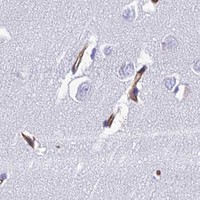


Comprehensive Human Protein Atlas IHC scoring per tissue (reliability: Uncertain — Antibody staining in cells/structures not annotated, view images.). Rows are taken directly from the HPA tissue chart — click any row's HPA link to view the source.
| Tissue | Cell type | Level | Evidence | Source |
|---|---|---|---|---|
| Liver | Hepatocytes | Medium | Protein (IHC) | HPA → |
| Tissue | Cell type | Level | Evidence | Source |
|---|---|---|---|---|
| Adipose tissue | Adipocytes | Not detected | Protein (IHC) | HPA → |
| Adrenal gland | Glandular cells | Not detected | Protein (IHC) | HPA → |
| Appendix | Glandular cells | Not detected | Protein (IHC) | HPA → |
| Bone marrow | Hematopoietic cells | Not detected | Protein (IHC) | HPA → |
| Breast | Adipocytes | Not detected | Protein (IHC) | HPA → |
Troubleshoot NINJ1 staining in paraffin sections by checking retrieval, antibody specificity, cellular localisation, and the reliability of each comparison tissue.
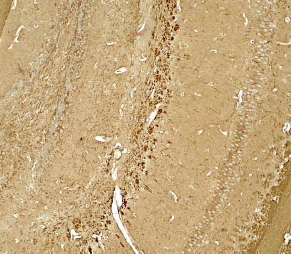
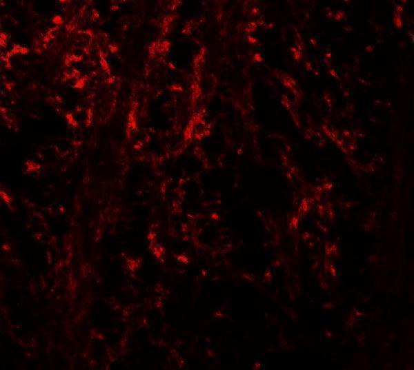
A08377 has IHC and IF images from mouse brain tissue (catalog image captions). Human, mouse, and rat are listed as reactive species (catalog: reactivity); the supplied images show mouse tissue only (catalog image captions).
A08377 will render with an IHC-P image of mouse brain tissue stained at 5 μg/mL (A08377 IHC image caption; catalog: applications). Its IF image also shows mouse brain tissue, stained at 20 μg/mL (A08377 IF image caption).
Which to pick: For paraffin-section tissue IHC, choose A08377: IHC-P is listed and its own image shows mouse brain tissue at 5 μg/mL (catalog: applications; A08377 IHC image caption); the fixative is unreported (A08377 IHC image caption). For IF, A08377 has a mouse brain tissue image at 20 μg/mL (A08377 IF image caption); ICC validation is unreported (catalog: applications and image captions). For human or rat samples, A08377 lists both species as reactive, while the supplied IHC and IF images show mouse tissue only (catalog: reactivity; A08377 image captions); clonality is unreported (catalog: clone).