NQO1 / NAD(P)H dehydrogenase [quinone] 1 · IHC design guide

Design Immunohistochemistry for NQO1

Plan paraffin-section NQO1 IHC around the cytoplasmic staining seen in most tissues (HPA tissue IHC). This guide covers cell-type controls (HPA tissue IHC) and the catalog antibody’s 2–5 μg/ml IHC range (datasheet A00494-2).

Evidence assembled Oct 2026 · For research use; verify linked source records and product datasheet before use
Immunohistochemistry protocol sheet for NQO1 (IHC for NQO1): expected localisation Cytoplasmic staining in most tissues (HPA tissue IHC), antibody A00494-2, validated IHC image, and IHC protocol steps
Printable NQO1 IHC protocol sheet — expected localisation Cytoplasmic staining in most tissues (HPA tissue IHC), antibody A00494-2, controls and protocol steps. Open the full NQO1 IHC guide →

NQO1 Immunohistochemistry Experimental Design Guide

Expected localisation, validated protocols, controls and antibodies — the at-a-glance facts below, then the full design guide.

Must know before staining
Expected localisation Cytoplasmic staining in most tissues (HPA tissue IHC)
Staining pattern High cytoplasmic staining in gallbladder glandular cells (HPA tissue IHC)
Antigen retrieval EDTA pH 8.0 HIER, heat-mediated (datasheet A00494-2)
Positive control ⓘ Gallbladder+4 more · see all
Negative control ⓘ Bone marrow+4 more · see all
Important caveats
Reasons your staining may differ from the expected pattern.
Fixation Keep fixation consistent across sections (standard IHC practice; not target-specific)
Caveat Staining varies by cell type; marrow hematopoietic cells are undetected (HPA tissue IHC)
Regulation Expression regulation unreported (UniProt)
Isoform / epitope 3 isoforms; verify epitope coverage; no transmembrane segment (UniProt)
Section 1

Recommended NQO1 IHC & IF Protocols

The catalog antibody’s IHC-P protocol (datasheet: A00494-2) is presented alongside three published NQO1 IHC protocols (PMC3743184; PMC6433262; PMC2377272).

Recommended immunohistochemistry (IHC-P) protocol parameters
SampleParaffin-embedded human colon cancer tissue; fixative not specified (datasheet A00494-2)
FixationImage fixative and duration unreported (datasheet A00494-2); verify before use.
Sectioning4–5 µm sections on charged slides (standard)
DeparaffinisationXylene, graded ethanol series to water (standard)
Antigen retrievalHeat retrieval: EDTA pH 8.0 (datasheet A00494-2); 20 min, 95–100 °C (standard)
Peroxidase block3% H2O2, 10 min, room temperature (standard)
Blocking10% goat serum (datasheet A00494-2)
Primary antibodyRabbit anti-NQO1, 2-5 μg/ml (datasheet A00494-2)
Primary incubationOvernight at 4 °C (datasheet A00494-2)
DetectionHRP-conjugated secondary, DAB chromogen (datasheet A00494-2)
CounterstainHematoxylin, blue, dehydrate and mount (standard)
Expected resultNQO1-positive staining in glandular cells of gallbladder (HPA tissue IHC: High). HPA tissue profile: Cytoplasmic expression in most tissues. No signal in the no-primary control.
💡Decision noteStart with heat-mediated EDTA pH 8.0 retrieval for the catalog antibody (datasheet: A00494-2). Published protocols used different retrieval conditions with different antibodies (PMC3743184; PMC6433262; PMC2377272).
Section 2

What Is the Expected NQO1 Staining Pattern?

NQO1 is a cytosolic protein with no transmembrane segment (UniProt P15559). In paraffin-section IHC, expect predominantly cytoplasmic staining in many tissues (HPA tissue IHC: cytoplasmic expression in most tissues). Gallbladder glandular cells and kidney glomerular cells are useful high-staining examples (HPA tissue IHC: High). The tissue pattern has Enhanced reliability because antibody staining is consistent with RNA expression (HPA tissue IHC: Enhanced).

What am I looking at on my slide?
Distinct cytoplasmic chromogen in gallbladder glandular cells or kidney glomerular cells.This matches two High-staining cell populations (HPA tissue IHC: Gallbladder and Kidney, High) and the expected cytosolic location (UniProt P15559; HPA subcellular: Cytosol). Compare cells within the same section before assigning a tissue-wide score (general IHC practice).
Predominantly nuclear, membranous, or extracellular staining replaces cytoplasmic staining.These compartments conflict with the annotated cytosolic location and lack of a transmembrane segment (UniProt P15559; HPA subcellular: Cytosol). Treat the pattern as suspect; inspect morphology, detection controls, and the antibody's IHC validation before interpreting it as NQO1 (general IHC practice).
Strong staining appears in cells listed as undetected, such as bone-marrow hematopoietic cells.HPA reports NQO1 as Not detected in those cells (HPA tissue IHC: Bone marrow, hematopoietic cells). Cross-reactivity or endogenous chromogen-generating activity is possible (general IHC practice); verify the stained cell identity and use an appropriate detection control.
Chromogen coats the section broadly, obscuring boundaries between cells.Uniform haze cannot establish the cell-restricted cytoplasmic pattern reported by HPA (HPA tissue IHC: cytoplasmic expression in most tissues). Review the no-primary control, blocking, washes, and chromogen development; these checks address background in chromogenic IHC generally (general IHC practice).
No staining appears in gallbladder glandular cells or kidney glomerular cells.An absent signal conflicts with HPA's High staining in those specific cells (HPA tissue IHC: Gallbladder and Kidney, High). Confirm that the expected cells are present, then review the catalog antibody's IHC-P instructions and the run controls before calling the specimen negative (general IHC practice).
💡Expected NQO1 appearanceA convincing positive shows predominantly cytoplasmic chromogen in the relevant cells, with High staining possible in gallbladder glandular or kidney glomerular cells; dominant nuclear, membrane, or uniform section-wide color is suspect (HPA tissue IHC: High and cytoplasmic profile; UniProt P15559).
How each factor affects the staining
Cell-specific reference patternUse the named cell population, not the organ name alone: stomach glandular cells are Medium, while liver cholangiocytes are Not detected (HPA tissue IHC). A mixed-cell section can therefore contain staining and unstained cells without contradicting the reference.
Antibody validationTwo listed antibodies have Enhanced IHC status (HPA antibodies: HPA007308 and CAB012421). HPA's tissue profile also has Enhanced reliability, indicating agreement between staining and RNA data (HPA tissue IHC); neither designation validates every compartment or every specimen.
Isoforms and antigen scopeUniProt lists 3 NQO1 isoforms (UniProt P15559). The supplied record does not locate the catalog antibody's epitope or establish which isoforms it detects, so do not explain a discrepant IHC pattern by isoform preference without antibody-specific evidence.
IF/ICC cross-check?HPA ICC-IF reports an enhanced cytosolic location and lists images from A-431, U-251MG, and U2OS cells (HPA subcellular: Cytosol, enhanced). This supports the compartment call; it does not establish an IHC-P retrieval condition or tissue intensity.
Detection chemistryEndogenous enzyme activity can create chromogen independent of primary-antibody binding (general chromogenic IHC practice). Include a no-primary control and check the detection system's recommended blocking steps; the supplied HPA and UniProt records do not identify an NQO1-specific interference.
Why is my staining missing, weak or wrong?
SituationLikely causeNext action
Expected High-staining cells are blank.The expected cell population may be absent from the section, or the IHC run may have failed; HPA reports High staining specifically in gallbladder glandular and kidney glomerular cells (HPA tissue IHC).Locate those cells on the counterstained section and check a suitable positive control. Review the catalog antibody's IHC-P instructions and the run record before changing conditions (general IHC practice).
Staining is mainly nuclear.A nuclear-dominant pattern conflicts with cytosolic NQO1 (UniProt P15559; HPA subcellular: Cytosol).Check the no-primary control and cellular boundaries, then compare with the antibody's IHC validation images where available (general IHC practice). Do not score nuclear color as the expected NQO1 pattern.
An HPA-undetected cell population stains strongly.Unexpected primary binding or detection background is possible (general IHC practice); HPA lists bone-marrow hematopoietic cells as Not detected (HPA tissue IHC).Confirm cell identity and inspect a no-primary control. Record the result as a discrepancy until the stain can be distinguished from background (general IHC practice).
Every cell and empty area looks brown.Widespread color obscures HPA's cytoplasmic tissue pattern (HPA tissue IHC); excess chromogen development or detection background is possible (general IHC practice).Compare the no-primary control, inspect washes and detection steps, and adjust development within the detection system's instructions (general IHC practice).
A presumed negative organ contains positive cells.HPA's negative calls can be cell-specific: liver cholangiocytes are Not detected, while the payload does not label every liver cell negative (HPA tissue IHC).Identify and score the actual stained cell type before comparing with HPA. Avoid treating a cell-specific Not detected entry as a whole-organ negative control (HPA tissue IHC).
Bronchial cilia stain, but cell bodies are less conspicuous.HPA reports Medium staining in bronchial ciliated cells at the cilia axoneme (HPA tissue IHC), an exception to its broad cytoplasmic tissue summary.Document the ciliary location separately and compare it with the bronchus reference. Do not force this observation into a uniform cell-body score (HPA tissue IHC).

Sample controls for NQO1 IHC & IF

🧪Run gallbladder first and expect staining in glandular cells (HPA: High in gallbladder glandular cells). Use heart muscle cardiomyocytes as the negative tissue (HPA: Not detected in cardiomyocytes); on the gallbladder slide, assess adjacent non-glandular cells for background staining, but do not assume they are NQO1-negative without separate validation (HPA: gallbladder result specifies glandular cells).
Positive control tissue: Gallbladder (Glandular cells, HPA High)
Negative control tissue: Bone marrow (HPA Not detected)
ICC-IF cell lines (HPA subcellular resource): HPA ICC-IF images show NQO1 in A-431, U-251MG, U2OS, U2OS, siRNA 1 (10x), U2OS, siRNA 1 (40x), U2OS, siRNA 2 (10x), U2OS, siRNA 2 (40x), U2OS, scrambled (10x), U2OS, scrambled (40x), with annotated localisation: Cytosol (enhanced) (HPA subcellular).
Technical controls: Include a no-primary, secondary-only section, a concentration-matched rabbit IgG isotype control, and an NQO1 knockout biological control where available (caption: rabbit primary; standard IHC practice). Block endogenous peroxidase and check for pigment that could resemble DAB staining in gallbladder sections (caption: HRP/DAB detection; standard IHC practice).
⚠️Feasibility: The selected A00494-2 paraffin-section caption does not report the fixative or a target-specific fixation window (selected-SKU IHC caption). It uses heat retrieval in EDTA, pH 8.0, but does not establish that retrieval is required or that frozen sections or IF are easier (selected-SKU IHC caption). Check gallbladder pigment when interpreting chromogenic staining (standard IHC practice).

HPA tissue IHC evidence for NQO1

Comprehensive Human Protein Atlas IHC scoring per tissue (reliability: Enhanced — High consistency between antibody staining and RNA expression data.). Rows are taken directly from the HPA tissue chart — click any row's HPA link to view the source.

Positive expression · recommended positive controls

TissueCell typeLevelEvidenceSource
Gallbladder Glandular cells High Protein (IHC) HPA →
Kidney Cells in glomeruli High Protein (IHC) HPA →
Stomach Glandular cells Medium Protein (IHC) HPA →
Adipose tissue Adipocytes Medium Protein (IHC) HPA →
Adrenal gland Glandular cells Medium Protein (IHC) HPA →

Undetected expression · recommended negative controls

TissueCell typeLevelEvidenceSource
Bone marrow Hematopoietic cells Not detected Protein (IHC) HPA →
Heart muscle Cardiomyocytes Not detected Protein (IHC) HPA →
Liver Cholangiocytes Not detected Protein (IHC) HPA →
Lymph node Germinal center cells Not detected Protein (IHC) HPA →
Ovary Follicle cells Not detected Protein (IHC) HPA →
Section 3

Advanced NQO1 IHC Tips

Use the cytosolic pattern expected for NQO1 to assess staining, while checking each cell population separately (UniProt P15559; HPA tissue IHC).

What retrieval should I start with when NQO1 staining is weak in paraffin sections?
Start with heat-mediated antigen retrieval in EDTA at pH 8.0 (datasheet A00494-2). The catalog antibody's paraffin-section example used that retrieval before 10% goat-serum blocking and an overnight incubation at 4°C with 2 μg/ml primary antibody (datasheet A00494-2). If staining remains weak, check that sections were fully deparaffinised and compare retrieval heating and cooling conditions on adjacent sections while holding antibody concentration and detection constant (standard IHC practice). Assess the resulting cytoplasmic signal in intact cells against a matched section lacking primary antibody, because stronger overall DAB colour alone does not establish improved NQO1 detection (HPA tissue IHC; standard IHC practice).
How should I troubleshoot NQO1 staining when fixation history is uncertain?
The catalog example identifies a paraffin-embedded human colon cancer section but does not state its fixative, so target-specific fixation sensitivity is unknown (datasheet A00494-2). Record the available fixation history and compare sections with similar processing before attributing a weak result to biology (standard IHC practice). Run the documented EDTA pH 8.0 retrieval and, where practical, include an internal cell population or a separately processed reference section showing interpretable cytoplasmic staining (datasheet A00494-2; HPA tissue IHC; standard IHC practice). If fixation histories differ, evaluate morphology and staining together, and report the processing uncertainty rather than assigning a target-specific fixation effect that has not been established (standard IHC practice).
How do I assess NQO1 staining that appears nuclear or membranous?
Expect predominantly cytoplasmic, specifically cytosolic, NQO1 signal; the protein has no annotated transmembrane segment (UniProt P15559; HPA subcellular; HPA tissue IHC). Inspect intact cells at higher magnification to distinguish cytoplasmic DAB from colour over nuclei, cell borders or extracellular deposits (standard IHC practice). Compare the same compartments on a section lacking primary antibody, and check whether strong signal persists where morphology is damaged or tissue edges are prominent (standard IHC practice). If a distinct nuclear or membranous pattern remains, describe it separately from expected cytosolic staining and seek independent confirmation before calling it NQO1 localisation (UniProt P15559; HPA subcellular; standard IHC practice).
Could isoforms or epitope masking explain patchy NQO1 IHC?
NQO1 has 3 annotated isoforms, but the supplied catalog caption does not identify the antibody epitope or establish isoform selectivity (UniProt P15559; datasheet A00494-2). Its annotated chain spans residues 1–274, with no signal peptide, propeptide or glycosylation site, and phosphoserine is annotated at residue 82 (UniProt P15559). Those annotations alone cannot show whether processing or phosphorylation alters this antibody's binding in paraffin sections (UniProt P15559; standard IHC interpretation). When staining varies between areas, first compare retrieval, morphology and cell composition under matched detection conditions, then use an independently characterised epitope or orthogonal measurement if an isoform-specific claim matters (standard IHC practice).
How can I investigate an unexpected IHC pattern with multiplex IF?
Use IF/ICC as a separate follow-up assay: the supplied catalog example demonstrates paraffin-section chromogenic IHC, while HPA reports cytosolic localisation in ICC/IF images (datasheet A00494-2; HPA subcellular). Pair NQO1 with a marker of the cell population being scored, choosing that marker to identify cells rather than assuming all neighbouring cells express NQO1 (HPA tissue IHC; standard IF practice). Select fluorophores after checking tissue autofluorescence and include single-colour and no-primary controls to assess channel overlap and background (standard IF practice). Because NQO1 is cytosolic and has no transmembrane segment, test permeabilisation that permits antibody access to the cytosol without losing morphology; establish fixation and antibody conditions for IF independently (UniProt P15559; HPA subcellular; standard IF practice).
What should I check when DAB background obscures cytoplasmic NQO1 staining?
First compare a no-primary section and inspect whether colour follows tissue edges, damaged areas or specific intact cells (standard IHC practice). The catalog example used 10% goat serum, 2 μg/ml primary antibody overnight at 4°C, and an anti-rabbit peroxidase secondary for 30 minutes at 37°C (datasheet A00494-2). Include a peroxidase-blocking step, adequate washes and a controlled DAB development time as general chromogenic IHC measures; these steps do not by themselves validate NQO1 specificity (standard IHC practice). If background persists, vary one condition at a time and judge improvement by preserved cytoplasmic contrast in intact cells, since HPA describes cytoplasmic expression across most tissues (HPA tissue IHC; standard IHC practice).
How should I score heterogeneous NQO1 staining across a tissue section? ⚠ ANSWER MARKED FOR VERIFICATION
Define the cell population and regions to be analysed before scoring, because HPA reports NQO1 staining in particular cells at different levels across tissues (HPA tissue IHC; standard IHC practice). For cytoplasmic DAB, record the percentage of positive cells and an intensity-based H-score using the same thresholds for every section (HPA subcellular; standard IHC practice). If counting discrete positive cells, report density per mm² of viable analysed tissue and normalise counts to that measured area; for percentage or H-score, use the same eligible-cell denominator across samples (standard IHC practice). Exclude folds, necrosis and edges by a predefined rule, and keep acquisition, counterstain and DAB assessment consistent across groups (standard IHC practice).
How can I distinguish true NQO1 positivity from an IHC artefact?
A plausible NQO1-positive cell shows cytoplasmic signal with preserved morphology, consistent with UniProt cytosolic localisation and the HPA tissue profile (UniProt P15559; HPA tissue IHC). Consider cell identity: HPA reports high staining in gallbladder glandular cells and kidney glomerular cells, but no detected staining in bone-marrow hematopoietic cells or heart cardiomyocytes (HPA tissue IHC). Treat isolated nuclear deposits, tissue-edge colour and signal over necrotic areas cautiously, and compare them with a no-primary section (HPA subcellular; standard IHC practice). Check for endogenous peroxidase contribution with an appropriate control before interpreting DAB as antibody signal; a positive-looking area requires localisation, cell-context and control agreement (standard IHC practice).
Boster reagents

Best NQO1 / NAD(P)H dehydrogenase [quinone] 1 IHC Antibodies

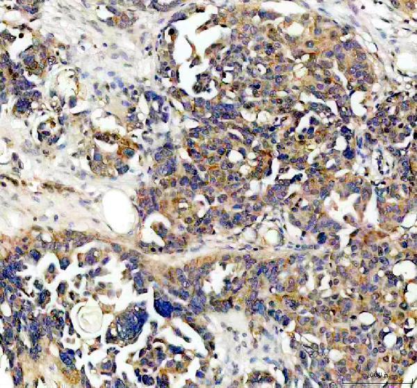
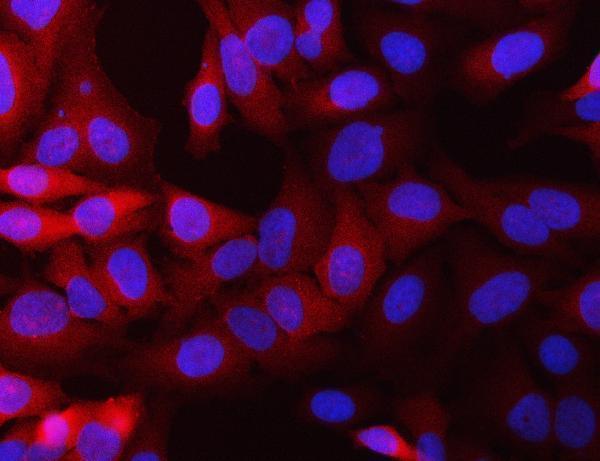
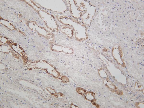
Catalog antibodies have IHC images from human colon and ovarian cancers, mouse stomach, and human, mouse and rat kidneys (IHC image captions); IF images show HeLa and MCF-7 cells (IF image captions).

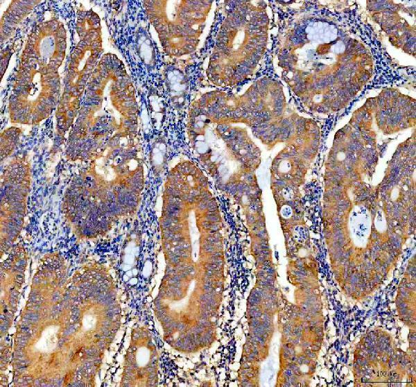
Real IHC data IHC analysis of NQO1 using anti-NQO1 antibody (A00494-2). NQO1 was detected in a paraffin-embedded section of human colon cancer tissue. Heat mediated antigen retrieval was performed in EDTA buffer (pH 8.0, epitope retrieval solution). The tissue section was blocked with 10% goat serum. The tissue section was then incubated with 2 μg/ml rabbit anti-NQO1 Antibody (A00494-2) overnight at 4°C. Peroxidase Conjugated Goat Anti-rabbit IgG was used as secondary antibody and incubated for 30 minutes at 37°C. The tissue section was developed using HRP Conjugated Rabbit IgG Super Vision Assay Kit (Catalog # SV0002) with DAB as the chromogen.
Anti-NQO1 Antibody ®
Cat # A00494-2
Real IHC data IHC analysis of NQO1 using anti-NQO1 antibody (M00494). NQO1 was detected in a paraffin-embedded section of mouse stomach tissue. Heat mediated antigen retrieval was performed in EDTA buffer (pH 8.0, epitope retrieval solution). The tissue section was blocked with 10% goat serum. The tissue section was then incubated with 1:200 rabbit anti-NQO1 Antibody (M00494) overnight at 4°C. Peroxidase Conjugated Goat Anti-rabbit IgG was used as secondary antibody and incubated for 30 minutes at 37°C. The tissue section was developed using HRP Conjugated Rabbit IgG Super Vision Assay Kit (Catalog # SV0002) with DAB as the chromogen.
Anti-NQO1/Dt Diaphorase Rabbit Monoclonal Antibody
Cat # M00494
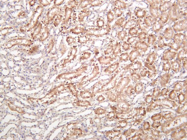
Real IHC data Rat kidney was stained with anti-NQO1 rabbit antibody
Anti-NQO1 Rabbit Monoclonal Antibody
Cat # M00494-3

A00494-2 has paraffin section IHC images from human colon and ovarian cancers and an IF image from HeLa cells (A00494-2 image captions); M00494 has a paraffin section IHC image from mouse stomach and an IF image from MCF-7 cells (M00494 image captions). M00494-3 has IHC images from human, mouse and rat kidneys, while IF is listed as an application without an IF image (M00494-3 catalog and image captions).

Which to pick: For human paraffin section IHC, choose A00494-2: its captions document EDTA retrieval at pH 8.0 and 2 μg/ml primary antibody (A00494-2 IHC image captions). For IF/ICC, A00494-2 has both applications listed and an IF image from HeLa cells (A00494-2 catalog and IF image caption). For IHC across human, mouse and rat, M00494-3 has kidney images from all three species; M00494 also lists all three species, but its IHC image shows mouse stomach only (M00494-3 and M00494 catalog and IHC image captions). The IHC captions describe paraffin sections but do not report the fixative (IHC image captions).

Each figure is that product's own IHC / IF validation image from its datasheet.

References

  1. UniProt Consortium. UniProt entry P15559 (NQO1_HUMAN, NAD(P)H dehydrogenase [quinone] 1).
  2. Human Protein Atlas. NQO1 tissue IHC expression (reliability: Enhanced).
  3. Human Protein Atlas. NQO1 subcellular location (ICC-IF): Localized to the cytosol..
  4. Human Protein Atlas. NQO1 antibody validation summary (2 antibodies).
  5. Selective Elimination of NRF2-Activated Cells by Competition With Neighboring Cells in the Esophageal Epithelium. Cellular and molecular gastroenterology and hepatology 2023 — PMC9672893.
  6. NAD(P)H: quinone oxidoreductase 1 and NRH:quinone oxidoreductase 2 polymorphisms in papillary thyroid microcarcinoma: correlation with phenotype. Yonsei medical journal 2013 — PMC3743184.
  7. The NRF2 transcriptional target NQO1 has low mRNA levels in TP53-mutated endometrial carcinomas. PloS one 2019 — PMC6433262.
  8. ATBF1 and NQO1 as candidate targets for allelic loss at chromosome arm 16q in breast cancer: absence of somatic ATBF1 mutations and no role for the C609T NQO1 polymorphism. BMC cancer 2008 — PMC2377272.
  9. PubMed PMID:2843525 — UniProt-cited evidence.
  10. PubMed PMID:1657151 — UniProt-cited evidence.
  11. PubMed PMID:14702039 — UniProt-cited evidence.