This website uses cookies to ensure you get the best experience on our website.


- Table of Contents
Plan paraffin-section NR5A2 IHC around widespread nuclear staining (HPA tissue IHC) and the catalog antibody’s 2–5 μg/ml IHC range (datasheet A01332-1). Compare staining with appropriate controls while accounting for its low consistency with RNA expression (HPA tissue IHC).

Expected localisation, validated protocols, controls and antibodies — the at-a-glance facts below, then the full design guide.
| Expected localisation | Nuclear staining in tissue sections (HPA tissue IHC) | |
| Staining pattern | Widespread nuclear staining across tissue cell types (HPA tissue IHC) | |
| Antigen retrieval | EDTA pH 8.0 HIER, heat-mediated (datasheet A01332-1) | |
| Positive control | Adipose tissue+4 more · see all | |
| Negative control | None in HPA (detected in all 45 tissues); use no-primary + isotype controls |
| Fixation | Keep fixation conditions consistent across sections (standard IHC practice; not target-specific) | |
| Caveat | Low consistency between staining and RNA expression (HPA tissue IHC) | |
| Regulation | Abundant in pancreas; lower in liver (UniProt) | |
| Isoform / epitope | 4 isoforms; epitope coverage is unspecified (UniProt) |
The catalog antibody protocol uses EDTA pH 8.0 heat retrieval (datasheet A01332-1). These 3 articles provide published NR5A2 IHC workflows (PMC6489909; PMC4696807; PMC3573348).
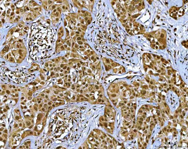
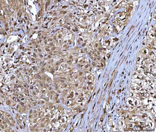
| Sample | Paraffin-embedded human parotid acinar cell carcinoma tissue; fixative not specified (datasheet A01332-1) |
| Fixation | Image fixative and duration unreported (datasheet A01332-1); verify before use. |
| Sectioning | 4–5 µm sections on charged slides (standard) |
| Deparaffinisation | Xylene, graded ethanol series to water (standard) |
| Antigen retrieval | Heat retrieval: EDTA pH 8.0 (datasheet A01332-1); 20 min, 95–100 °C (standard) |
| Peroxidase block | 3% H2O2, 10 min, room temperature (standard) |
| Blocking | 10% goat serum (datasheet A01332-1) |
| Primary antibody | Rabbit anti-NR5A2, 2-5 μg/ml (datasheet A01332-1) |
| Primary incubation | Overnight at 4 °C (datasheet A01332-1) |
| Detection | HRP-conjugated secondary, DAB chromogen (datasheet A01332-1) |
| Counterstain | Hematoxylin, blue, dehydrate and mount (standard) |
| Expected result | NR5A2-positive staining in adipocytes of adipose tissue (HPA tissue IHC: High). HPA tissue profile: Ubiquitous nuclear expression. No signal in the no-primary control. |
NR5A2 is a nuclear and chromosome-associated protein with no transmembrane segment (UniProt O00482: location and topology). In paraffin-section IHC, expect nuclear staining in cells documented by HPA, including adipocytes, glandular cells and hematopoietic cells (HPA: High in these cell types). HPA describes ubiquitous nuclear expression, but rates its tissue IHC evidence Approved with low consistency between antibody staining and RNA expression (HPA: tissue IHC reliability and profile).
| Distinct chromogenic signal overlaps nuclei in HPA-documented cells, while surrounding tissue remains relatively clear. | This fits the expected compartment (UniProt O00482: nucleus; HPA: ubiquitous nuclear expression). HPA reports High staining in adipocytes and in glandular cells from several tissues (HPA: tissue IHC). Judge the signal against nuclear morphology and controls; HPA's Approved rating carries a low staining–RNA consistency caveat (HPA: reliability description). |
| Strong staining is predominantly cytoplasmic, membranous or extracellular, with little nuclear signal. | Treat this as a possible staining artefact or off-target signal because NR5A2 is nuclear/chromosome-associated and has no transmembrane segment (UniProt O00482: location and topology). Check whether the apparent signal follows tissue pigment, section edges or detection background using routine IHC controls (standard IHC practice). |
| Chromogen appears mainly in a cell population outside the HPA-documented examples, especially without convincing nuclear staining. | Consider antibody cross-reactivity or endogenous detection activity (standard IHC practice). Cell identity alone cannot establish an error: HPA describes ubiquitous nuclear expression and supplies no negative cell-type list (HPA: tissue IHC profile). Compare localisation and appropriate detection controls before calling the population NR5A2-positive (UniProt O00482: nucleus; standard IHC practice). |
| Weak, diffuse colour covers many compartments or obscures nuclear boundaries. | Diffuse background does not establish NR5A2 expression; the expected readout is nuclear (UniProt O00482: location; HPA: nuclear IHC profile). Review background controls, blocking, antibody concentration and chromogen development as general IHC variables (standard IHC practice). HPA's High tissue calls should not be used to excuse indiscriminate colour (HPA: tissue IHC levels). |
| No nuclear signal appears in an HPA-reported High cell population, such as adipocytes in adipose tissue. | The run may lack a detectable positive signal, but one negative section does not establish biological absence (HPA: High in adipocytes; HPA: low staining–RNA consistency). First assess tissue preservation and the assay's positive and detection controls (standard IHC practice). Interpret any remaining negative result in light of HPA's reliability caveat (HPA: tissue IHC reliability). |
| Subcellular location | Score nuclear localisation as the principal IHC pattern (UniProt O00482: nucleus and chromosome; HPA: ubiquitous nuclear expression). No transmembrane segment or signal peptide is listed, so a membrane or secreted pattern has no support here (UniProt O00482: topology and processing). |
| Tissue and cell context | HPA reports High IHC staining in adipocytes, adrenal/appendix/breast glandular cells, marrow hematopoietic cells, bronchial respiratory epithelium, caudate glia and cerebellar granular-layer cells (HPA: tissue IHC). These examples guide slide review; the supplied HPA extract lists no negative tissues or cells (HPA: tissue IHC). |
| Confidence in the observed pattern | The HPA tissue IHC assessment is Approved, while antibody staining has low consistency with RNA expression (HPA: reliability description). The listed antibodies HPA005455 and HPA017067 are IHC Approved; neither is marked Enhanced in the supplied record (HPA: antibody validation). Avoid treating any single stained population as independently confirmed by RNA (HPA: reliability description). |
| Isoforms and epitope coverage | UniProt lists four NR5A2 isoforms (UniProt O00482: isoforms 1–4). The supplied record gives no antibody epitope or isoform-specific IHC validation, so it cannot establish which isoforms contribute to a stain (UniProt O00482: isoforms; HPA: supplied antibody validation). |
| IF/ICC Q&A: should puncta be expected? | HPA's ICC-IF summary places NR5A2 in nuclear speckles but labels the main location uncertain (HPA: subcellular ICC-IF). For this paraffin-section IHC guide, assess nuclear staining; do not require resolvable speckles for a positive chromogenic result (UniProt O00482: nucleus; HPA: tissue IHC profile). |
| Situation | Likely cause | Next action |
|---|---|---|
| Nuclear signal is absent from HPA-reported High adipocytes or glandular cells. | A weak or failed IHC run is possible; HPA's staining–RNA consistency caveat also limits certainty for a single specimen (HPA: High tissue calls and reliability description). | Check a documented positive section, detection reagents and counterstained nuclear morphology; review antigen-retrieval and antibody-incubation settings as general workflow variables (standard IHC practice). Do not infer NR5A2-specific retrieval sensitivity from these sources (HPA: tissue IHC; UniProt O00482). |
| Most colour lies outside nuclei. | Non-nuclear staining conflicts with the reported NR5A2 location and may reflect nonspecific staining or detection background (UniProt O00482: nucleus; standard IHC practice). | Compare with a no-primary control and a documented positive section; assess where chromogen falls relative to nuclei (standard IHC practice; HPA: nuclear IHC profile). |
| Unexpected cells stain strongly. | Cross-reactivity or endogenous detection activity is possible, but HPA reports ubiquitous nuclear expression and provides no negative cell-type list (standard IHC practice; HPA: tissue IHC profile). | Confirm cell identity and nuclear localisation; use an appropriate detection control before assigning NR5A2 expression (standard IHC practice; UniProt O00482: nucleus). |
| Diffuse background makes individual nuclei hard to score. | Blocking, antibody concentration or chromogen development may contribute to background in chromogenic IHC (standard IHC practice). | Check background controls and optimise those general assay variables so nuclear boundaries can be judged (standard IHC practice; HPA: nuclear IHC profile). |
| Two slides show different proportions of positive cells. | HPA's Approved tissue IHC assessment includes low consistency between antibody staining and RNA expression; the extract does not provide a universal positive-cell threshold (HPA: reliability and tissue IHC). | Compare equivalent cell populations, section quality and control performance before interpreting a biological difference (standard IHC practice). Report the observed nuclear distribution rather than an unsupported cutoff (UniProt O00482: nucleus; HPA: tissue IHC). |
| ICC-IF shows punctate nuclei while chromogenic IHC appears uniformly nuclear. | HPA calls nuclear speckles in ICC-IF uncertain, whereas its tissue IHC profile is broadly nuclear (HPA: subcellular ICC-IF and tissue IHC). | Assess each image by its own stated evidence: nuclear localisation supports IHC interpretation, and speckle resolution is not required for chromogenic scoring (UniProt O00482: nucleus; HPA: tissue IHC profile and ICC-IF uncertainty). |


Comprehensive Human Protein Atlas IHC scoring per tissue (reliability: Approved — Low consistency between antibody staining and RNA expression data.). Rows are taken directly from the HPA tissue chart — click any row's HPA link to view the source.
| Tissue | Cell type | Level | Evidence | Source |
|---|---|---|---|---|
| Adipose tissue | Adipocytes | High | Protein (IHC) | HPA → |
| Adrenal gland | Glandular cells | High | Protein (IHC) | HPA → |
| Appendix | Glandular cells | High | Protein (IHC) | HPA → |
| Bone marrow | Hematopoietic cells | High | Protein (IHC) | HPA → |
| Breast | Glandular cells | High | Protein (IHC) | HPA → |
| Tissue | Cell type | Level | Evidence | Source |
|---|---|---|---|---|
| None in HPA: NR5A2 is detected in all 45 scored tissues. Use a no-primary (secondary-only) and an isotype control instead. | ||||
Troubleshoot NR5A2 staining in paraffin sections by checking retrieval, nuclear localisation, controls, and scoring before interpreting chromogenic signal.
One human-reactive anti-NR5A2 antibody has real chromogenic IHC data from paraffin-embedded human carcinoma sections; no IF/ICC data are supplied (catalog applications and image captions).
A01332-1 will render with its own IHC figure from a paraffin-embedded human parotid acinar cell carcinoma section (catalog figure caption). Additional catalog captions show IHC staining in paraffin-embedded human bladder urothelial carcinoma and colorectal adenocarcinoma sections (catalog image captions).
Which to pick: For human tissue IHC, choose A01332-1: its paraffin-section caption reports EDTA retrieval at pH 8.0 and 2 μg/ml primary antibody; the fixative is unreported (catalog figure caption). No IF/ICC or cross-species option is documented, because A01332-1 lists IHC and human reactivity only for those choices (catalog applications and reactivity). A01332-1 is a rabbit antibody, and its clonality is unreported (catalog host and clone fields).