This website uses cookies to ensure you get the best experience on our website.


- Table of Contents
Plan OVOL1 chromogenic IHC in paraffin sections using the catalog antibody's 1:100–1:300 dilution range (datasheet A11495-1). Assess the reported cytoplasmic tissue staining (HPA tissue IHC) alongside the annotated nuclear location (UniProt), and interpret results cautiously because tissue staining reliability is uncertain (HPA tissue IHC).

Expected localisation, validated protocols, controls and antibodies — the at-a-glance facts below, then the full design guide.
| Expected localisation | Cytoplasmic tissue staining (HPA tissue IHC); nuclear location (UniProt) | |
| Staining pattern | Cytoplasmic staining in squamous epithelial cells (HPA tissue IHC) | |
| Antigen retrieval | Tris-EDTA pH 9.0 HIER, heat-mediated (datasheet A11495-1) | |
| Positive control | Cervix+4 more · see all | |
| Negative control | Adipose tissue+4 more · see all |
| Fixation | Keep paraffin-section fixation consistent across samples (standard IHC practice; not target-specific) | |
| Caveat | Presumed off-target binding; IHC/RNA concordance is very low (HPA tissue IHC) | |
| Regulation | Expression varies by tissue (UniProt) | |
| Isoform / epitope | No isoforms or processing annotated; epitope effects unknown (UniProt) |
The catalog antibody uses heat-mediated Tris-EDTA retrieval at pH 9.0 (datasheet: A11495-1). The published IHC protocols below cover salivary tumors, skin lesions, and pancreatic tissues (PMC12603596; PMC7141138; PMC12304111).
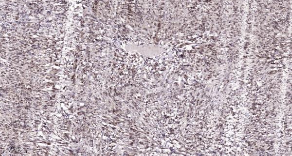
| Sample | Paraffin-embedded human Colon cancer tissue; fixative not specified (datasheet A11495-1) |
| Fixation | Image fixative and duration unreported (datasheet A11495-1); verify before use. |
| Sectioning | 4–5 µm sections on charged slides (standard) |
| Deparaffinisation | Xylene, graded ethanol series to water (standard) |
| Antigen retrieval | Heat retrieval: Tris-EDTA pH 9.0 (datasheet A11495-1); 20 min, 95–100 °C (standard) |
| Peroxidase block | 3% H2O2, 10 min, room temperature (standard) |
| Blocking | 10% normal serum of the secondary host, 30 min, room temperature (standard) |
| Primary antibody | Rabbit anti-OVOL1, 1:100-1:300 (datasheet A11495-1) |
| Primary incubation | Overnight at 4 °C (standard) |
| Detection | HRP-polymer secondary, DAB chromogen 5–10 min (standard) |
| Counterstain | Hematoxylin, blue, dehydrate and mount (standard) |
| Expected result | OVOL1-positive staining in squamous epithelial cells of cervix (HPA tissue IHC: High). HPA tissue profile: Cytoplasmic expression in several tissues, including squamous epithelial. No signal in the no-primary control. |
OVOL1 is annotated as nuclear and has no transmembrane segment (UniProt O14753: subcellular location and topology). In paraffin section IHC, assess signal in squamous epithelial cells of cervix, esophagus, or vagina, and glandular cells of prostate, where HPA reports high staining (HPA: tissue IHC). HPA describes the tissue staining as cytoplasmic but rates its reliability Uncertain because antibody staining and RNA expression have very low consistency and presumed off-target binding was observed (HPA: tissue IHC).
| Defined nuclear staining in squamous epithelial cells, with limited surrounding background. | This fits OVOL1's nuclear annotation and the cell types HPA scores High in cervix, esophagus, and vagina (UniProt O14753: subcellular location; HPA: tissue IHC). Treat it as a plausible result requiring validation: the HPA tissue IHC reliability is Uncertain (HPA: tissue IHC). |
| Cytoplasmic staining dominates, with little or no convincing nuclear signal. | The compartment conflicts with UniProt's nuclear annotation and HPA's approved nucleoplasmic ICC-IF location (UniProt O14753: subcellular location; HPA: subcellular). HPA also reports cytoplasmic tissue staining, but flags that tissue result as Uncertain; cytoplasmic signal alone cannot establish OVOL1 specificity (HPA: tissue IHC). |
| Strong signal appears mainly outside the expected cells, or in a negative comparison cell population. | Check for cross-reactivity or endogenous detection activity before assigning that signal to OVOL1. HPA scores cervical, esophageal, and vaginal squamous epithelial cells High, while adipocytes in adipose tissue are Not detected (HPA: tissue IHC). Those are cell-specific observations, not a guarantee about every cell in either tissue. |
| Colour spreads across cells and stroma without clear cell boundaries or a nuclear pattern. | Diffuse staining cannot support a compartment call. Assess background with a no-primary control, then review blocking, washing, and detection conditions as general IHC practice. The need for a clear nuclear call follows the UniProt nuclear annotation; HPA tissue IHC remains Uncertain (UniProt O14753: subcellular location; HPA: tissue IHC). |
| No signal appears in squamous epithelial cells of an HPA High tissue. | First check section quality, detection, and the antibody's IHC-P conditions as general IHC practice. HPA scores those cells High in cervix, esophagus, and vagina, but its Uncertain reliability means a blank section does not by itself establish absent OVOL1 or a failed assay (HPA: tissue IHC). |
| Compartment agreement | UniProt places OVOL1 in the nucleus, and HPA approves nucleoplasm as the main ICC-IF location (UniProt O14753: subcellular location; HPA: subcellular). HPA tissue IHC instead describes cytoplasmic expression and rates that staining Uncertain, so report the observed compartment explicitly rather than treating either image type as proof of tissue IHC specificity (HPA: tissue IHC). |
| Cell and tissue choice | HPA scores squamous epithelial cells High in cervix, esophagus, and vagina; prostate glandular cells are also High, while oral mucosal squamous epithelial cells are Medium (HPA: tissue IHC). Assess the named cell population within a section. HPA's Uncertain tissue reliability limits how strongly any one positive or negative comparison can validate the assay (HPA: tissue IHC). |
| Antibody validation limit | The listed antibody, HPA003984, is Uncertain for IHC and Approved for ICC (HPA: antibodies). HPA attributes its Uncertain tissue assessment to very low agreement between staining and RNA expression and to presumed off-target binding (HPA: tissue IHC). Use morphology, compartment, controls, and independent evidence when interpreting IHC signal. |
| Topology and processing | OVOL1 has no transmembrane segment, signal peptide, propeptide, or annotated glycosylation sites; its annotated chain spans residues 1–267 (UniProt O14753: topology, processing, glycosylation). These annotations provide no basis to predict a membrane pattern or a shed-product pattern. They also do not establish an antigen retrieval condition or OVOL1-specific fixation sensitivity. |
| IF/ICC Q&A | Q: What compartment should an IF/ICC result show? A: HPA lists nucleoplasm as the approved main ICC-IF location, consistent with UniProt's nuclear annotation (HPA: subcellular; UniProt O14753: subcellular location). This answers localisation only; IF/ICC procedure selection belongs to its separate guide. |
| Situation | Likely cause | Next action |
|---|---|---|
| Cytoplasmic signal is stronger than nuclear signal in a candidate positive section. | The observed compartment conflicts with nuclear and approved nucleoplasmic annotations, although HPA describes cytoplasmic tissue staining with Uncertain reliability (UniProt O14753: subcellular location; HPA: subcellular and tissue IHC). | Record nuclear and cytoplasmic scores separately. Check a no-primary control and seek independent evidence before calling cytoplasmic staining specific; these are general IHC interpretation steps. |
| The expected squamous epithelial cells show no visible chromogenic signal. | HPA scores those cells High in cervix, esophagus, and vagina, but its tissue IHC reliability is Uncertain (HPA: tissue IHC). A technical failure or a weakly reproducible reference pattern remains possible. | Confirm that the section and detection controls worked, then review the chosen antibody's IHC-P instructions, including its stated retrieval and dilution conditions if provided. Do not infer a target-specific fixation effect. |
| Staining is widespread across cells or connective tissue. | Diffuse background can obscure cell and compartment boundaries; it does not match the discrete nuclear location annotated for OVOL1 (UniProt O14753: subcellular location). | As general IHC practice, compare a no-primary control and review blocking, washing, detection reagent activity, and counterstain before scoring positive cells. |
| A comparison section stains in adipocytes expected to be negative. | HPA reports adipocytes in adipose tissue as Not detected, but the overall tissue IHC assessment is Uncertain (HPA: tissue IHC). Unexpected staining could reflect nonspecific antibody binding or endogenous detection activity. | Check the cell identity and a no-primary control; localise any residual signal before treating the comparison as evidence about OVOL1. Apply the HPA result to the specified cell population only. |
| Nuclear and cytoplasmic calls differ between IHC and ICC-IF. | HPA reports cytoplasmic tissue IHC with Uncertain reliability, while its approved ICC-IF location is nucleoplasm (HPA: tissue IHC and subcellular). The records do not identify a mechanism for the difference. | Report each application's observed compartment and validation status separately. For IHC, assess cell type, background controls, and independent evidence before assigning specificity. |
| A weak signal appears in a tissue expected to be negative from UniProt. | UniProt reports no expression in intestine, peripheral blood lymphocytes, and ovary, while the supplied HPA IHC negatives refer to specific cells in other tissues; those records are not interchangeable (UniProt O14753: tissue specificity; HPA: tissue IHC). | Identify the stained cell population and check background controls before interpreting the signal. State which source and cell population define the comparison; do not extend a cell-specific HPA result to a whole organ. |
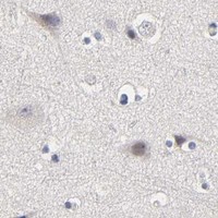


Comprehensive Human Protein Atlas IHC scoring per tissue (reliability: Uncertain — Very low consistency between antibody staining and RNA expression data. Presumed off target binding observed and disregarded.). Rows are taken directly from the HPA tissue chart — click any row's HPA link to view the source.
| Tissue | Cell type | Level | Evidence | Source |
|---|---|---|---|---|
| Cervix | Squamous epithelial cells | High | Protein (IHC) | HPA → |
| Esophagus | Squamous epithelial cells | High | Protein (IHC) | HPA → |
| Prostate | Glandular cells | High | Protein (IHC) | HPA → |
| Vagina | Squamous epithelial cells | High | Protein (IHC) | HPA → |
| Caudate | Glial cells | Medium | Protein (IHC) | HPA → |
| Tissue | Cell type | Level | Evidence | Source |
|---|---|---|---|---|
| Adipose tissue | Adipocytes | Not detected | Protein (IHC) | HPA → |
| Appendix | Glandular cells | Not detected | Protein (IHC) | HPA → |
| Breast | Adipocytes | Not detected | Protein (IHC) | HPA → |
| Colon | Endothelial cells | Not detected | Protein (IHC) | HPA → |
| Duodenum | Glandular cells | Not detected | Protein (IHC) | HPA → |
Use the catalog antibody’s paraffin-section IHC conditions as a starting point, then judge OVOL1 staining by compartment, cell type, and controls.
The catalog lists human, mouse, and rat IHC reactivity across its OVOL1 antibodies; A11495-1 also lists IF and has a human paraffin-section IHC figure (catalog: applications/reactivity; A11495-1 image caption).
A11495-1 is the SKU that will render; it lists human and mouse reactivity for IHC and IF (A11495-1 catalog: applications/reactivity). Its figure shows IHC on paraffin-embedded human colon cancer; the fixative is unreported (A11495-1 image caption).
Which to pick: For tissue IHC, choose A11495-1 when a human paraffin-section example is useful; its own figure documents that preparation, with no fixative reported (A11495-1 image caption). For IF/ICC, A11495-1 lists IF at 1:50, but supplies no IF figure or ICC-specific validation (A11495-1 catalog: applications/dilution/image alts). For rat IHC, A11495 lists rat reactivity but has no IHC figure; both catalog antibodies are rabbit polyclonals (A11495 catalog: applications/reactivity/image alts/host/dilution_raw; A11495-1 catalog: host/dilution_raw).