PAQR7 / Membrane progestin receptor alpha · IHC design guide

Design Immunohistochemistry for PAQR7

PAQR7 shows cytoplasmic staining across several tissues, including high staining in colon glandular cells (HPA tissue IHC). This guide helps plan paraffin-section fixation, controls and chromogenic scoring around that observed pattern.

Evidence assembled Oct 2026 · For research use; verify linked source records and product datasheet before use
Immunohistochemistry protocol sheet for PAQR7 (IHC for PAQR7): expected localisation Cytoplasmic staining (HPA tissue IHC); cell membrane annotation (UniProt), antibody A11720, validated IHC image, and IHC protocol steps
Printable PAQR7 IHC protocol sheet — expected localisation Cytoplasmic staining (HPA tissue IHC); cell membrane annotation (UniProt), antibody A11720, controls and protocol steps. Open the full PAQR7 IHC guide →

PAQR7 Immunohistochemistry Experimental Design Guide

Expected localisation, validated protocols, controls and antibodies — the at-a-glance facts below, then the full design guide.

Must know before staining
Expected localisation Cytoplasmic staining (HPA tissue IHC); cell membrane annotation (UniProt)
Staining pattern Cytoplasmic staining in several tissue types (HPA tissue IHC)
Antigen retrieval Tris-EDTA pH 9.0 HIER, heat-mediated (datasheet A11720)
Positive control ⓘ Cerebellum+4 more · see all
Negative control ⓘ Adipose tissue+4 more · see all
Important caveats
Reasons your staining may differ from the expected pattern.
Fixation Keep fixation consistent across sections (standard IHC practice; not target-specific)
Caveat Antibody staining and RNA show low consistency (HPA tissue IHC)
Regulation RNA is tissue enhanced in testis (HPA tissue RNA)
Isoform / epitope No annotated isoforms; map epitopes to cytoplasmic or extracellular loops (UniProt)
Section 1

Recommended PAQR7 IHC & IF Protocols

Compare the catalog antibody’s IHC-P protocol (datasheet A11720) with the published PAQR7 chromogenic staining conditions below (PMC4908248).

Recommended immunohistochemistry (IHC-P) protocol parameters
SampleParaffin-embedded human liver cancer tissue; fixative not specified (datasheet A11720)
FixationImage fixative and duration unreported (datasheet A11720); verify before use.
Sectioning4–5 µm sections on charged slides (standard)
DeparaffinisationXylene, graded ethanol series to water (standard)
Antigen retrievalHeat retrieval: Tris-EDTA pH 9.0 (datasheet A11720); 20 min, 95–100 °C (standard)
Peroxidase block3% H2O2, 10 min, room temperature (standard)
Blocking10% normal serum of the secondary host, 30 min, room temperature (standard)
Primary antibodyRabbit anti-PAQR7, 1:50 recommended; image 1:200 (datasheet A11720)
Primary incubationOvernight at 4 °C (standard)
DetectionHRP-polymer secondary, DAB chromogen 5–10 min (standard)
CounterstainHematoxylin, blue, dehydrate and mount (standard)
Expected resultPAQR7-positive staining in purkinje cells of cerebellum (HPA tissue IHC: High). HPA tissue profile: Cytoplasmic expression in several different tissue types. No signal in the no-primary control.
💡Decision noteStart with heat-mediated Tris-EDTA retrieval at pH 9.0 (datasheet A11720); the article’s pretreatment and primary dilution are referenced in Supplemental Table 2 but absent from the excerpt (PMC4908248).
Section 2

What Is the Expected PAQR7 Staining Pattern?

PAQR7 is a seven-pass cell-membrane receptor (UniProt Q86WK9 topology), while HPA describes cytoplasmic tissue IHC staining and approved cytosolic ICC-IF localisation (HPA: tissue IHC; HPA: subcellular). In paraffin sections, look for staining in the specific cells HPA reports, such as cerebellar Purkinje cells and colonic glandular cells (HPA: High). Interpret the pattern cautiously: HPA rates its IHC antibody Approved but reports low consistency with RNA expression and pending external verification (HPA: reliability).

What am I looking at on my slide?
Purkinje cells or colonic glandular cells stain strongly, with a cytoplasmic appearance.This matches HPA's high cell-specific tissue IHC observations and its overall cytoplasmic profile (HPA: cerebellum and colon, High; HPA: tissue profile). Record the stained cell type and compartment; HPA's images do not by themselves establish the distribution of every epitope within a cell.
Only nuclei stain, or signal follows a compartment unrelated to the reported cytoplasmic or membrane patterns.Treat this as a possible artefact and review the counterstain and controls. Nuclear localisation is absent from the supplied annotations (HPA: cytoplasmic tissue IHC and cytosolic ICC-IF; UniProt Q86WK9: cell membrane). Neither source establishes that every unexpected signal is nonspecific.
Strong signal appears chiefly in adipocytes, cardiomyocytes or skeletal myocytes.These cell types are reported as not detected in HPA tissue IHC (HPA: adipose tissue, heart muscle and skeletal muscle). Check whether the signal tracks tissue structures, endogenous detection activity or background before attributing it to PAQR7; a discrepant result needs independent validation.
Most cells and extracellular spaces show similar diffuse chromogen, obscuring cell boundaries.This does not reproduce HPA's cell-specific high staining examples (HPA: Purkinje cells; colon glandular cells). In routine IHC, inspect a no-primary control and the detection reagents for background, then reassess blocking, washes and primary concentration (standard IHC practice).
No signal is visible in a section expected to provide a positive comparison.First verify that the comparison contains the relevant cells: HPA reports high staining in cerebellar Purkinje cells and duodenal glandular cells (HPA: High). A blank result may reflect a failed staining run or sample differences; the supplied sources give no PAQR7-specific fixation explanation.
💡Expected PAQR7 appearanceA convincing positive shows stronger, cell-associated cytoplasmic staining in an HPA high cell population, such as Purkinje or colonic glandular cells (HPA: tissue IHC, High); uniform haze, isolated nuclear signal or dominant staining of HPA not-detected cell types warrants control-based review (HPA: tissue IHC and ICC-IF; standard IHC practice).
How each factor affects the staining
Compartment evidenceUniProt assigns PAQR7 to the cell membrane and annotates seven transmembrane segments (UniProt Q86WK9 topology). HPA reports cytoplasmic tissue IHC and approved cytosolic ICC-IF localisation (HPA: tissue IHC; HPA: subcellular). Preserve this discrepancy when scoring; neither source resolves it for a given paraffin section.
Cell selection for comparisonsHPA reports High staining in Purkinje cells, several glandular cell populations, kidney glomerular cells and chondrocytes (HPA: tissue IHC). It reports Not detected for adipocytes, cardiomyocytes and skeletal myocytes (HPA: tissue IHC). Compare named cell populations, since a tissue name alone can hide cell-specific differences.
Validation confidenceThe listed antibody HPA046936 is IHC Approved, while HPA flags low agreement between staining and RNA expression and pending external verification (HPA: antibody status; HPA: reliability). Treat a matching image as support for an expected pattern, then use controls when interpreting an individual experiment.
Topology and epitope accessThe annotated chain spans residues 1–346, with alternating cytoplasmic and extracellular loops and seven membrane segments (UniProt Q86WK9 topology). An antibody's epitope and its availability after processing are unspecified here; the topology cannot determine retrieval conditions or predict PAQR7-specific fixation sensitivity.
Why is my staining missing, weak or wrong?
SituationLikely causeNext action
Expected high cell population is blank.The control tissue may lack the named cells, or the IHC run may have failed (HPA: cell-specific High examples; standard IHC practice).Confirm the cell type on the counterstained section, then check a known-working run control and reagent sequence (standard IHC practice). Do not infer a PAQR7-specific fixation effect from a blank slide.
Signal is diffuse across tissue and empty areas.Background from primary or detection reagents can obscure cell-specific staining (standard IHC practice); this appearance differs from HPA's named high cell populations (HPA: tissue IHC).Inspect a no-primary control, review blocking and washes, and titrate the primary within the antibody's validated IHC conditions (standard IHC practice). No dilution is supplied here.
A supposedly negative tissue shows strong staining.The apparent signal may arise from another cell type, cross-reactivity or endogenous detection activity (standard IHC practice). HPA's Not detected calls apply to specified cells, such as adipocytes and cardiomyocytes (HPA: tissue IHC).Identify the stained cell on the counterstain, examine a no-primary control and compare a separate positive tissue (standard IHC practice; HPA: named cell populations).
Staining is exclusively nuclear.That compartment conflicts with the supplied membrane and cytoplasmic annotations (UniProt Q86WK9: cell membrane; HPA: tissue IHC and subcellular).Check counterstain separation and no-primary staining, then reassess the result with an independent control (standard IHC practice). Report the discrepancy rather than assigning nuclear PAQR7 localisation.
Membrane staining is weak, but cytoplasmic staining is visible.UniProt assigns a membrane location, whereas HPA describes cytoplasmic tissue IHC and cytosolic ICC-IF (UniProt Q86WK9; HPA: tissue IHC and subcellular). The supplied evidence does not explain the difference.Score the observed compartment explicitly and compare the appropriate HPA cell type. Avoid requiring a crisp membrane rim as the sole acceptance criterion for paraffin IHC.
IF/ICC question: should a cytosolic image invalidate the IHC result?HPA approves cytosolic ICC-IF localisation in A-431, U-251MG and U2OS images, while its tissue IHC profile is cytoplasmic (HPA: subcellular; HPA: tissue IHC).Assess the paraffin result against tissue IHC cell-type evidence and controls. Treat the IF/ICC image as context for localisation, with assay-specific design covered in its own guide.

Sample controls for PAQR7 IHC & IF

🧪Run colon first: glandular cells must stain (HPA: High in colon glandular cells). Use skeletal muscle myocytes as the negative tissue (HPA: Not detected in skeletal muscle myocytes); on the colon slide, compare glandular staining with adjacent non-glandular cells, which should show only background under the negative controls rather than being assumed PAQR7-negative.
Positive control tissue: Cerebellum (Purkinje cells, HPA High)
Negative control tissue: Adipose tissue (HPA Not detected)
ICC-IF cell lines (HPA subcellular resource): HPA ICC-IF images show PAQR7 in A-431, U-251MG, U2OS, with annotated localisation: Cytosol (approved) (HPA subcellular).
Technical controls: Include no-primary (secondary-only) and host-species-, immunoglobulin-class-, and clonality-matched isotype controls, plus PAQR7 knockout tissue as a biological negative where available (standard IHC controls). Quench endogenous peroxidase and check for staining from inflammatory cells in colon sections (standard chromogenic IHC practice).
⚠️Feasibility: A target-specific fixation window or fixation effect is unreported, and the selected A11720 paraffin-section caption does not state a fixative (caption: fixative not stated). The caption uses Tris-EDTA at pH 9.0 for retrieval and a 1:200 primary dilution overnight at 4°C; it does not establish that retrieval is required (caption: retrieval and dilution). There is no matched evidence that frozen sections or IF are easier; interpret any IF signal cautiously because HPA reports cytosolic ICC-IF localization while UniProt places PAQR7 at the cell membrane (HPA: Cytosol approved; UniProt Q86WK9: cell membrane).

HPA tissue IHC evidence for PAQR7

Comprehensive Human Protein Atlas IHC scoring per tissue (reliability: Approved — Low consistency between antibody staining and RNA expression data. Pending external verification.). Rows are taken directly from the HPA tissue chart — click any row's HPA link to view the source.

Positive expression · recommended positive controls

TissueCell typeLevelEvidenceSource
Cerebellum Purkinje cells High Protein (IHC) HPA →
Colon Glandular cells High Protein (IHC) HPA →
Duodenum Glandular cells High Protein (IHC) HPA →
Epididymis Glandular cells High Protein (IHC) HPA →
Kidney Cells in glomeruli High Protein (IHC) HPA →

Undetected expression · recommended negative controls

TissueCell typeLevelEvidenceSource
Adipose tissue Adipocytes Not detected Protein (IHC) HPA →
Cervix Glandular cells Not detected Protein (IHC) HPA →
Heart muscle Cardiomyocytes Not detected Protein (IHC) HPA →
Ovary Ovarian stroma cells Not detected Protein (IHC) HPA →
Skeletal muscle Myocytes Not detected Protein (IHC) HPA →
Section 3

Advanced PAQR7 IHC Tips

Troubleshoot PAQR7 staining by evaluating retrieval, cellular compartment, controls and scoring together (datasheet A11720; UniProt Q86WK9; HPA tissue IHC).

How should I adjust retrieval when PAQR7 staining is weak in paraffin sections?
Start with heat-mediated Tris-EDTA pH 9.0 retrieval for paraffin sections (datasheet A11720). The selected image used primary antibody at 1:200, 4°C overnight, and secondary antibody at 1:200 for 45 minutes at room temperature; it does not report a retrieval time (datasheet A11720). If staining is weak, vary heating and cooling conditions on matched sections, and consider another buffer only as a documented fallback (standard IHC practice). Compare morphology and a no-primary control at each condition so stronger DAB does not mask cellular boundaries or amplify background (standard IHC practice). Record the heat cycle used for every batch (standard IHC practice).
Could fixation explain absent PAQR7 staining in my paraffin sections?
Target-specific fixation sensitivity is unknown: the selected paraffin-section image does not state its fixative (datasheet A11720). Document the specimen’s actual fixative and fixation interval before attributing a negative result to PAQR7 biology (standard IHC practice). Compare sections with similar processing histories using the stated Tris-EDTA pH 9.0 retrieval and 1:200 primary dilution (datasheet A11720; standard IHC practice). Check preservation of tissue architecture and whether a no-primary control remains clear before changing antibody concentration (standard IHC practice). A difference between processing batches warrants a controlled comparison; it does not establish a PAQR7-specific fixation effect (standard IHC practice).
How should I interpret cytoplasmic staining for a membrane receptor?
PAQR7 is annotated at the cell membrane with 7 transmembrane segments (UniProt Q86WK9 topology and localisation). Tissue IHC nevertheless reports cytoplasmic expression across several tissue types, while subcellular ICC/IF lists approved cytosolic localisation (HPA tissue IHC; HPA subcellular). Score crisp membrane-associated and diffuse cytoplasmic patterns separately rather than treating either pattern alone as proof of specificity (standard IHC practice; UniProt Q86WK9 localisation; HPA tissue IHC). Compare the same cell types, retrieval condition and no-primary control across sections, since diffuse chromogen can obscure compartment boundaries (standard IHC practice). Resolve consequential compartment disagreements with an independent antibody or orthogonal expression evidence (standard IHC practice).
What can epitope topology tell me about inconsistent PAQR7 staining?
The record annotates one chain spanning residues 1–346, 7 transmembrane segments and no alternative isoforms (UniProt Q86WK9). Cytoplasmic regions include residues 1–72 and 191–237, whereas extracellular regions include 94–103 and 259–276 (UniProt Q86WK9 topology). The supplied caption gives no antibody epitope, so topology cannot identify which side this antibody recognises (datasheet A11720; UniProt Q86WK9 topology). Ask for an epitope map before interpreting compartment-dependent staining or selecting an epitope-specific control (standard IHC practice). No glycosylation sites or modified residues are annotated; that absence does not establish why a particular section stains weakly (UniProt Q86WK9).
How can I assess PAQR7 by multiplex IF alongside this IHC study?
Treat IF as a separate application and pair PAQR7 with a marker identifying the expected cell population, such as colon glandular cells reported as high by tissue IHC (HPA tissue IHC; standard IF practice). Choose a spectrally separated, preferably far-red PAQR7 fluorophore when tissue autofluorescence is prominent, and inspect single-channel controls before merging images (standard IF practice). If the antibody epitope is cytoplasmic, use controlled permeabilisation; if it is extracellular, assess staining without permeabilisation first (UniProt Q86WK9 topology; standard IF practice). The epitope is unspecified in the supplied caption, so neither choice is established for the catalog antibody (datasheet A11720). Compare membrane and cytosolic signals explicitly when relating IF to IHC (UniProt Q86WK9 localisation; HPA subcellular).
How do I distinguish PAQR7 signal from chromogenic background?
Inspect a no-primary section to identify detection-system or tissue-derived colour before scoring the PAQR7 stain (standard IHC practice). Include a peroxidase block before chromogenic development and assess DAB deposition as general IHC workflow steps, not PAQR7-specific validation (standard IHC practice). Titrate primary antibody around the caption’s 1:200 condition while holding retrieval, incubation and development constant (datasheet A11720; standard IHC practice). Check whether colour concentrates at section edges, damaged areas or folds, where processing artefacts can mislead interpretation (standard IHC practice). The selected liver-cancer image reports paraffin embedding but no fixative, so its staining cannot establish how fixation affects background (datasheet A11720).
What scoring method captures PAQR7 intensity and cellular distribution? ⚠ ANSWER MARKED FOR VERIFICATION
Define the cell population and compartment before scoring, and report membrane-associated and cytoplasmic staining separately (UniProt Q86WK9 localisation; HPA tissue IHC; standard IHC practice). For intensity, calculate an H-score as the sum of percentages at intensities 0–3 multiplied by their intensity grades, yielding 0–300 (standard IHC practice). Also report the percentage of positive cells; for spatially restricted cells, report positive-cell density per mm² of evaluable tissue (standard IHC practice). Normalise counts to viable cells or measured evaluable area, and exclude folds and necrosis consistently (standard IHC practice). Keep retrieval, chromogen exposure, threshold and counterstain comparable across sections before comparing scores (standard IHC practice).
When is PAQR7 staining convincing rather than an artefact?
A convincing result should recur in the same cell population and compartment across comparable sections and exceed the no-primary control (standard IHC practice). Check membrane-associated staining against PAQR7’s membrane annotation, while recording cytoplasmic signal separately because HPA reports cytoplasmic tissue staining and approved cytosolic ICC/IF localisation (UniProt Q86WK9 localisation; HPA tissue IHC; HPA subcellular). High staining in colon glandular cells and absent staining in adipocytes offer reported comparison patterns, but tissue identity alone cannot validate an antibody (HPA tissue IHC; standard IHC practice). Reject edge effects, necrotic deposits and endogenous-enzyme signal as evidence of expression (standard IHC practice). Interpret disagreements cautiously because HPA reports low consistency between staining and RNA expression, pending external verification (HPA tissue IHC).
Boster reagents

Best PAQR7 / Membrane progestin receptor alpha IHC Antibodies

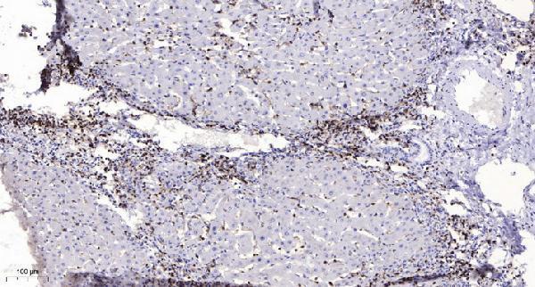
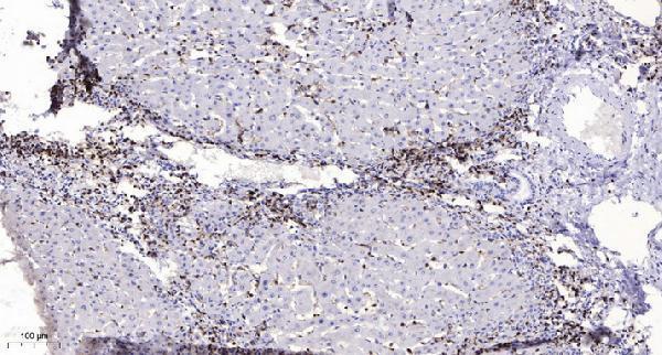
Two anti-PAQR7 antibodies have IHC images from paraffin-embedded human liver cancer (A11720 and A30835 image captions); both list human and mouse reactivity, and A30835 also lists rat (catalog).

Real IHC data Immunohistochemical analysis of paraffin-embedded human liver cancer. 1, Antibody was diluted at 1:200 (4° overnight). 2, Tris-EDTA,pH9.0 was used for antigen retrieval. 3,Secondary antibody was diluted at 1:200 (room temperature, 45min).
Anti-mPR alpha PAQR7 Antibody
Cat # A11720
Real IHC data Immunohistochemical analysis of paraffin-embedded human liver cancer. 1, Antibody was diluted at 1:200(4° overnight). 2, Tris-EDTA,pH9.0 was used for antigen retrieval. 3,Secondary antibody was diluted at 1:200(room temperature, 45min).
Anti-MPRA Antibody
Cat # A30835

A11720 lists IHC and human/mouse reactivity, with an IHC image from paraffin-embedded human liver cancer (catalog; A11720 image caption). A30835 lists IHC and human/mouse/rat reactivity, with an IHC image from paraffin-embedded human liver cancer (catalog; A30835 image caption).

Which to pick: For paraffin-section tissue IHC, either rabbit polyclonal antibody has its own human liver cancer IHC image (A11720 and A30835 image captions); the fixative is unreported in both captions. Choose A30835 when rat reactivity is needed; A11720 lists human and mouse reactivity (catalog). Neither SKU lists IF/ICC or provides an IF image, so there is no payload-supported IF/ICC pick (catalog).

Each figure is that product's own IHC / IF validation image from its datasheet.

References

  1. UniProt Consortium. UniProt entry Q86WK9 (PAQR7_HUMAN, Membrane progestin receptor alpha).
  2. Human Protein Atlas. PAQR7 tissue IHC expression (reliability: Approved).
  3. Human Protein Atlas. PAQR7 subcellular location (ICC-IF): Localized to the cytosol..
  4. Human Protein Atlas. PAQR7 antibody validation summary (1 antibodies).
  5. Expression of Progesterone Receptor Membrane Component 1 (PGRMC1), Progestin and AdipoQ Receptor 7 (PAQPR7), and Plasminogen Activator Inhibitor 1 RNA-Binding Protein (PAIRBP1) in Glioma Spheroids In Vitro. BioMed research international 2016 — PMC4908248.
  6. Emerging roles of CCM genes during tumorigenesis with potential application as novel biomarkers across major types of cancers. Oncology reports 2020 — PMC7160551.
  7. Sex steroids regulate skin pigmentation through nonclassical membrane-bound receptors. eLife 2016 — PMC4863824.
  8. PubMed PMID:12601167 — UniProt-cited evidence.
  9. PubMed PMID:16044242 — UniProt-cited evidence.
  10. PubMed PMID:16710414 — UniProt-cited evidence.