This website uses cookies to ensure you get the best experience on our website.


- Table of Contents
Plan PIAS4 chromogenic IHC on paraffin sections using round or early spermatids as a high-staining reference (HPA tissue IHC). The catalog antibody starts at 5 μg/mL (datasheet); interpret cytoplasmic tissue staining alongside the nuclear/PML-body annotation (HPA tissue IHC; UniProt).

Expected localisation, validated protocols, controls and antibodies — the at-a-glance facts below, then the full design guide.
| Expected localisation | Cytoplasmic in tissue (HPA tissue IHC); nuclear/PML body (UniProt) | |
| Staining pattern | Cytoplasmic staining; high in round/early spermatids (HPA tissue IHC) | |
| Antigen retrieval | Tris-EDTA pH 9.0 HIER, 95–98 °C, 20 min (rule: nuclear antigen) | |
| Positive control | Testis | |
| Negative control | Adipose tissue+4 more · see all |
| Fixation | Keep fixation consistent across sections (standard IHC practice; not target-specific); Matched tissue-IHC evidence does not establish the fixation claim. Validate the specimen-specific method before use. (selected-SKU IHC image A02994) | |
| Caveat | Tissue staining is cytoplasmic despite nuclear annotation (HPA tissue IHC; UniProt) | |
| Regulation | Highest expression in testis (UniProt) | |
| Isoform / epitope | No listed isoforms; mature chain spans residues 2–510 (UniProt) |
The catalog antibody's IHC-P protocol is accompanied by two published PIAS4 IHC workflows (PMC12853826; PMC11954794).
| Sample | Tissue sections; selected-image fixative not specified (standard IHC workflow) |
| Fixation | Image fixative and duration unreported (datasheet A02994); verify before use. |
| Sectioning | 4–5 µm sections on charged slides (standard) |
| Deparaffinisation | Xylene, graded ethanol series to water (standard) |
| Antigen retrieval | Heat-induced epitope retrieval in Tris-EDTA buffer, pH 9.0, 20 min at 95–98 °C (standard rule: nuclear antigen) |
| Peroxidase block | 3% H2O2, 10 min, room temperature (standard) |
| Blocking | 10% normal serum of the secondary host, 30 min, room temperature (standard) |
| Primary antibody | Rabbit anti-PIAS4, 5 μg/mL (datasheet A02994) |
| Primary incubation | Overnight at 4 °C (standard) |
| Detection | HRP-polymer secondary, DAB chromogen 5–10 min (standard) |
| Counterstain | Hematoxylin, blue, dehydrate and mount (standard) |
| Expected result | PIAS4-positive staining in round or early spermatids of testis (HPA tissue IHC: High). HPA tissue profile: Cytoplasmic expression in several tissues, including testis. No signal in the no-primary control. |
PIAS4 is a nuclear protein with no transmembrane segment (UniProt Q8N2W9). For paraffin-section IHC, expect the strongest documented staining in testicular round or early spermatids, although HPA describes cytoplasmic staining across several tissues (HPA: testis High; tissue IHC profile). HPA rates the tissue IHC evidence Enhanced while describing only medium consistency with RNA expression (HPA: reliability description).
| Strong cytoplasmic staining in round or early spermatids on a testis section. | This matches the reported high testis signal and cytoplasmic tissue IHC profile (HPA: testis High; tissue IHC profile). Record the stained cell type and compartment separately; a nuclear location is also reported for PIAS4 (UniProt Q8N2W9). |
| Nuclear staining, possibly appearing as discrete foci, accompanies staining in the expected cells. | A nuclear signal is biologically plausible: PIAS4 localizes to the nucleus and a subset of PML bodies (UniProt Q8N2W9). HPA’s nucleoplasm result comes from ICC-IF, so it does not establish the appearance or required intensity of nuclear foci in chromogenic IHC (HPA: subcellular ICC-IF). |
| The apparent positive signal is confined to cell membranes, with no expected spermatid staining. | An exclusively membranous pattern conflicts with the reported nucleus and PML-body location and lack of a transmembrane segment (UniProt Q8N2W9). Treat it as suspect; review morphology, controls and detection background before assigning PIAS4 positivity (general IHC practice). |
| Strong staining dominates HPA-listed negative cell populations, such as adipocytes or bone-marrow hematopoietic cells. | These populations were reported as not detected (HPA: adipocytes and bone-marrow hematopoietic cells). Unexpected signal may reflect cross-reactivity or endogenous detection activity; it is not, by itself, proof of either cause (general IHC practice). |
| Weak, widespread color obscures cell boundaries, or testis round or early spermatids show no signal. | Diffuse background makes compartment scoring unreliable (general IHC practice). Absent signal in the reported high-staining testis population calls for a technical review, but HPA’s Enhanced rating and medium RNA–staining consistency do not guarantee positivity in every specimen (HPA: testis High; reliability description). |
| Cell type and tissue | Testis round or early spermatids are the supplied high-staining benchmark (HPA: testis High). HPA also reports several low or undetected populations; do not score an entire tissue as uniformly positive or negative (HPA: tissue IHC). |
| Compartment evidence | HPA describes cytoplasmic tissue IHC, whereas UniProt places PIAS4 in the nucleus and PML bodies (HPA: tissue IHC profile; UniProt Q8N2W9). Keep assay-specific observations distinct when judging a slide. |
| Antibody validation | HPA010598 is IHC Enhanced; HPA076008 has no listed IHC rating (HPA: antibody validation). An Enhanced rating supports the reported pattern but does not authenticate every stained structure (HPA: reliability description). |
| Topology and processing | PIAS4 has no transmembrane segment or signal peptide, and its annotated chain is residues 2–510 (UniProt Q8N2W9). These annotations provide no basis for expecting a membrane or secreted staining pattern. |
| Retrieval and fixation evidence | No target-specific retrieval condition or fixation sensitivity is supplied (UniProt Q8N2W9; HPA: tissue IHC). Evaluate retrieval as a general paraffin IHC variable without claiming a PIAS4-specific effect (general IHC practice). |
| IF/ICC comparison | HPA supports nucleoplasm and approves an additional centrosome location, while both listed antibodies have Uncertain ICC ratings (HPA: subcellular ICC-IF; antibody validation). Use that evidence to interpret IF, not to prescribe an IF protocol. |
| Situation | Likely cause | Next action |
|---|---|---|
| No signal in testis round or early spermatids. | The expected high-staining population is absent from the result (HPA: testis High); the available evidence does not identify a PIAS4-specific fixation or retrieval cause. | Check that the relevant cells are present, then review primary-antibody application, detection reagents and a known-positive control; assess retrieval using a standard paraffin IHC workflow (general IHC practice). |
| Weak brown color covers most cells or extracellular space. | Diffuse deposition can obscure the reported cell-specific pattern (HPA: testis High; general IHC practice); appearance alone does not identify its source. | Compare a section processed without primary antibody, then inspect blocking, washes and chromogen development before adjusting working conditions (general IHC practice). |
| Strong color appears in adipocytes or bone-marrow hematopoietic cells. | HPA reports these cell populations as not detected (HPA: tissue IHC). Cross-reactivity and endogenous detection activity are possibilities, not established explanations (general IHC practice). | Check the no-primary control and the tissue distribution on the same run; confirm any unexpected positive call with an independently validated antibody where available (general IHC practice). |
| Staining appears only on cell membranes. | Exclusive membrane staining is inconsistent with the annotated nuclear location and absence of a transmembrane segment (UniProt Q8N2W9). | Recheck cell boundaries against the counterstain and inspect background controls; withhold a PIAS4-positive interpretation until the pattern is independently supported (general IHC practice). |
| Nuclear staining differs from the cytoplasmic tissue IHC profile. | The sources describe different assay readouts: cytoplasmic tissue IHC and nuclear or PML-body localization (HPA: tissue IHC profile; UniProt Q8N2W9). | Report the observed compartment and cell type. Compare with the reported testis pattern and antibody validation; do not require visible PML foci in chromogenic sections (HPA: testis High; HPA010598 IHC Enhanced; general IHC practice). |
| What should an IF/ICC result show? | Nucleoplasm is the supported main location and centrosome an approved additional location, but the listed ICC antibody ratings are Uncertain (HPA: subcellular ICC-IF; antibody validation). | Interpret nuclear signal as compatible with PIAS4 localization, assess any centrosome signal cautiously, and use appropriate IF controls before claiming specificity (HPA: subcellular ICC-IF; general IF practice). |


Comprehensive Human Protein Atlas IHC scoring per tissue (reliability: Enhanced — Medium consistency between antibody staining and RNA expression data.). Rows are taken directly from the HPA tissue chart — click any row's HPA link to view the source.
| Tissue | Cell type | Level | Evidence | Source |
|---|---|---|---|---|
| Testis | Round or early spermatids | High | Protein (IHC) | HPA → |
| Tissue | Cell type | Level | Evidence | Source |
|---|---|---|---|---|
| Adipose tissue | Adipocytes | Not detected | Protein (IHC) | HPA → |
| Bone marrow | Hematopoietic cells | Not detected | Protein (IHC) | HPA → |
| Caudate | Glial cells | Not detected | Protein (IHC) | HPA → |
| Cerebellum | Cells in granular layer | Not detected | Protein (IHC) | HPA → |
| Duodenum | Glandular cells | Not detected | Protein (IHC) | HPA → |
Troubleshoot PIAS4 staining in paraffin sections by checking retrieval, cell identity and subcellular pattern before interpreting chromogenic signal.
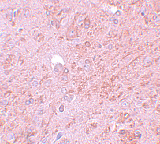
A02994 has pictured PIAS4 IHC staining in rat brain; Human, Mouse and Rat reactivity is listed, with no IF image supplied (A02994 image caption; catalog reactivity; IF image alts).
A02994 is listed for IHC-P and shows PIAS4 staining in rat brain at 5 μg/mL (catalog applications; A02994 image caption). Human and Mouse reactivity is listed, but neither has a supplied IHC image (catalog reactivity; A02994 image alts).
Which to pick: For tissue IHC, choose A02994: its own caption shows rat brain staining at 5 μg/mL, while its application list specifies IHC-P; the caption does not report the fixative or section processing (A02994 image caption; catalog applications). For IF/ICC, A02994 has a suggested IF starting concentration of 20 μg/mL, but no IF image or IF/ICC validation is supplied (catalog dilution; IF image alts; catalog applications). For cross-species work, A02994 lists Human, Mouse and Rat reactivity; its host is Rabbit and clonality is unreported (catalog reactivity; host; clone).