PLEC / Plectin · IHC design guide

Design Immunohistochemistry for PLEC

Plan PLEC staining in paraffin sections around its membranous and cytoplasmic tissue pattern (HPA tissue IHC). Use the IHC-validated antibody and compare staining within defined cell populations, including strongly stained lung macrophages and colon glandular cells (HPA tissue IHC).

Evidence assembled Oct 2026 · For research use; verify linked source records and product datasheet before use
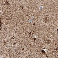
Immunohistochemistry protocol sheet for PLEC (IHC for PLEC): expected localisation Membranous and cytoplasmic staining (HPA tissue IHC), antibody PB9430, validated IHC image, and IHC protocol steps
Printable PLEC IHC protocol sheet — expected localisation Membranous and cytoplasmic staining (HPA tissue IHC), antibody PB9430, controls and protocol steps. Open the full PLEC IHC guide →

PLEC Immunohistochemistry Experimental Design Guide

Expected localisation, validated protocols, controls and antibodies — the at-a-glance facts below, then the full design guide.

Must know before staining
Expected localisation Membranous and cytoplasmic staining (HPA tissue IHC)
Staining pattern Membranous and cytoplasmic staining in almost all cells (HPA tissue IHC)
Antigen retrieval EDTA pH 8.0 HIER, heat-mediated (datasheet PB9430)
Positive control ⓘ Cervix+4 more · see all
Negative control ⓘ None in HPA (detected in all 45 tissues); use no-primary + isotype controls
Important caveats
Reasons your staining may differ from the expected pattern.
Fixation Keep fixation consistent across paraffin sections. (standard IHC practice; not target-specific)
Caveat Lung macrophages stain strongly; score by cell type (HPA tissue IHC)
Regulation Highest in muscle and heart (UniProt)
Isoform / epitope 9 isoforms; check epitope coverage across isoforms (UniProt)
Section 1

Recommended PLEC IHC & IF Protocols

The catalog antibody’s IHC-P protocol (datasheet: PB9430) is paired with a published Plectin-1 staining method for IPMN tissue (PMC3806105).

Recommended immunohistochemistry (IHC-P) protocol parameters
SampleParaffin-embedded human intestine cancer tissue; fixative not specified (datasheet PB9430)
FixationImage fixative and duration unreported (datasheet PB9430); verify before use.
Sectioning4–5 µm sections on charged slides (standard)
DeparaffinisationXylene, graded ethanol series to water (standard)
Antigen retrievalHeat retrieval: EDTA pH 8.0 (datasheet PB9430); 20 min, 95–100 °C (standard)
Peroxidase block3% H2O2, 10 min, room temperature (standard)
Blocking10% goat serum (datasheet PB9430)
Primary antibodyRabbit anti-PLEC, 2-5μg/ml (datasheet PB9430)
Primary incubationOvernight at 4 °C (datasheet PB9430)
DetectionHRP-conjugated secondary, DAB chromogen (datasheet PB9430)
CounterstainHematoxylin, blue, dehydrate and mount (standard)
Expected resultPLEC-positive staining in glandular cells of cervix (HPA tissue IHC: High). HPA tissue profile: Membranous and cytoplasmic expression in almost all cells. No signal in the no-primary control.
💡Decision noteStart with heat-mediated EDTA pH 8.0 retrieval for the catalog antibody (datasheet: PB9430). To reproduce the published method, use its boiling retrieval step (PMC3806105).
Section 2

What Is the Expected PLEC Staining Pattern?

PLEC is a cytoskeletal protein associated with cytoplasm, hemidesmosomes and podosomes; it has no transmembrane segment (UniProt Q15149 subcellular location; topology). In paraffin sections, expect cytoplasmic and membrane-associated staining across many cell types, with strong staining in several glandular epithelia, urothelium and lung macrophages (HPA tissue IHC: Enhanced; membranous and cytoplasmic profile). HPA reports medium consistency between antibody staining and RNA expression (HPA tissue IHC: reliability description).

What am I looking at on my slide?
Cytoplasmic staining with a peripheral or junction-associated accent in expected cells.This fits PLEC's cytoskeletal and hemidesmosomal locations (UniProt Q15149 subcellular location) and HPA's membranous and cytoplasmic tissue profile (HPA tissue IHC). A peripheral outline alone does not establish surface expression: PLEC has no transmembrane segment (UniProt Q15149 topology).
Predominantly nuclear staining, with little cytoplasmic signal.A nuclear-dominant pattern conflicts with the reported cytoskeletal, cytoplasmic and junctional locations (UniProt Q15149 subcellular location; HPA tissue IHC profile). Treat it as suspect staining and check controls and antibody conditions before assigning it to PLEC (general IHC practice).
Strong staining chiefly in a cell population reported as low, while expected high-staining cells are weak.This disagrees with the sampled HPA cell-type pattern; for example, HPA reports low staining in oral squamous epithelial cells and high staining in colon glandular cells (HPA tissue IHC). Consider cross-reactivity or endogenous chromogen activity, but do not call a low-staining population PLEC-negative (general IHC practice; HPA tissue IHC).
Uniform haze covers tissue and spaces between cells.Diffuse background obscures the cell-associated pattern HPA reports (HPA tissue IHC profile). Review background in reagent controls and compare areas with and without tissue before scoring individual cells (general IHC practice).
No staining in a section expected to contain strongly positive cells.A blank result in colon glandular cells, for example, conflicts with HPA's high staining report (HPA: colon glandular cells, High). Check that the expected cells are present and that the staining run worked; one blank section cannot establish biological absence (general IHC practice).
💡Expected PLEC appearanceCall positive when appropriate cells show clear cytoplasmic staining, often with a peripheral accent, above local background; HPA reports high staining in colon glandular cells, urothelial cells and lung macrophages (HPA tissue IHC), while nuclear-dominant staining or uniform haze is suspect against the reported locations (UniProt Q15149 subcellular location; HPA tissue IHC profile).
How each factor affects the staining
Compartment and topologyPLEC links cytoskeletal filaments and anchors them at junctions (UniProt Q15149 function). Interpret an apparent membrane rim alongside cytoplasmic staining: UniProt places PLEC at hemidesmosomes but lists no transmembrane segment (UniProt Q15149 subcellular location; topology).
Cell and tissue selectionHPA reports high staining in cervix, colon and gallbladder glandular cells, lung macrophages and urinary bladder urothelial cells; it also reports low staining in several sampled cell populations (HPA tissue IHC). Choose a control by its named cell population, since a tissue label alone does not identify the cells to score (general IHC practice).
Evidence strengthThe HPA tissue profile is rated Enhanced, with medium consistency between antibody staining and RNA expression (HPA tissue IHC: reliability; reliability description). The three listed HPA antibodies have Enhanced IHC status (HPA antibodies: HPA025967, HPA029906, CAB003847). Use those observations as reference patterns, while assessing the antibody used in the experiment on its own controls (general IHC practice).
Isoforms and epitope coverageUniProt lists nine PLEC isoforms (UniProt Q15149 isoforms). The supplied records give no epitope position or isoform coverage for the experimental antibody, so an unusual cell pattern cannot be assigned to a particular isoform from these data alone (UniProt Q15149 isoforms; HPA antibodies).
Why is my staining missing, weak or wrong?
SituationLikely causeNext action
Expected high-staining cells are blank.The relevant cells may be absent from the section, or a run step may have failed (general IHC practice).Confirm cell identity and inspect a same-run positive control; if it also fails, review the validated IHC antibody instructions and the retrieval, detection and counterstain steps (general IHC practice). HPA identifies colon glandular cells as High (HPA tissue IHC).
Brown signal appears in the no-primary control.Endogenous detection activity or nonspecific detection-reagent binding can produce chromogen without primary antibody (general IHC practice).Check the detection system's blocking and control procedure, then score only signal exceeding the no-primary background (general IHC practice). HPA reports high PLEC staining in lung macrophages, so assess those cells against the control rather than dismissing them by cell type (HPA tissue IHC).
Nuclei dominate the staining.This does not match the reported cytoplasmic and junctional locations (UniProt Q15149 subcellular location; HPA tissue IHC profile).Compare with the no-primary control and an expected positive cell population, then review antibody dilution and detection conditions under the validated IHC procedure (general IHC practice).
A reported low-staining cell population appears strongest.Cross-reactivity or local detection background is possible; HPA low means some staining was observed, not none (HPA tissue IHC; general IHC practice).Check the same-run control and compare the named cells with a reported High population, such as colon glandular cells (HPA tissue IHC); avoid scoring the whole tissue as one cell type (general IHC practice).
Diffuse haze makes cell boundaries hard to judge.Excess background can obscure the cell-associated pattern reported for PLEC (HPA tissue IHC profile; general IHC practice).Review reagent controls, blocking, washes and detection conditions; score only interpretable areas with a clear cell-associated signal (general IHC practice).
Q: Should an IF/ICC image show the same chromogenic outline as IHC?A: HPA places PLEC mainly in intermediate filaments and cytosol by ICC-IF, with additional focal-adhesion localization (HPA subcellular: supported filaments and focal adhesions; approved cytosol).Compare IF/ICC compartment localization with those HPA observations and assess the antibody's ICC status separately: HPA029906 is Supported, HPA025967 is Uncertain, and CAB003847 has no ICC status listed (HPA antibodies).

Sample controls for PLEC IHC & IF

🧪Run colon first and require staining in its glandular cells (HPA: High in colon glandular cells). HPA detects PLEC in all 45 scored tissues, so there is no HPA-supported negative tissue; use no-primary and isotype controls, and treat cells without specific cytoskeletal staining on the positive slide as local background rather than proven PLEC-negative cells (HPA: no negative tissue rows; UniProt Q15149 subcellular location).
Positive control tissue: Cervix (Glandular cells, HPA High)
Negative control tissue: None in HPA: PLEC is detected in all 45 scored tissues. Use a no-primary (secondary-only) and an isotype control instead.
ICC-IF cell lines (HPA subcellular resource): HPA ICC-IF images show PLEC in A-431, U-251MG, U2OS, with annotated localisation: Intermediate filaments (supported), Cytosol (approved) (HPA subcellular).
Technical controls: Include a no-primary, secondary-only control, a concentration-matched rabbit isotype control matched to the primary antibody’s clonality where known, and a PLEC knockout specimen if available as a biological negative (PB9430 caption: rabbit primary; standard IHC controls). Block endogenous peroxidase and inspect inflammatory cells for misleading DAB signal in colon sections (standard chromogenic IHC practice).
⚠️Feasibility: No target-specific fixation window or fixation effect is reported in the supplied evidence, and the selected PB9430 paraffin-section caption does not state its fixative (PB9430 caption). Heat retrieval in EDTA at pH 8.0 was used in that paraffin-section example; the evidence does not establish whether retrieval is required for every specimen or whether frozen sections or IF are easier (PB9430 caption). In colon, inflammatory-cell peroxidase can create misleading chromogenic signal, so interpret glandular-cell staining against the controls (HPA: High in colon glandular cells; standard chromogenic IHC practice).

HPA tissue IHC evidence for PLEC

Comprehensive Human Protein Atlas IHC scoring per tissue (reliability: Enhanced — Medium consistency between antibody staining and RNA expression data.). Rows are taken directly from the HPA tissue chart — click any row's HPA link to view the source.

Positive expression · recommended positive controls

TissueCell typeLevelEvidenceSource
Cervix Glandular cells High Protein (IHC) HPA →
Colon Glandular cells High Protein (IHC) HPA →
Gallbladder Glandular cells High Protein (IHC) HPA →
Lung Macrophages High Protein (IHC) HPA →
Urinary bladder Urothelial cells High Protein (IHC) HPA →

Undetected expression · recommended negative controls

TissueCell typeLevelEvidenceSource
None in HPA: PLEC is detected in all 45 scored tissues. Use a no-primary (secondary-only) and an isotype control instead.
Section 3

Advanced PLEC IHC Tips

Troubleshoot PLEC staining by checking retrieval, tissue processing, expected localisation and controls before comparing signal across sections.

Which retrieval conditions should I start with for paraffin PLEC IHC?
Start with heat-mediated retrieval in EDTA pH 8.0 for paraffin sections (datasheet PB9430). The pictured PB9430 section was incubated with 2 μg/ml primary antibody overnight at 4°C, providing a documented starting point after retrieval (caption PB9430). If staining is weak, compare retrieval duration on adjacent sections while holding antibody concentration and chromogenic development constant (standard IHC practice). Check whether stronger staining appears in expected cytoplasmic or junction-associated regions, rather than simply increasing diffuse background (UniProt Q15149 localisation; standard IHC practice). Record heating, cooling and section loss for each condition so that retrieval effects remain interpretable (standard IHC practice).
Could fixation explain weak or uneven PLEC staining?
PLEC-specific fixation sensitivity is unknown from the supplied evidence; the PB9430 paraffin caption does not state a fixative (caption PB9430). Record the actual fixative and fixation interval for each specimen, then compare sections processed under matched conditions (standard IHC practice). If staining varies, assess tissue preservation and run the same retrieval, antibody incubation and DAB development across adjacent sections (standard IHC practice). The documented PB9430 workflow used EDTA pH 8.0 retrieval and 2 μg/ml primary antibody overnight at 4°C (caption PB9430). Do not assign a fixation effect to PLEC from expression patterns or protein modifications alone (HPA tissue IHC; UniProt Q15149 modified residues).
Where should I expect PLEC signal in tissue sections?
Expect predominantly cytoplasmic and junction-associated staining, consistent with PLEC at the cytoskeleton and hemidesmosomes (UniProt Q15149 localisation). HPA describes membranous and cytoplasmic expression in almost all cells; its tissue-IHC reliability is Enhanced, with medium consistency against RNA data (HPA tissue IHC). Interpret a linear boundary signal alongside cytoplasmic staining and tissue architecture, since PLEC has no transmembrane segment (UniProt Q15149 topology and localisation). A purely nuclear pattern deserves scrutiny against controls and neighbouring cells because it does not match the supplied localisation evidence (UniProt Q15149 localisation; HPA tissue IHC). Compare like cell types across sections before judging a compartment shift (standard IHC practice).
How do isoforms and epitope location affect a negative PLEC result?
PLEC has 9 listed isoforms, so an antibody's epitope determines which variants it can recognise (UniProt Q15149 isoforms; standard IHC practice). Check the antibody's stated immunogen or epitope against the isoform sequence before treating absent staining as absent PLEC (standard IHC practice). The supplied record places CH domains at residues 179–282 and 295–400, and an SH3 domain at 941–998 (UniProt Q15149 domains). These positions do not identify the PB9430 epitope, and the supplied caption does not establish isoform specificity (UniProt Q15149 domains; caption PB9430). Compare a negative region with an appropriate positive tissue control processed in the same run (standard IHC practice).
How should I extend the PLEC assessment to multiplex IF?
Treat multiplex IF as a separate assay: the supplied PB9430 evidence documents chromogenic staining of a paraffin section, without an IF procedure (caption PB9430). Pair PLEC with a validated marker for the expected cell type and assess overlap within individual cells (HPA tissue IHC; standard IF practice). Choose spectrally separated fluorophores after checking tissue autofluorescence in an unstained section, and include single-marker controls when separating channels (standard IF practice). Because PLEC has no transmembrane segment and localises to cytosol and intermediate filaments, test permeabilisation for access to the intracellular epitope (UniProt Q15149 topology; HPA subcellular; standard IF practice). Document the fixation and permeabilisation used rather than transferring conditions from the IHC caption (caption PB9430; standard IF practice).
What should I check when PLEC DAB staining is diffuse?
First compare the stained section with a no-primary control to assess nonspecific detection and endogenous peroxidase signal (standard IHC practice). Use a peroxidase block and inspect whether background persists outside the expected cytoplasmic or junction-associated pattern (standard IHC practice; UniProt Q15149 localisation). The PB9430 caption reports 10% goat-serum blocking, 2 μg/ml primary antibody and a 30-minute secondary incubation at 37°C (caption PB9430). If diffuse signal remains, compare shorter primary exposure or lower concentration on adjacent sections while keeping retrieval and DAB development matched (standard IHC practice). Score only interpretable cellular staining after checking folds, damaged edges and pigment (standard IHC practice).
How should I score PLEC across comparable tissue sections? ⚠ ANSWER MARKED FOR VERIFICATION
Define the cell population and expected cytoplasmic or junction-associated compartment before scoring PLEC (UniProt Q15149 localisation; standard IHC practice). For a cellular readout, report the percentage of positive cells or an H-score that combines staining intensity and positive-cell fraction (standard IHC practice). Normalise those measures to eligible cells in the same annotated region, rather than total section area when cellularity differs (standard IHC practice). If counting discrete positive cells, report density per mm² of viable tissue and apply the same exclusion rules to every section (standard IHC practice). Keep retrieval, DAB development and imaging conditions consistent across the comparison (standard IHC practice).
How can I distinguish true PLEC staining from artefact?
A credible PLEC result follows cellular cytoplasm or junction-associated structures and agrees with tissue morphology (UniProt Q15149 localisation; HPA tissue IHC). Compare candidate staining with a same-run positive control; HPA reports high signal in colon glandular cells and lung macrophages (HPA tissue IHC). Treat isolated nuclear staining, damaged-edge staining and signal concentrated in necrotic areas as suspect until controls support them (UniProt Q15149 localisation; standard IHC practice). Use a no-primary control and peroxidase blocking to evaluate DAB signal from endogenous enzyme activity (standard IHC practice). HPA's Enhanced tissue-IHC reliability still carries medium consistency with RNA expression, so interpret discordant cells cautiously (HPA tissue IHC).
Boster reagents

Best PLEC / Plectin IHC Antibodies

PB9430 has paraffin-section IHC images from human intestine and lung cancer, mouse intestine, and rat intestine, plus IF data from U20S cells (catalog image captions).

Real IHC data IHC analysis of Plectin using anti-Plectin antibody (PB9430). Plectin was detected in a paraffin-embedded section of human intestine cancer tissue. Heat mediated antigen retrieval was performed in EDTA buffer (pH 8.0, epitope retrieval solution). The tissue section was blocked with 10% goat serum. The tissue section was then incubated with 2 μg/ml rabbit anti-Plectin Antibody (PB9430) overnight at 4°C. Peroxidase Conjugated Goat Anti-rabbit IgG was used as secondary antibody and incubated for 30 minutes at 37°C. The tissue section was developed using HRP Conjugated Rabbit IgG Super Vision Assay Kit (Catalog # SV0002) with DAB as the chromogen.
Anti-Plectin Antibody ®
Cat # PB9430

PB9430 is the SKU with a rendered card; its IHC images show paraffin sections of human intestine and lung cancer, mouse intestine, and rat intestine (PB9430 IHC image captions). Its IF image shows U20S cells (PB9430 IF image caption), and IF and ICC are listed applications (PB9430 catalog applications).

Which to pick: Choose PB9430 for tissue IHC: its paraffin-section caption reports EDTA retrieval at pH 8.0 and primary antibody at 2 μg/ml (PB9430 IHC image caption); the fixative is unreported (PB9430 IHC image caption). Choose PB9430 for IF/ICC because both are listed applications and it has a cell IF image (PB9430 catalog applications; PB9430 IF image caption). For cross-species IHC, PB9430 has human, mouse, and rat paraffin-section images (PB9430 IHC image captions).

Each figure is that product's own IHC / IF validation image from its datasheet.

References

  1. UniProt Consortium. UniProt entry Q15149 (PLEC_HUMAN, Plectin).
  2. Human Protein Atlas. PLEC tissue IHC expression (reliability: Enhanced).
  3. Human Protein Atlas. PLEC subcellular location (ICC-IF): Mainly localized to the cytosol and intermediate filaments. In addition localized to the focal adhesion sites..
  4. Human Protein Atlas. PLEC antibody validation summary (3 antibodies).
  5. Integrin alpha1 beta1 promotes interstitial fibrosis in a mouse model of polycystic kidney disease. bioRxiv : the preprint server for biology 2024 — PMC11526950.
  6. Epstein-Barr virus-driven molecular pathogenesis of primary pulmonary lymphoepithelial carcinoma. Frontiers in oncology 2025 — PMC12380799.
  7. Plectin-1 is a biomarker of malignant pancreatic intraductal papillary mucinous neoplasms. Journal of gastrointestinal surgery : official journal of the Society for Surgery of the Alimentary Tract 2009 — PMC3806105.
  8. Plectin promotes an aggressive phenotype and represses cytotoxic T cell activity in pancreatic cancer. Frontiers in immunology 2026 — PMC13526953.
  9. PubMed PMID:8633055 — UniProt-cited evidence.
  10. PubMed PMID:8698233 — UniProt-cited evidence.
  11. PubMed PMID:14672974 — UniProt-cited evidence.