PNP / Purine nucleoside phosphorylase · IHC design guide

Design Immunohistochemistry for PNP

Plan paraffin-section PNP staining using epididymal glandular cells and kidney collecting ducts as positive controls (HPA tissue IHC). Expect cytoplasmic signal and score red blood cells separately because they are also PNP-positive (HPA tissue IHC).

Evidence assembled Oct 2026 · For research use; verify linked source records and product datasheet before use
Immunohistochemistry protocol sheet for PNP (IHC for PNP): expected localisation Cytoplasmic staining in most tissues (HPA tissue IHC), antibody A00988-1, validated IHC image, and IHC protocol steps
Printable PNP IHC protocol sheet — expected localisation Cytoplasmic staining in most tissues (HPA tissue IHC), antibody A00988-1, controls and protocol steps. Open the full PNP IHC guide →

PNP Immunohistochemistry Experimental Design Guide

Expected localisation, validated protocols, controls and antibodies — the at-a-glance facts below, then the full design guide.

Must know before staining
Expected localisation Cytoplasmic staining in most tissues (HPA tissue IHC)
Staining pattern Cytoplasmic; high in epididymal glands and kidney ducts (HPA tissue IHC)
Antigen retrieval Citrate pH 6 HIER, heat-mediated (datasheet A00988-1)
Positive control ⓘ Bone marrow+4 more · see all
Negative control ⓘ Adipose tissue+4 more · see all
Important caveats
Reasons your staining may differ from the expected pattern.
Fixation Keep fixation consistent across sections (standard IHC practice; not target-specific). Selected-image fixative and duration unreported (datasheet A00988-1); verify before use.
Caveat PNP-positive red blood cells can complicate cell scoring (HPA tissue IHC)
Regulation Tissue-enhanced RNA: marrow, epididymis (HPA tissue RNA)
Isoform / epitope No annotated isoforms; one cytosolic chain, 1–289 (UniProt)
Section 1

Recommended PNP IHC & IF Protocols

The catalog antibody protocol uses citrate pH 6 heat retrieval (datasheet A00988-1). The published IHC protocols below cover prostate and breast tissue (PMC3261671; PMC12774560).

Recommended immunohistochemistry (IHC-P) protocol parameters
SampleParaffin-embedded mouse kidney tissues; fixative not specified (datasheet A00988-1)
FixationImage fixative and duration unreported (datasheet A00988-1); verify before use.
Sectioning4–5 µm sections on charged slides (standard)
DeparaffinisationXylene, graded ethanol series to water (standard)
Antigen retrievalHeat retrieval: Citrate pH 6, 20 min (datasheet A00988-1)
Peroxidase block3% H2O2, 10 min, room temperature (standard)
Blocking10% goat serum (datasheet A00988-1)
Primary antibodyRabbit anti-PNP, 0.5-1μg/ml (datasheet A00988-1)
Primary incubationOvernight at 4 °C (datasheet A00988-1)
DetectionStreptavidin-biotin complex (SABC), DAB chromogen (datasheet A00988-1)
CounterstainHematoxylin, blue, dehydrate and mount (standard)
Expected resultPNP-positive staining in hematopoietic cells of bone marrow (HPA tissue IHC: High). HPA tissue profile: Cytoplasmic expression in most tissues, highly abundant in epididymis, kidney and the gastrointestinal tract. Distinct positivity in red blood cells. No signal in the no-primary control.
💡Decision noteStart with citrate pH 6 heat retrieval for the catalog antibody (datasheet A00988-1). The breast tissue study specifies microwave retrieval for 15 min (PMC12774560).
Section 2

What Is the Expected PNP Staining Pattern?

PNP should appear mainly in the cytoplasm, consistent with its cytosolic location and lack of a transmembrane segment (UniProt P00491: cytosol; no transmembrane segment). Expect strong staining in selected cells of bone marrow, epididymis, kidney, gut, placenta and spleen, with distinct red blood cell positivity (HPA tissue IHC: High in listed cells; red blood cells positive). HPA rates its tissue IHC evidence Enhanced (HPA tissue IHC: reliability Enhanced).

What am I looking at on my slide?
Strong cytoplasmic staining in epididymal glandular cells or kidney collecting ducts, with little nuclear signal.This fits the reported High staining in those cell populations and PNP's cytosolic location (HPA tissue IHC: High; UniProt P00491: cytosol). Score the named cells separately from neighboring structures, since a tissue-wide average can hide the expected cell-specific pattern (general IHC practice).
Predominantly nuclear staining, or a sharp cell-surface rim without convincing cytoplasmic staining.That compartment conflicts with the reported cytosolic location and absence of a transmembrane segment (UniProt P00491: cytosol; no transmembrane segment). Treat the pattern as suspect until it is reproduced with appropriate staining controls and a separately validated antibody (general IHC practice).
Strong staining in adipocytes, esophageal squamous cells or cardiomyocytes.Those specific cells are reported as Not detected, making prominent staining unexpected (HPA tissue IHC: adipocytes, esophageal squamous cells and cardiomyocytes Not detected). Consider cross-reactivity or endogenous chromogenic activity; check the cell identity and detection controls before assigning the signal to PNP (general IHC practice).
Diffuse color across cells and extracellular areas, without a discernible cytoplasmic pattern.A field-wide haze does not match the reported cellular cytoplasmic pattern (HPA tissue IHC: cytoplasmic expression in most tissues). Review no-primary and detection controls for background, then assess blocking, washing and detection conditions as general IHC variables (general IHC practice).
No cellular signal in an otherwise evaluable epididymis, kidney or bone marrow section.Absence in the relevant glandular, collecting-duct or hematopoietic cells conflicts with their reported High staining (HPA tissue IHC: High in those cells). Check that the expected cells are present and that retrieval, antibody incubation and chromogenic detection worked; the supplied sources give no PNP-specific fixation sensitivity.
💡Expected PNP appearanceCall a section positive when its expected cells show clear cytoplasmic staining, potentially strong in HPA High populations and distinct in red blood cells; isolated nuclear or cell-surface signal is suspect (HPA tissue IHC: High populations; red blood cells positive; UniProt P00491: cytosol, no transmembrane segment).
How each factor affects the staining
Compartment and topology (UniProt P00491: cytosol; no transmembrane segment)Interpret cell-body cytoplasmic color as the expected localization; a membrane rim alone does not establish PNP staining (UniProt P00491: cytosol; no transmembrane segment). Use the counterstain to distinguish cytoplasm from nuclei (general IHC practice).
Cell-specific tissue distribution (HPA tissue IHC: High and Not detected entries)Compare like cells within a section: kidney collecting ducts are High, whereas adipocytes are Not detected (HPA tissue IHC). HPA's negative designation applies to the named cell population, not every cell in that organ (HPA tissue IHC: cell-level entries).
Antibody evidence (HPA tissue IHC: reliability Enhanced; HPA antibody HPA001625: IHC Enhanced)The reported IHC pattern has supporting validation, but it does not make every new staining run specific (HPA tissue IHC: reliability Enhanced; HPA antibody HPA001625: IHC Enhanced). Interpret the run alongside its own controls (general IHC practice).
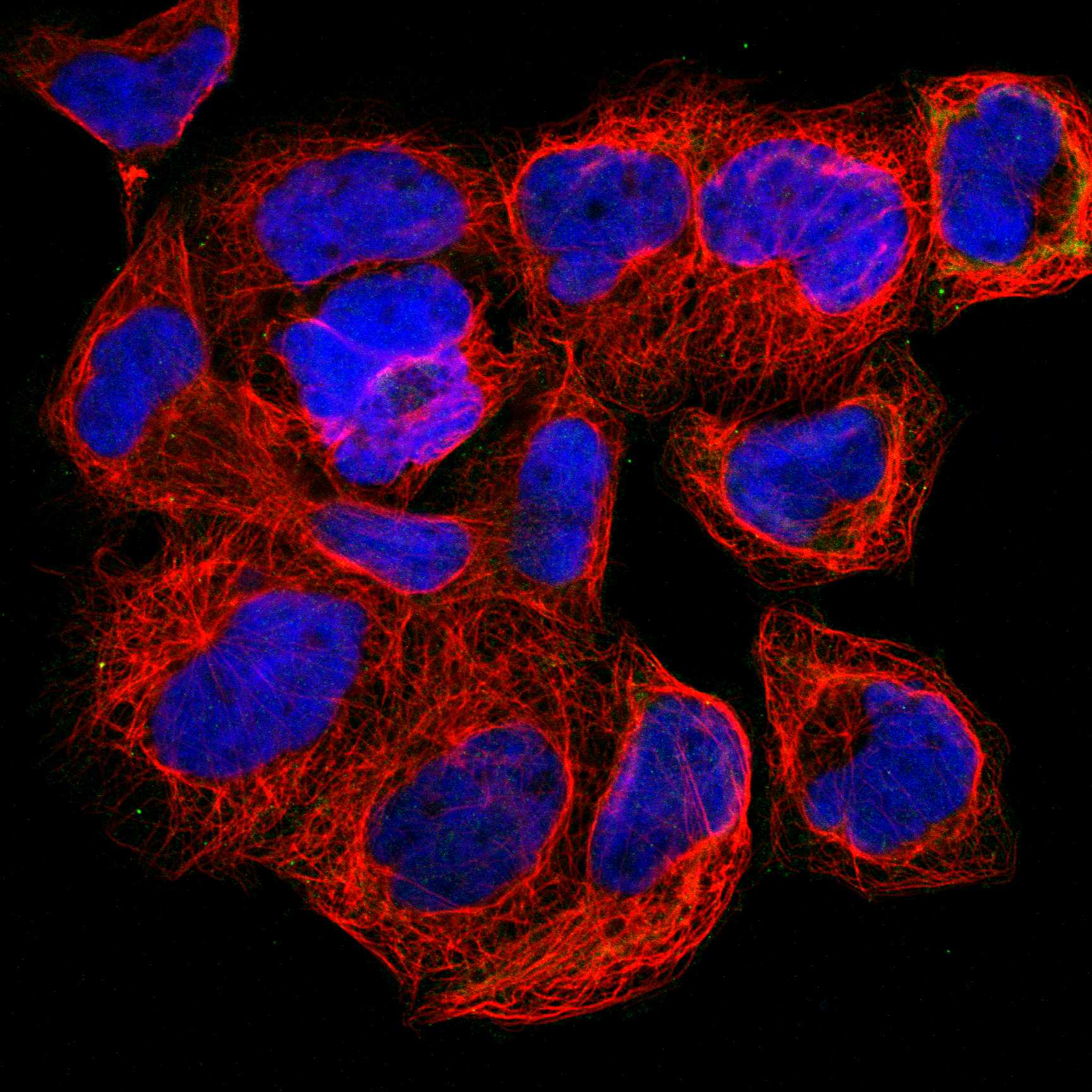
IF/ICC question: Where should fluorescence appear? (HPA subcellular ICC-IF: cytosol supported)Expect cytosolic fluorescence: HPA reports supported cytosol localization in ICC-IF (HPA subcellular ICC-IF: cytosol supported). That observation can inform localization checks, while this section's tissue intensity calls come from HPA tissue IHC.
Why is my staining missing, weak or wrong?
SituationLikely causeNext action
The expected positive cells are unstained.The sampled section may lack the named cells, or a general IHC step may have failed; PNP-specific fixation sensitivity is unreported in the supplied sources.Confirm the cell population against the counterstain and compare a known High population, such as epididymal glandular cells (HPA tissue IHC: High). Then check retrieval, antibody incubation and detection controls (general IHC practice).
Only nuclei stain.Nuclear dominance conflicts with PNP's cytosolic assignment (UniProt P00491: cytosol).Verify the compartment at higher magnification, review the counterstain and no-primary control, and seek concordant cytoplasmic staining before scoring PNP positive (general IHC practice; UniProt P00491: cytosol).
A sharp membrane outline dominates.A surface-only pattern conflicts with the reported cytosolic location and lack of a transmembrane segment (UniProt P00491: cytosol; no transmembrane segment).Check whether any cell-body cytoplasm stains and compare the result with an expected positive cell population; withhold a positive call based on the rim alone (HPA tissue IHC: High populations; general IHC practice).
Chromogen appears in an HPA Not detected cell population.Cross-reactivity or endogenous detection activity is possible; the HPA designation is specific to the listed cells (HPA tissue IHC: Not detected entries).Confirm cell identity, inspect no-primary and detection controls, and evaluate whether blocking and washing reduce the unexpected color (general IHC practice).
The entire section has diffuse color.General staining background can obscure the cellular cytoplasmic distribution reported for PNP (HPA tissue IHC: cytoplasmic expression in most tissues).Compare a no-primary control, then review blocking, washing, antibody concentration and chromogen development as general IHC variables (general IHC practice). Do not score diffuse haze as cellular positivity.
Red blood cells stain strongly while nearby nucleated cells are weak.Distinct red blood cell positivity is part of the reported pattern, so it does not by itself establish nonspecific staining (HPA tissue IHC: red blood cells positive; UniProt P00491: expressed in red blood cells).Identify red blood cells by morphology and score the neighboring named cell populations separately (general IHC practice). Compare their staining with HPA's cell-specific levels rather than treating red blood cell color as a tissue-wide result (HPA tissue IHC: cell-level entries).

Sample controls for PNP IHC & IF

🧪Run kidney first: collecting duct cells should stain strongly (HPA: High in kidney collecting ducts). Use adipose tissue as the negative tissue, with adipocytes expected to lack detectable staining (HPA: Not detected in adipocytes); on the kidney slide, count cells outside collecting ducts as internal negatives only when they are visibly unstained, since their PNP status is not specified by the supplied HPA row.
Positive control tissue: Bone marrow (Hematopoietic cells, HPA High)
Negative control tissue: Adipose tissue (HPA Not detected)
ICC-IF cell lines (HPA subcellular resource): HPA ICC-IF images show PNP in A-431, RPTEC/TERT1, U2OS, with annotated localisation: Cytosol (supported) (HPA subcellular).
Technical controls: Include a no-primary, secondary-only control and a concentration-matched rabbit IgG isotype control (selected-SKU caption: rabbit primary antibody); use PNP-knockout material as a biological negative when available (standard IHC practice). Check endogenous peroxidase and biotin background in kidney, particularly with the caption’s biotin–streptavidin DAB detection (selected-SKU caption: SABC with DAB).
⚠️Feasibility: A target-specific fixation window and fixation effect are unreported; the selected-SKU paraffin-section caption does not state a fixative (selected-SKU caption: fixative unreported). The caption uses citrate retrieval at pH 6 for 20 minutes, but retrieval dependency is unreported (selected-SKU caption: citrate retrieval); no matched evidence establishes that frozen sections or IF are easier. Endogenous biotin or peroxidase may complicate kidney DAB staining with the reported detection system, so interpret staining against the background controls (selected-SKU caption: kidney tissue and SABC with DAB; standard IHC practice).

HPA tissue IHC evidence for PNP

Comprehensive Human Protein Atlas IHC scoring per tissue (reliability: Enhanced — High consistency between antibody staining and RNA expression data. External characterization data supports antibody staining.). Rows are taken directly from the HPA tissue chart — click any row's HPA link to view the source.

Positive expression · recommended positive controls

TissueCell typeLevelEvidenceSource
Bone marrow Hematopoietic cells High Protein (IHC) HPA →
Colon Enterocytes - Microvilli High Protein (IHC) HPA →
Duodenum Endocrine cells High Protein (IHC) HPA →
Epididymis Glandular cells High Protein (IHC) HPA →
Kidney Collecting ducts High Protein (IHC) HPA →

Undetected expression · recommended negative controls

TissueCell typeLevelEvidenceSource
Adipose tissue Adipocytes Not detected Protein (IHC) HPA →
Adrenal gland Glandular cells Not detected Protein (IHC) HPA →
Caudate Glial cells Not detected Protein (IHC) HPA →
Cerebral cortex Endothelial cells Not detected Protein (IHC) HPA →
Esophagus Squamous epithelial cells Not detected Protein (IHC) HPA →
Section 3

Advanced PNP IHC Tips

Use the catalog antibody’s paraffin-section conditions as the IHC starting point, then judge staining by cell type, cytosolic localisation and matched controls.

Which antigen retrieval conditions should I use for PNP in paraffin sections?
Start with heat-mediated citrate retrieval at pH 6 for 20 minutes (datasheet A00988-1; selected IHC caption). Let sections cool consistently, then compare a retrieved section with a matched section processed without retrieval to assess whether staining depends on that step (standard IHC practice). The catalog antibody’s illustrated workflow used 10% goat serum blocking and 1 μg/ml primary antibody overnight at 4°C, so keep those conditions fixed while assessing retrieval (selected IHC caption). Evaluate signal in kidney collecting ducts alongside tissue background, because that cell population is reported as highly positive (HPA: High in kidney collecting ducts). If staining remains weak, optimise heating within the citrate pH 6 workflow before testing another buffer as a documented fallback (standard IHC practice).
How should I assess fixation when PNP staining is unexpectedly weak?
Target-specific fixation sensitivity is unknown: the selected paraffin-section caption does not report a fixative (selected IHC caption). Record each specimen’s fixative and processing history, and compare sections processed the same way before attributing a signal difference to PNP abundance (standard IHC practice). Keep section thickness, citrate retrieval at pH 6, and primary antibody conditions consistent across that comparison (datasheet A00988-1; standard IHC practice). Include a known positive cell population, such as kidney collecting ducts, on the same run to check assay performance (HPA: High in kidney collecting ducts; standard IHC practice). If fixation histories differ, report that limitation with the staining result instead of assigning a PNP-specific fixation effect (standard IHC practice).
What subcellular staining pattern supports a PNP IHC result?
Expect predominantly cytoplasmic staining, consistent with PNP’s cytosolic localisation and absence of a transmembrane segment (UniProt P00491; HPA: cytoplasmic expression in most tissues). Assess the pattern within identified cells, because red blood cells can also stain distinctly and may occupy spaces beside the cells being scored (HPA: distinct positivity in red blood cells). In kidney, examine collecting ducts as an expected positive population rather than treating every nearby structure as equivalent (HPA: High in kidney collecting ducts). Diffuse nuclear-only or crisp membrane-only staining warrants review of morphology, controls and background before assignment to PNP (UniProt P00491 localisation; standard IHC practice). Compare the questioned pattern with a matched no-primary control and the same tissue’s cytoplasmic signal (standard IHC practice).
Could an isoform or epitope difference explain discordant PNP staining?
The supplied record annotates 0 isoforms and a single 1–289 PNP chain, so an annotated isoform switch does not explain discordance here (UniProt P00491). The record lists N-terminal acetylmethionine and phosphoserine at position 251, but provides no antibody epitope map or evidence that either modification changes staining (UniProt P00491; selected IHC caption). Check the catalog antibody’s epitope documentation if available, and avoid assigning a specific residue or modification to an unexplained IHC pattern (standard IHC practice). Compare sections under the stated citrate pH 6 retrieval and 1 μg/ml primary conditions before interpreting discordance (datasheet A00988-1; selected IHC caption). Document differences in processing and cell composition alongside staining intensity (standard IHC practice).
How can I investigate a PNP IHC pattern with multiplex IF?
Use IF as a separate assay and pair PNP with a marker that identifies the cell population under investigation, such as a collecting-duct marker when examining the reported kidney-positive population (HPA: High in kidney collecting ducts; standard IF practice). Choose spectrally separated fluorophores and place the weaker signal in a channel less affected by the specimen’s autofluorescence, checking single-stain and unstained controls (standard IF practice). PNP is cytosolic with no transmembrane segment, so permit access across the plasma membrane with a controlled permeabilisation step while checking that cell morphology remains interpretable (UniProt P00491; standard IF practice). Compare cytosolic overlap within the identified cells rather than fluorescence from adjacent red blood cells, which can contain PNP (HPA: distinct positivity in red blood cells). Do not carry the IHC caption’s paraffin retrieval conditions into IF without validating that IF preparation (selected IHC caption; standard IF practice).
How do I distinguish PNP signal from chromogenic background?
Inspect matched no-primary and secondary-only controls for nonspecific detection, and check endogenous peroxidase blocking before interpreting DAB deposits (standard chromogenic IHC practice). The illustrated assay used a biotinylated secondary, streptavidin–biotin detection and DAB, making detection-reagent background worth assessing in that workflow (selected IHC caption; standard IHC practice). Compare the location of deposits with cell boundaries and the expected cytoplasmic pattern, especially where red blood cells are present (UniProt P00491 localisation; HPA: distinct positivity in red blood cells). Reduce excessive staining by optimising blocking, washes, primary concentration and chromogen development one variable at a time (standard IHC practice). Treat signal persisting in reagent controls as background until resolved, even if its intensity resembles a positive cell (standard IHC practice).
What is a defensible way to quantify PNP in tissue IHC? ⚠ ANSWER MARKED FOR VERIFICATION
Define the cell population and region before scoring, then record the percentage of positive cells and staining intensity for cytoplasmic PNP (UniProt P00491 localisation; standard IHC practice). An H-score can combine intensity categories 0–3 with their positive-cell percentages to yield 0–300, provided the same thresholds are applied across sections (standard IHC practice). For spatial comparisons, report positive-cell density per mm² of viable tissue or the fraction positive within a specified cell population (standard IHC practice). Normalise to the number of evaluable cells or viable tissue area, and score red blood cells separately from resident tissue cells (HPA: distinct positivity in red blood cells; standard IHC practice). Keep acquisition, counterstain assessment and scoring rules consistent across the comparison (standard IHC practice).
Which findings would make a strong PNP stain biologically unconvincing?
A credible PNP result should show cytoplasmic signal in identifiable cells and agree with the sampled tissue’s expected cell pattern (UniProt P00491 localisation; HPA: cytoplasmic expression in most tissues). Kidney collecting ducts and red blood cells are documented positive populations, whereas adipocytes are reported as not detected (HPA: High in kidney collecting ducts; HPA: distinct positivity in red blood cells; HPA: Not detected in adipocytes). Question isolated nuclear or membrane-only staining, section-edge accentuation, and deposits concentrated in necrotic areas (UniProt P00491 localisation; standard IHC practice). Check no-primary controls and endogenous peroxidase blocking when a DAB signal follows blood-rich areas or persists outside intact cells (standard chromogenic IHC practice). Resolve doubtful cases against morphology, matched processing controls and another validated measure of PNP expression where available (standard IHC practice).
Boster reagents

Best PNP / Purine nucleoside phosphorylase IHC Antibodies

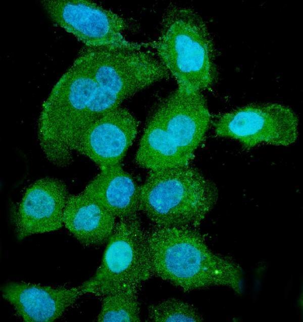
Anti-PNP antibodies have IHC images from paraffin sections of mouse and rat kidney and human cancer tissues (A00988-1 and M00988 IHC captions), plus an A431 cell IF image (M00988 IF caption).

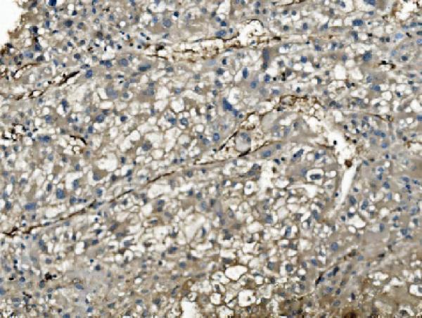
Real IHC data IHC analysis of PNP using anti-PNP antibody (A00988-1). PNP was detected in paraffin-embedded section of mouse kidney tissues. Heat mediated antigen retrieval was performed in citrate buffer (pH6, epitope retrieval solution) for 20 mins. The tissue section was blocked with 10% goat serum. The tissue section was then incubated with 1μg/ml rabbit anti-PNP Antibody (A00988-1) overnight at 4°C. Biotinylated goat anti-rabbit IgG was used as secondary antibody and incubated for 30 minutes at 37°C. The tissue section was developed using Strepavidin-Biotin-Complex (SABC)(Catalog # SA1022) with DAB as the chromogen.
Anti-Nucleoside phosphorylase/PNP Antibody ®
Cat # A00988-1
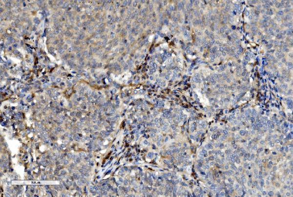
Real IHC data IHC analysis of PNP using anti-PNP antibody (M00988). PNP was detected in paraffin-embedded section of human lung cancer tissue. Heat mediated antigen retrieval was performed in EDTA buffer (pH8.0, epitope retrieval solution). The tissue section was blocked with 10% goat serum. The tissue section was then incubated with 2μg/ml mouse anti-PNP Antibody (M00988) overnight at 4°C. Biotinylated goat anti-mouse IgG was used as secondary antibody and incubated for 30 minutes at 37°C. The tissue section was developed using Strepavidin-Biotin-Complex (SABC) (Catalog # SA1021) with DAB as the chromogen.
Anti-PNP Antibody ® (monoclonal, 7G5)
Cat # M00988

A00988-1 will render with an IHC image of mouse kidney; its other IHC captions show rat kidney and human liver and renal cancer (A00988-1 IHC captions). M00988 will render with an IHC image of human lung cancer; its other captions show human liver cancer by IHC and A431 cells by IF (M00988 IHC and IF captions).

Which to pick: For tissue IHC across human, mouse, and rat, choose A00988-1 (catalog: IHC application and human, mouse, and rat reactivity; A00988-1 IHC captions: paraffin sections). For human tissue IHC or IF/ICC, choose monoclonal M00988, clone 7G5 (catalog: human reactivity and IHC, IF, and ICC applications; M00988 IHC captions: paraffin sections; M00988 IF caption: A431 cells). The fixative is unreported for both antibodies’ paraffin section images (A00988-1 and M00988 IHC captions).

Each figure is that product's own IHC / IF validation image from its datasheet.

References

  1. UniProt Consortium. UniProt entry P00491 (PNPH_HUMAN, Purine nucleoside phosphorylase).
  2. Human Protein Atlas. PNP tissue IHC expression (reliability: Enhanced).
  3. Human Protein Atlas. PNP subcellular location (ICC-IF): Localized to the cytosol..
  4. Human Protein Atlas. PNP antibody validation summary (1 antibodies).
  5. Tumour suppressors miR-1 and miR-133a target the oncogenic function of purine nucleoside phosphorylase (PNP) in prostate cancer. British journal of cancer 2012 — PMC3261671.
  6. PNP as a Metabolic and Prognostic Driver of Breast Cancer Aggressiveness: Insights from Patient Tissue and Cell Models. Oncology research 2025 — PMC12774560.
  7. Paraneoplastic pemphigus associated with nonhuman papillomavirus-related tonsillar squamous cell carcinoma: A case report. Medicine 2024 — PMC11384057.
  8. Nanoparticle delivery of an AKT/PDK1 inhibitor improves the therapeutic effect in pancreatic cancer. International journal of nanomedicine 2014 — PMC4263440.
  9. PubMed PMID:6087295 — UniProt-cited evidence.
  10. PubMed PMID:3029074 — UniProt-cited evidence.
  11. PubMed PMID:12928150 — UniProt-cited evidence.