This website uses cookies to ensure you get the best experience on our website.


- Table of Contents
Plan PNPLA8 chromogenic IHC on paraffin sections with the IHC-validated antibody, starting at 1:50 (datasheet A30583). Expect cytoplasmic tissue staining (HPA tissue IHC); bronchial respiratory epithelium stains strongly, while vaginal squamous epithelium is not detected (HPA tissue IHC).

Expected localisation, validated protocols, controls and antibodies — the at-a-glance facts below, then the full design guide.
| Expected localisation | Cytoplasm (HPA tissue IHC); ER, mitochondrial and peroxisomal membranes (UniProt) | |
| Staining pattern | Cytoplasmic staining in glandular, respiratory epithelial and neuronal cells (HPA tissue IHC) | |
| Antigen retrieval | Tris-EDTA pH 9.0 HIER, heat-mediated (datasheet A30583) | |
| Positive control | Adrenal gland+4 more · see all | |
| Negative control | Vagina |
| Fixation | Keep paraffin-section fixation consistent across samples (standard IHC practice; not target-specific) | |
| Caveat | Tissue staining and RNA show medium consistency (HPA tissue IHC) | |
| Regulation | Expression regulation is not annotated (UniProt) | |
| Isoform / epitope | 3 isoforms; check epitope coverage and membrane orientation (UniProt) |
The catalog antibody’s IHC-P protocol uses Tris-EDTA retrieval (datasheet A30583). Two published PNPLA8 tissue microarray protocols provide citrate-retrieval alternatives (PMC10989391; PMC10683240).
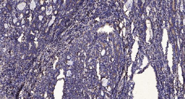
| Sample | Paraffin-embedded human Gastric adenocarcinoma tissue; fixative not specified (datasheet A30583) |
| Fixation | Image fixative and duration unreported (datasheet A30583); verify before use. |
| Sectioning | 4–5 µm sections on charged slides (standard) |
| Deparaffinisation | Xylene, graded ethanol series to water (standard) |
| Antigen retrieval | Heat retrieval: Tris-EDTA pH 9.0 (datasheet A30583); 20 min, 95–100 °C (standard) |
| Peroxidase block | 3% H2O2, 10 min, room temperature (standard) |
| Blocking | 10% normal serum of the secondary host, 30 min, room temperature (standard) |
| Primary antibody | Rabbit anti-PNPLA8, 1:50 recommended; image 1:200 (datasheet A30583) |
| Primary incubation | Overnight at 4 °C (standard) |
| Detection | HRP-polymer secondary, DAB chromogen 5–10 min (standard) |
| Counterstain | Hematoxylin, blue, dehydrate and mount (standard) |
| Expected result | PNPLA8-positive staining in glandular cells of adrenal gland (HPA tissue IHC: High). HPA tissue profile: General cytoplasmic expression. No signal in the no-primary control. |
PNPLA8 IHC should show general cytoplasmic staining in glandular cells of several organs, bronchial respiratory epithelium, and selected brain cells (HPA tissue IHC). PNPLA8 is annotated on endoplasmic reticulum, mitochondrial, and peroxisomal membranes, with one transmembrane segment at residues 475–495 (UniProt Q9NP80 topology). HPA rates the tissue staining Approved, with medium consistency between staining and RNA expression (HPA tissue IHC).
| Clear cytoplasmic staining in bronchial respiratory epithelial cells or colon glandular cells. | This fits the reported High staining in those cell types (HPA tissue IHC). Score the relevant cells and their cytoplasmic signal; ordinary chromogenic IHC does not resolve which PNPLA8-associated organelle produced it (UniProt Q9NP80 subcellular location; general IHC practice). |
| Staining appears mainly extracellular or in glandular lumens, without convincing cellular cytoplasmic signal. | That distribution does not match HPA’s general cytoplasmic IHC profile (HPA tissue IHC). Inspect a no-primary control and section quality before assigning it to PNPLA8; deposited chromogen or nonspecific staining can mislead interpretation (general IHC practice). |
| Strong staining appears in vaginal squamous epithelium or broadly across unrelated cell types. | Vaginal squamous epithelial cells are reported as Not detected (HPA tissue IHC). Treat unexpected staining as unconfirmed: cross-reactivity or endogenous detection activity are possibilities, and morphology alone cannot distinguish them (general IHC practice). |
| Diffuse colour covers the section, obscuring boundaries between positive cells and surrounding tissue. | A section-wide haze prevents comparison with HPA’s cell-specific observations (HPA tissue IHC). Check the no-primary control, detection background, blocking, washing, and exposure to chromogen before scoring intensity (general IHC practice). |
| A bronchus or colon section shows no convincing signal in the expected epithelial or glandular cells. | That conflicts with the reported High staining in those cells (HPA tissue IHC), but a single negative slide cannot establish absent PNPLA8. Check tissue identity and run a documented positive section alongside it; then assess the IHC workflow (general IHC practice). |
| Membrane location and light-microscopy resolution | PNPLA8 has one transmembrane segment at 475–495 and is annotated on three intracellular membrane systems (UniProt Q9NP80 topology and subcellular location). HPA describes the tissue IHC appearance as general cytoplasmic expression; chromogenic IHC alone cannot assign an individual organelle (HPA tissue IHC; general IHC practice). |
| Tissue selection and validation strength | Adrenal, appendix, colon, and duodenal glandular cells, bronchial epithelium, and selected brain cells are reported High; vaginal squamous cells are Not detected (HPA tissue IHC). The listed antibody, HPA020083, is IHC Approved; no IHC Enhanced designation is supplied (HPA antibodies). |
| Isoforms and unknown epitope | UniProt lists 3 PNPLA8 isoforms (UniProt Q9NP80). The supplied evidence does not identify the IHC antibody’s epitope or establish which isoforms it detects, so isoform-specific staining or loss of signal cannot be inferred from this record. |
| Does IF/ICC predict the IHC compartment? | HPA ICC-IF reports approved nucleoplasm and cytosol locations, plus Golgi localization (HPA subcellular ICC-IF). HPA tissue IHC instead summarizes a general cytoplasmic pattern (HPA tissue IHC); evaluate a nuclear-only IHC result with controls rather than treating either assay’s compartment call as interchangeable. |
| Situation | Likely cause | Next action |
|---|---|---|
| Expected positive epithelial cells are blank. | The result conflicts with HPA’s High bronchial or colon cell staining, but the failed step is undetermined (HPA tissue IHC). | Confirm the cell type on the section; compare a documented positive section processed in the same IHC run, then review retrieval, antibody incubation, and detection as general IHC practice. |
| The no-primary control develops brown signal. | Primary-antibody-independent staining suggests detection-system background, including possible endogenous enzyme activity (general chromogenic IHC practice). | Resolve the control signal before interpreting PNPLA8; review the detection system, endogenous-activity control, washing, and chromogen development (general IHC practice). |
| Vaginal squamous cells stain strongly. | Those cells are listed as Not detected (HPA tissue IHC); the unexpected signal could reflect nonspecific binding or detection background (general IHC practice). | Check cell identity and no-primary staining, then compare with a documented HPA-positive tissue before assigning PNPLA8 positivity (HPA tissue IHC; general IHC practice). |
| All compartments look uniformly stained. | Uniform haze does not permit the cell-specific, general cytoplasmic assessment reported for PNPLA8 (HPA tissue IHC). | Use the no-primary control to assess background; review blocking, washes, antibody concentration, and chromogen development as general IHC practice, without assuming a PNPLA8-specific fixation effect. |
| Only nuclei stain in tissue IHC. | HPA ICC-IF reports nucleoplasm localization, while its tissue IHC summary is general cytoplasmic expression (HPA subcellular ICC-IF; HPA tissue IHC). The assays do not establish the cause of this slide’s discrepancy. | Check the cytoplasmic signal in a documented positive tissue and examine no-primary staining; record nuclear-only IHC as unresolved until controlled (general IHC practice). |
| A negative result is attributed to fixation. | The supplied HPA tissue IHC and UniProt records provide no target-specific evidence that fixation changes PNPLA8 staining (HPA tissue IHC; UniProt Q9NP80). | Keep fixation sensitivity unassigned; first verify a positive control and routine retrieval and detection performance under the same IHC conditions (general IHC practice). |
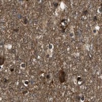


Comprehensive Human Protein Atlas IHC scoring per tissue (reliability: Approved — Medium consistency between antibody staining and RNA expression data.). Rows are taken directly from the HPA tissue chart — click any row's HPA link to view the source.
| Tissue | Cell type | Level | Evidence | Source |
|---|---|---|---|---|
| Adrenal gland | Glandular cells | High | Protein (IHC) | HPA → |
| Appendix | Glandular cells | High | Protein (IHC) | HPA → |
| Bronchus | Respiratory epithelial cells | High | Protein (IHC) | HPA → |
| Caudate | Neuronal cells | High | Protein (IHC) | HPA → |
| Cerebellum | Cells in granular layer | High | Protein (IHC) | HPA → |
| Tissue | Cell type | Level | Evidence | Source |
|---|---|---|---|---|
| Vagina | Squamous epithelial cells | Not detected | Protein (IHC) | HPA → |
Use the catalog antibody’s paraffin-section IHC conditions as a starting point, then judge PNPLA8 staining against tissue morphology and controls.
A30583 has IHC data from paraffin-embedded human gastric adenocarcinoma (A30583 image caption). Its listed reactivity covers human and mouse (A30583 catalog).
A30583 is listed for IHC and has an IHC image from paraffin-embedded human gastric adenocarcinoma (A30583 catalog; A30583 image caption). It is listed as human- and mouse-reactive; the supplied image shows human tissue only (A30583 catalog; A30583 image caption).
Which to pick: For tissue IHC, choose A30583: it is a rabbit polyclonal listed for IHC, with a paraffin-section example; the fixative is unreported (A30583 catalog; A30583 image caption). For IF/ICC, A30583 has neither a listed IF application nor an IF image (A30583 catalog). For cross-species work, A30583 lists human and mouse reactivity, while its supplied IHC image shows human tissue only (A30583 catalog; A30583 image caption).