This website uses cookies to ensure you get the best experience on our website.


- Table of Contents
Plan PRDM2 paraffin IHC with the catalog antibody at 2–5 μg/ml (datasheet: A03223-2). Use high-staining brain neuronal cells as a reference (HPA tissue IHC), and score nuclear and cytoplasmic staining separately (HPA tissue IHC; standard IHC practice).

Expected localisation, validated protocols, controls and antibodies — the at-a-glance facts below, then the full design guide.
| Expected localisation | Nuclear and cytoplasmic staining in most tissues (HPA tissue IHC) | |
| Staining pattern | Most tissues show nuclear and cytoplasmic cell staining (HPA tissue IHC) | |
| Antigen retrieval | EDTA pH 8.0 HIER, heat-mediated (datasheet A03223-2) | |
| Positive control | Caudate+4 more · see all | |
| Negative control | Ovary+3 more · see all |
| Fixation | Keep fixation conditions consistent across sections. (standard IHC practice; not target-specific) | |
| Caveat | Staining and RNA expression show low consistency (HPA tissue IHC) | |
| Regulation | Isoform 1 exceeds isoform 3 in testis (UniProt) | |
| Isoform / epitope | 5 isoforms; epitope coverage is undetermined (UniProt) |
The catalog antibody uses heat-mediated EDTA pH 8.0 retrieval (datasheet A03223-2). The published lung tumor IHC protocol below adds a tissue-specific example (PMC12327139: IHC methods).
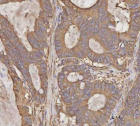
| Sample | Paraffin-embedded human rectum adenocarcinoma tissue; fixative not specified (datasheet A03223-2) |
| Fixation | Image fixative and duration unreported (datasheet A03223-2); verify before use. |
| Sectioning | 4–5 µm sections on charged slides (standard) |
| Deparaffinisation | Xylene, graded ethanol series to water (standard) |
| Antigen retrieval | Heat retrieval: EDTA pH 8.0 (datasheet A03223-2); 20 min, 95–100 °C (standard) |
| Peroxidase block | 3% H2O2, 10 min, room temperature (standard) |
| Blocking | 10% goat serum (datasheet A03223-2) |
| Primary antibody | Rabbit anti-PRDM2, 2-5 μg/ml (datasheet A03223-2) |
| Primary incubation | Overnight at 4 °C (datasheet A03223-2) |
| Detection | HRP-conjugated secondary, DAB chromogen (datasheet A03223-2) |
| Counterstain | Hematoxylin, blue, dehydrate and mount (standard) |
| Expected result | PRDM2-positive staining in neuronal cells of caudate (HPA tissue IHC: High). HPA tissue profile: Nuclear and cytoplasmic expression in most tissues. No signal in the no-primary control. |
PRDM2 is a nuclear protein with no transmembrane segment (UniProt Q13029). In tissue IHC, HPA reports nuclear and cytoplasmic staining in most tissues, with high staining in neurons of the caudate, cerebral cortex and hippocampus (HPA tissue IHC). Use these cells as reference positives while allowing for tissue dependent intensity. HPA rates the tissue staining Approved but reports low consistency between antibody staining and RNA expression (HPA tissue IHC).
| Neuronal nuclei stain strongly in caudate, cerebral cortex or hippocampus, with some cytoplasmic staining. | This fits the highest listed tissue IHC signals and HPA’s nuclear and cytoplasmic profile (HPA tissue IHC: High in neuronal cells; nuclear and cytoplasmic expression). Interpret the nuclear component alongside PRDM2’s nuclear localization (UniProt Q13029), while keeping HPA’s low staining–RNA consistency in mind (HPA tissue IHC). |
| Adrenal or breast glandular cells, or kidney tubular cells, show moderate staining. | Medium staining in these specific cell populations is consistent with the tissue observations (HPA tissue IHC: Medium in adrenal and breast glandular cells and kidney tubular cells). Compare the cell type and compartment as well as intensity; a medium result here need not resemble the high neuronal examples (HPA tissue IHC). |
| A sharp membrane rim dominates, or staining appears mainly in a cell population outside the listed pattern. | A membrane dominant pattern conflicts with the reported nuclear and cytoplasmic tissue profile and lack of a transmembrane segment (HPA tissue IHC; UniProt Q13029 topology). Staining in an unexpected cell type warrants checks for cross reactivity or endogenous detection activity; HPA’s tissue list is a guide, not proof that every unlisted cell is negative (HPA tissue IHC). |
| Diffuse color covers cells and surrounding tissue without a clear cellular pattern. | Treat this as background until the detection controls separate tissue staining from reagent or endogenous activity (general IHC practice). It cannot be assigned to PRDM2 merely because cytoplasmic expression is reported (HPA tissue IHC: nuclear and cytoplasmic expression). Check whether recognizable nuclei and the expected neuronal cell population remain distinguishable (HPA tissue IHC: High in neuronal cells). |
| No staining appears in neuronal cells of a caudate, cerebral cortex or hippocampus section. | This disagrees with the listed high staining observations (HPA tissue IHC: High in neuronal cells). First check section quality, retrieval, primary antibody use and detection with appropriate controls (general IHC practice). A single blank section does not establish absent PRDM2, particularly given the reported low consistency between antibody staining and RNA expression (HPA tissue IHC). |
| Tissue and cell selection (HPA tissue IHC) | Caudate, cerebral cortex and hippocampus neuronal cells are listed High; adrenal and breast glandular cells and kidney tubular cells are Medium (HPA tissue IHC). Ovarian stroma, splenic red pulp, thyroid glandular cells and vaginal squamous cells are listed Not detected in their specified populations (HPA tissue IHC). |
| Strength of tissue evidence (HPA tissue IHC; HPA antibody validation) | The tissue profile is Approved, and antibody HPA005809 is IHC Approved, but the tissue report flags low consistency between staining and RNA expression (HPA tissue IHC; HPA antibodies). Treat a matching pattern as supportive evidence and investigate discordant specimens with controls rather than treating either readout as definitive. |
| Antigen retrieval in paraffin sections (general IHC practice) | If a reference positive section is weak, review the established IHC-P retrieval and detection conditions before interpreting it as biologically negative (general IHC practice). The supplied UniProt and HPA records do not report PRDM2 specific retrieval or fixation sensitivity; no retrieval condition or target specific effect can be inferred from them. |
| Isoforms and antibody epitope (UniProt Q13029) | UniProt lists five PRDM2 isoforms and says isoform 1 is expressed in testis at a much higher level than isoform 3 (UniProt Q13029). Antibody epitope coverage is not supplied, so a staining difference cannot be assigned to an isoform without independent epitope information. UniProt lists no signal peptide or propeptide and one chain spanning residues 1–1718 (UniProt Q13029). |
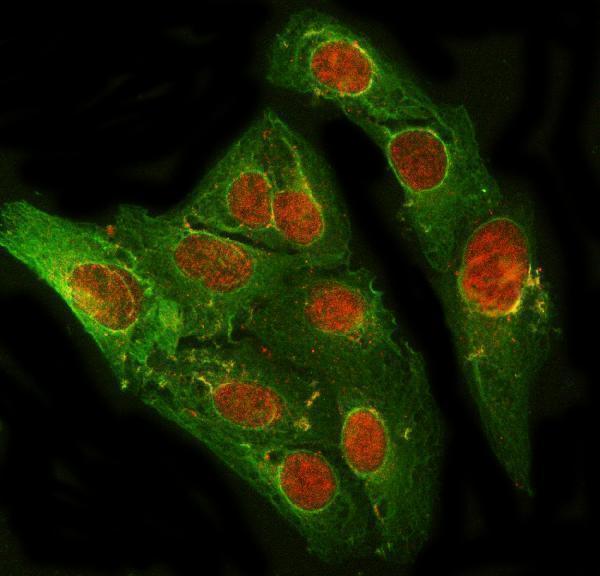
| What should IF/ICC show? (HPA subcellular; HPA antibodies) | HPA reports nucleoplasm as Supported and Golgi apparatus as Approved in ICC-IF, with images from A-431, U-251MG and U2OS; antibody HPA005809 is ICC Supported (HPA subcellular; HPA antibodies). These ICC-IF observations help interpret compartment patterns but do not supply an IF/ICC protocol or establish that every tissue IHC section will show a distinct Golgi signal. |
| Situation | Likely cause | Next action |
|---|---|---|
| The chosen positive section has weak or absent neuronal staining. | The result conflicts with listed High neuronal staining in caudate, cerebral cortex and hippocampus (HPA tissue IHC); an IHC workflow failure is possible (general IHC practice). | Confirm the intended neuronal population, then review section quality, antigen retrieval, primary antibody application and detection controls (general IHC practice). Do not infer PRDM2 specific fixation sensitivity from the supplied records. |
| A sharp membrane outline is the dominant staining pattern. | A membrane dominant pattern does not fit nuclear PRDM2 without a transmembrane segment or HPA’s nuclear and cytoplasmic tissue profile (UniProt Q13029; HPA tissue IHC). | Recheck cell boundaries and counterstain, compare the same run with a reference positive section, and inspect primary omitted controls for detection background (general IHC practice). Treat a persistent membrane only pattern as unconfirmed. |
| Unexpected cells stain as strongly as, or more strongly than, the expected population. | Cross reactivity or endogenous detection activity may contribute (general IHC practice); HPA’s Approved tissue profile has low staining–RNA consistency (HPA tissue IHC). | Locate staining within the unexpected cells, compare with the specified HPA cell populations, and run appropriate primary omitted and detection controls (general IHC practice; HPA tissue IHC). Do not assign specificity from intensity alone. |
| Color is diffuse across tissue or appears outside cells. | Background from blocking, washing or the detection system can obscure a cellular pattern (general IHC practice). Cytoplasmic staining is reported for PRDM2, but a noncellular haze is not that pattern (HPA tissue IHC). | Use a primary omitted control, review blocking and washes, and, where relevant to the chosen chromogen system, assess endogenous enzyme activity (general IHC practice). Score only interpretable cellular staining. |
| A presumed negative tissue contains stained cells. | HPA’s Not detected calls apply to named populations, such as ovarian stroma cells or splenic red pulp cells, rather than every cell in the whole organ (HPA tissue IHC). | Identify the stained cell population before calling the control discordant. If the named population stains, check primary omitted and detection controls, then interpret the finding in light of HPA’s low staining–RNA consistency (general IHC practice; HPA tissue IHC). |
| ICC-IF shows a Golgi pattern while tissue IHC is read mainly as nuclear and cytoplasmic. | HPA reports Golgi apparatus as Approved and nucleoplasm as Supported in ICC-IF, while its tissue IHC summary is nuclear and cytoplasmic (HPA subcellular; HPA tissue IHC). | Keep the application and specimen type attached to each observation. Assess tissue IHC against its reported cell populations and compartments; use the ICC-IF localization as context, without requiring a visible Golgi pattern in paraffin sections (HPA tissue IHC; HPA subcellular). |
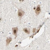
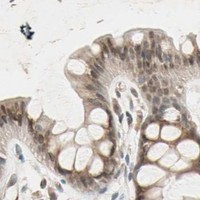
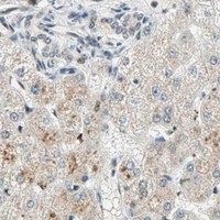
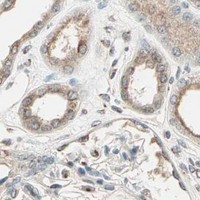


Comprehensive Human Protein Atlas IHC scoring per tissue (reliability: Approved — Low consistency between antibody staining and RNA expression data.). Rows are taken directly from the HPA tissue chart — click any row's HPA link to view the source.
| Tissue | Cell type | Level | Evidence | Source |
|---|---|---|---|---|
| Caudate | Neuronal cells | High | Protein (IHC) | HPA → |
| Cerebral cortex | Neuronal cells | High | Protein (IHC) | HPA → |
| Hippocampus | Neuronal cells | High | Protein (IHC) | HPA → |
| Adrenal gland | Glandular cells | Medium | Protein (IHC) | HPA → |
| Breast | Glandular cells | Medium | Protein (IHC) | HPA → |
| Tissue | Cell type | Level | Evidence | Source |
|---|---|---|---|---|
| Ovary | Ovarian stroma cells | Not detected | Protein (IHC) | HPA → |
| Spleen | Cells in red pulp | Not detected | Protein (IHC) | HPA → |
| Thyroid gland | Glandular cells | Not detected | Protein (IHC) | HPA → |
| Vagina | Squamous epithelial cells | Not detected | Protein (IHC) | HPA → |
Use the catalog antibody’s paraffin section IHC example as the starting point, and interpret compartmental staining alongside controls and the reported tissue profile.
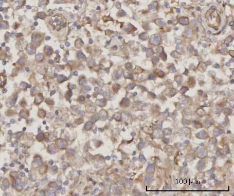
A03223-2 has IHC images from human rectum adenocarcinoma, human testicular seminoma, and mouse and rat testis paraffin sections (catalog IHC captions). Its IF/ICC image uses U2OS cells (catalog IF caption).
A03223-2 is listed for IHC and shown on paraffin sections of human rectum adenocarcinoma, human testicular seminoma, mouse testis, and rat testis (catalog applications; A03223-2 IHC captions). A03223-2 is also listed for IF/ICC and shown in U2OS cells (catalog applications; A03223-2 IF caption).
Which to pick: For tissue IHC, choose A03223-2 for paraffin sections; its listed concentration is 2–5 μg/ml, and its IHC images used 2 μg/ml after EDTA retrieval at pH 8.0 (catalog dilution; A03223-2 IHC captions). For IF/ICC, the same SKU has a U2OS cell image using 5 μg/ml (A03223-2 IF caption). For cross-species IHC, A03223-2 has human, mouse, and rat paraffin-section images; its host is rabbit, clonality is unreported, and the IHC captions do not report a fixative (catalog entry; A03223-2 IHC captions).