PRKCD / Protein kinase C delta type · IHC design guide

Design Immunohistochemistry for PRKCD

Plan paraffin-section PRKCD IHC with the IHC-validated antibody at 0.5–1 µg/mL (datasheet A00822-1). Use colon glandular cells as a positive tissue reference and assess cytoplasmic and membranous staining (HPA tissue IHC).

Evidence assembled Oct 2026 · For research use; verify linked source records and product datasheet before use
Immunohistochemistry protocol sheet for PRKCD (IHC for PRKCD): expected localisation Cytoplasmic and membranous staining (HPA tissue IHC), antibody A00822-1, validated IHC image, and IHC protocol steps
Printable PRKCD IHC protocol sheet — expected localisation Cytoplasmic and membranous staining (HPA tissue IHC), antibody A00822-1, controls and protocol steps. Open the full PRKCD IHC guide →

PRKCD Immunohistochemistry Experimental Design Guide

Expected localisation, validated protocols, controls and antibodies — the at-a-glance facts below, then the full design guide.

Must know before staining
Expected localisation Cytoplasmic and membranous staining (HPA tissue IHC)
Staining pattern General cytoplasmic and membranous staining (HPA tissue IHC)
Antigen retrieval Citrate pH 6 HIER, heat-mediated (datasheet A00822-1)
Positive control ⓘ Appendix+4 more · see all
Negative control ⓘ Soft tissue
Important caveats
Reasons your staining may differ from the expected pattern.
Fixation Keep fixation conditions consistent across sections. (standard IHC practice; not target-specific)
Caveat Activation can redistribute PRKCD (UniProt)
Regulation Some cancer cells highly express PRKCD (UniProt)
Isoform / epitope 2 isoforms; cleavage may separate N/C epitopes (UniProt)
Section 1

Recommended PRKCD IHC & IF Protocols

The catalog antibody uses citrate pH 6 retrieval (datasheet: A00822-1). The published FFPE PRKCD protocol uses EDTA retrieval (PMC6284509: IHC).

Recommended immunohistochemistry (IHC-P) protocol parameters
SampleParaffin-embedded human mammary cancer tissues; fixative not specified (datasheet A00822-1)
FixationImage fixative and duration unreported (datasheet A00822-1); verify before use.
Sectioning4–5 µm sections on charged slides (standard)
DeparaffinisationXylene, graded ethanol series to water (standard)
Antigen retrievalHeat retrieval: Citrate pH 6, 20 min (datasheet A00822-1)
Peroxidase block3% H2O2, 10 min, room temperature (standard)
Blocking10% goat serum (datasheet A00822-1)
Primary antibodyRabbit anti-PRKCD, 0.5-1μg/ml (datasheet A00822-1)
Primary incubationOvernight at 4 °C (datasheet A00822-1)
DetectionStreptavidin-biotin complex (SABC), DAB chromogen (datasheet A00822-1)
CounterstainHematoxylin, blue, dehydrate and mount (standard)
Expected resultPRKCD-positive staining in glandular cells of appendix (HPA tissue IHC: High). HPA tissue profile: General cytoplasmic and membranous expression. No signal in the no-primary control.
💡Decision noteStart with citrate pH 6 for the catalog antibody (datasheet: A00822-1); consider EDTA when adapting the published FFPE protocol (PMC6284509: IHC).
Section 2

What Is the Expected PRKCD Staining Pattern?

PRKCD staining is generally cytoplasmic and membranous in tissue IHC (HPA tissue IHC); PRKCD has no transmembrane segment and can also localize to the nucleus, mitochondrion and endomembrane system (UniProt Q05655). Expect staining in glandular, respiratory epithelial and hematopoietic cells among the HPA-reported positives (HPA tissue IHC). The HPA tissue profile has Enhanced reliability, with medium consistency between antibody staining and RNA expression (HPA tissue IHC).

What am I looking at on my slide?
Cytoplasmic staining, with some membranous staining, in appendix or colon glandular cells.This fits the reported general pattern and High staining in those cells (HPA tissue IHC). Judge staining in the relevant cells, because other cells in the section may differ (general IHC practice).
Staining is confined to extracellular material, without convincing cellular staining.This is outside the reported cellular locations (UniProt Q05655; HPA tissue IHC). Treat it as possible artefact and review morphology and detection controls (general IHC practice). Nuclear staining alone needs context because the nucleus is an annotated location (UniProt Q05655).
Strong staining appears in peripheral nerve within soft tissue.HPA reports PRKCD as not detected there (HPA tissue IHC). Check cell identification, antibody specificity and endogenous detection activity before calling it positive (general IHC practice); one HPA observation is not a universal negative standard.
Diffuse color obscures cell boundaries across the section.The pattern cannot establish cellular localization (general IHC practice). Examine a no-primary control for detection background, then review blocking, washes and antibody concentration (general IHC practice).
No staining appears in an appendix glandular-cell control.HPA reports High staining in those cells (HPA tissue IHC). Confirm that the expected cells are present, then check retrieval and detection conditions for the IHC-validated antibody (general IHC practice).
💡Expected PRKCD appearanceCall a section positive when relevant cells show interpretable cytoplasmic, often membranous, staining consistent with the HPA tissue level (HPA tissue IHC); diffuse extracellular color alone is a suspect result (general IHC practice).
How each factor affects the staining
Choice of positive tissueAppendix and colon glandular cells, bronchial respiratory epithelial cells, and bone-marrow hematopoietic cells are reported High (HPA tissue IHC). Confirm that the named cell population is present before scoring a control (general IHC practice).
Subcellular distributionThe tissue profile is generally cytoplasmic and membranous (HPA tissue IHC). UniProt also lists nuclear, perinuclear, mitochondrial and endomembrane locations, with translocation after activation or apoptotic stimulation (UniProt Q05655); localization alone does not prove activation.
Antibody recognitionUniProt lists 2 isoforms and regulatory and catalytic products from PRKCD processing (UniProt Q05655). The supplied record gives no epitope for the catalog antibody, so its recognition of individual forms cannot be predicted.
IHC evidenceFour listed HPA antibodies have Enhanced IHC validation (HPA antibodies). The tissue profile's Enhanced reliability includes medium staining–RNA consistency (HPA tissue IHC); compare morphology and controls when a result conflicts with the reported pattern (general IHC practice).
IF/ICC: what pattern is expected?HPA reports mainly cytosolic PRKCD, with additional endoplasmic-reticulum, Golgi and vesicular localization (HPA subcellular ICC-IF). This is an IF/ICC interpretation reference; tissue IHC remains the primary application here.
Why is my staining missing, weak or wrong?
SituationLikely causeNext action
Positive-control glandular cells are blank.The signal may have failed at retrieval, primary-antibody or detection steps (general IHC practice); appendix glandular cells are reported High (HPA tissue IHC).Confirm the cells and run morphology and detection checks; review the catalog antibody's IHC-P conditions before adjusting retrieval or dilution (general IHC practice).
All tissue compartments show similar diffuse color.Nonspecific background or detection activity may obscure the reported cytoplasmic and membranous pattern (general IHC practice; HPA tissue IHC).Inspect a no-primary control; review blocking, washes and antibody concentration, then score only resolved cellular staining (general IHC practice).
Soft-tissue peripheral nerve stains strongly.HPA reports that cell population as not detected (HPA tissue IHC); misidentified cells, cross-reactivity or detection background are possibilities (general IHC practice).Recheck anatomy and controls; seek independent antibody support before treating the signal as PRKCD (general IHC practice).
Only extracellular deposits show color.Extracellular-only staining conflicts with the listed cellular locations (UniProt Q05655; HPA tissue IHC).Review morphology and a no-primary control, and withhold a positive call unless cellular staining is resolved (general IHC practice).
A nuclear component accompanies cytoplasmic staining.The nucleus is a listed PRKCD location (UniProt Q05655), while HPA describes the general tissue pattern as cytoplasmic and membranous (HPA tissue IHC).Assess the cell type and controls; report the nuclear component descriptively without inferring activation from staining alone (general IHC practice).
A tissue expected to stain shows a weaker signal than anticipated.HPA levels describe observed cell populations, and its Enhanced tissue profile has medium staining–RNA consistency (HPA tissue IHC); section conditions may also affect interpretation (general IHC practice).Confirm the sampled cells and compare a known-positive section in the same run; review IHC-P detection conditions (general IHC practice). Target-specific fixation sensitivity is unreported in the supplied sources.

Sample controls for PRKCD IHC & IF

🧪Run appendix first: its glandular cells should stain (HPA: High in appendix glandular cells). Use peripheral nerve in soft tissue as the negative tissue compartment (HPA: Not detected in soft tissue peripheral nerve); on the appendix slide, assess adjacent nonglandular cells for background, but do not assume they lack PRKCD without validation (HPA: appendix result specifies glandular cells).
Positive control tissue: Appendix (Glandular cells, HPA High)
Negative control tissue: Soft tissue (HPA Not detected)
ICC-IF cell lines (HPA subcellular resource): HPA ICC-IF images show PRKCD in A-431, U2OS, U-251MG, KOLF2.1J, HeLa BAC 6264, HeLa BAC 6296, HeLa , with annotated localisation: Cytosol (enhanced) (HPA subcellular).
Technical controls: Include a no-primary (secondary-only) slide, a concentration-matched rabbit isotype control appropriate to the primary antibody’s clonality, and PRKCD-knockout tissue or cells as a biological negative (caption: rabbit primary; standard IHC practice). Block endogenous peroxidase and check for endogenous biotin signal in appendix when using the caption’s biotin-based DAB detection (caption: biotinylated secondary, SABC and DAB; standard IHC practice).
⚠️Feasibility: A target-specific fixation window or fixation effect is unreported in the supplied evidence, and the selected A00822-1 paraffin-section caption does not state a fixative (caption: fixative not stated). That IHC example uses 20 minutes of citrate retrieval at pH 6, but does not establish that retrieval is required for every specimen (caption: retrieval conditions). The evidence does not establish that frozen sections or IF are easier; for appendix, assess possible endogenous biotin background with the caption’s biotin-based detection (caption: SABC detection; standard IHC practice).

HPA tissue IHC evidence for PRKCD

Comprehensive Human Protein Atlas IHC scoring per tissue (reliability: Enhanced — Medium consistency between antibody staining and RNA expression data.). Rows are taken directly from the HPA tissue chart — click any row's HPA link to view the source.

Positive expression · recommended positive controls

TissueCell typeLevelEvidenceSource
Appendix Glandular cells High Protein (IHC) HPA →
Bone marrow Hematopoietic cells High Protein (IHC) HPA →
Bronchus Respiratory epithelial cells High Protein (IHC) HPA →
Colon Glandular cells High Protein (IHC) HPA →
Endometrium Glandular cells Medium Protein (IHC) HPA →

Undetected expression · recommended negative controls

TissueCell typeLevelEvidenceSource
Soft tissue Peripheral nerve Not detected Protein (IHC) HPA →
Section 3

Advanced PRKCD IHC Tips

Troubleshoot PRKCD staining in paraffin sections by checking retrieval, compartment patterns, controls and scoring against the documented IHC example (caption A00822-1; UniProt Q05655).

What retrieval should I start with if PRKCD staining is weak?
Start with heat-mediated retrieval in citrate buffer at pH 6 for 20 minutes (caption A00822-1; datasheet A00822-1). Compare weak sections with a concurrently processed positive control, then check retrieval temperature, buffer pH and section attachment before changing antibody concentration (standard IHC practice). The documented paraffin-section example used 1 μg/mL catalog antibody overnight at 4°C, so these conditions provide a practical comparison point (caption A00822-1). If staining remains weak, test a different retrieval buffer on serial sections as a fallback, recording signal and tissue damage side by side (standard IHC practice).
Can fixation explain a weak PRKCD result in paraffin sections?
The selected PRKCD paraffin-section caption does not report its fixative, so target-specific fixation sensitivity is unknown (caption A00822-1). Record the specimen’s actual fixative and fixation time, and compare similarly processed sections before attributing weak staining to PRKCD biology (standard IHC practice). Begin with the documented citrate pH 6, 20-minute heat retrieval and 1 μg/mL overnight antibody incubation to reduce other workflow differences (caption A00822-1). If matched specimens differ, assess morphology and an independent control stain alongside PRKCD; neither tissue-expression patterns nor protein modifications establish a fixation effect (standard IHC practice; HPA tissue IHC; UniProt Q05655).
Should I accept nuclear or membranous PRKCD staining as specific?
Assess compartment patterns within intact cells: PRKCD is reported in cytoplasm, perinuclear region, nucleus, membrane and mitochondria (UniProt Q05655). The tissue IHC profile describes general cytoplasmic and membranous expression, whereas cell imaging places PRKCD mainly in cytosol (HPA tissue IHC; HPA subcellular). Nuclear staining can therefore be plausible, but compare its distribution with cytoplasmic staining, cell morphology and a negative-reagent control on adjacent sections (UniProt Q05655; standard IHC practice). PRKCD can relocate after activation or apoptotic stimulation, so a compartment shift requires appropriate matched biological controls before interpretation (UniProt Q05655; standard IHC practice).
Could epitope choice change which PRKCD structures I detect?
Check the catalog antibody’s stated immunogen before assigning a staining pattern to a PRKCD isoform or cleavage product (standard IHC practice). PRKCD has 2 annotated isoforms and a full-length 1–676 chain; annotated regulatory and catalytic chains span 1–329 and 330–676, respectively (UniProt Q05655). Its C2 domain spans 1–106, while the kinase domain spans 349–603, making epitope location relevant to what an antibody could recognise (UniProt Q05655). Because the supplied caption gives no immunogen or epitope, compartment staining alone cannot establish which isoform or chain was detected; compare antibodies with documented epitopes if that distinction matters (caption A00822-1; standard IHC practice).
How should I adapt PRKCD localisation checks to multiplex IF?
Use IF controls and a marker for the expected cell type to confirm which cells carry PRKCD signal; tissue IHC reports high staining in appendix glandular cells (HPA tissue IHC; standard IF practice). Choose spectrally separated fluorophores and inspect an unstained section in each channel, especially where tissue autofluorescence could obscure a weak signal (standard IF practice). PRKCD has no transmembrane segment and is mainly cytosolic in cell imaging, so test permeabilisation appropriate for an intracellular epitope while preserving compartment structure (UniProt Q05655; HPA subcellular; standard IF practice). The pH 6, 20-minute retrieval comes from the selected chromogenic tissue example; establish IF conditions with IF controls rather than assuming that example validates multiplex staining (caption A00822-1; standard IF practice).
How can I separate diffuse PRKCD staining from chromogenic background?
Run a section without primary antibody to reveal detection-system background, and inspect tissue edges, folds and damaged regions before judging diffuse DAB staining (standard IHC practice). The documented example used 10% goat serum block, biotinylated secondary antibody and a streptavidin–biotin complex with DAB (caption A00822-1). Include a peroxidase block and check background from the biotin-based detection workflow as general chromogenic IHC controls (standard IHC practice). PRKCD may produce genuine cytoplasmic or membranous staining, so compare intact-cell patterns against the negative control and reduce antibody or detection exposure only after identifying the background source (HPA tissue IHC; standard IHC practice).
What should I score when comparing PRKCD across paraffin sections? ⚠ ANSWER MARKED FOR VERIFICATION
Define the cell population and compartment before scoring because PRKCD tissue staining is generally cytoplasmic and membranous, with other locations also reported (HPA tissue IHC; UniProt Q05655). For comparable regions, report the percentage of positive target cells and intensity as an H-score, or count positive cells per mm² when cell density is the question (standard IHC practice). Normalize positive counts to all evaluable cells of the same type, or normalize area-based counts to evaluable tissue area; exclude folds and necrosis consistently (standard IHC practice). Hold retrieval at citrate pH 6 for 20 minutes and keep imaging and scoring thresholds consistent across the comparison (caption A00822-1; standard IHC practice).
When is a strong PRKCD signal biologically credible?
A credible result follows intact-cell boundaries and a plausible compartment pattern, while edge-only staining, necrotic debris and signal in a no-primary control suggest artefact (UniProt Q05655; standard IHC practice). Check cell identity: high staining is reported in colon glandular cells and bone marrow hematopoietic cells, whereas peripheral nerve in soft tissue was not detected (HPA tissue IHC). Cytoplasmic or membranous staining fits the general tissue profile; an unexpected nuclear pattern merits matched controls because PRKCD localisation can change with stimulation (HPA tissue IHC; UniProt Q05655; standard IHC practice). If DAB appears in the negative control, investigate endogenous peroxidase or detection background before interpreting intensity as PRKCD abundance (standard IHC practice).
Boster reagents

Best PRKCD / Protein kinase C delta type IHC Antibodies

IHC examples cover human mammary cancer, tonsil and breast carcinoma (IHC captions), plus rat intestine (A00822-1 IHC caption); IF/ICC examples cover U20S and A549 cells (A00822-1 IF captions).

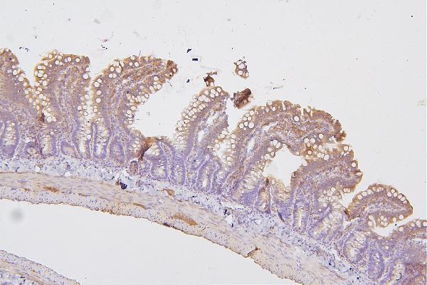
Real IHC data IHC analysis of PRKCD using anti-PRKCD antibody (A00822-1). PRKCD was detected in paraffin-embedded section of human mammary cancer tissues. Heat mediated antigen retrieval was performed in citrate buffer (pH6, epitope retrieval solution) for 20 mins. The tissue section was blocked with 10% goat serum. The tissue section was then incubated with 1μg/ml rabbit anti-PRKCD Antibody (A00822-1) overnight at 4°C. Biotinylated goat anti-rabbit IgG was used as secondary antibody and incubated for 30 minutes at 37°C. The tissue section was developed using Strepavidin-Biotin-Complex (SABC)(Catalog # SA1022) with DAB as the chromogen.
Anti-PKC delta/PRKCD Antibody ®
Cat # A00822-1
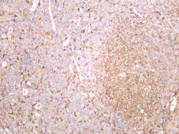
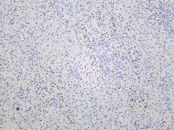
Real IHC data Human tonsil was stained with anti-PKC δ rabbit antibody
Anti-PKC δ Rabbit Monoclonal Antibody
Cat # M00822-3
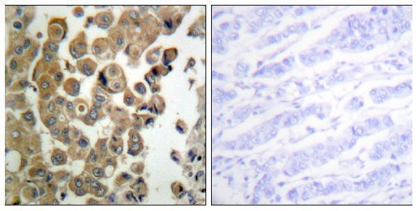
Real IHC data Immunohistochemistry analysis of paraffin-embedded human breast carcinoma, using PKC delta (Phospho-Thr505) Antibody. The picture on the right is blocked with the phospho peptide.
Anti-Phospho-PKC delta (T507) PRKCD Antibody
Cat # A00822T507

A00822-1 shows paraffin-section IHC in human mammary cancer and rat intestine (A00822-1 IHC captions); M00822-3 shows IHC staining in human tonsil and spleen (M00822-3 IHC captions). A00822T507 shows paraffin-section IHC in human breast carcinoma with a phospho-peptide blocking control (A00822T507 IHC caption).

Which to pick: For tissue IHC, choose A00822-1 for a documented paraffin-section procedure (A00822-1 IHC caption), M00822-3 for a monoclonal antibody illustrated in human tonsil and spleen (M00822-3 catalog and IHC captions), or A00822T507 for the phospho-targeted option with a blocking control (A00822T507 catalog and IHC caption). For IF/ICC, A00822-1 has illustrated cell staining and lists both applications (A00822-1 IF captions and applications). For cross-species IHC, A00822-1 has human and rat tissue examples, while all three list human, mouse and rat reactivity (IHC captions; catalog reactivity); the paraffin-section captions do not report the fixative (A00822-1 and A00822T507 IHC captions).

Each figure is that product's own IHC / IF validation image from its datasheet.

References

  1. UniProt Consortium. UniProt entry Q05655 (KPCD_HUMAN, Protein kinase C delta type).
  2. Human Protein Atlas. PRKCD tissue IHC expression (reliability: Enhanced).
  3. Human Protein Atlas. PRKCD subcellular location (ICC-IF): Mainly localized to the cytosol. In addition localized to the Golgi apparatus, endoplasmic reticulum and vesicles..
  4. Human Protein Atlas. PRKCD antibody validation summary (4 antibodies).
  5. Superbinder based phosphoproteomic landscape revealed PRKCD_pY313 mediates the activation of Src and p38 MAPK to promote TNBC progression. Cell communication and signaling : CCS 2024 — PMC10860301.
  6. miR-424 coordinates multilayered regulation of cell cycle progression to promote esophageal squamous cell carcinoma cell proliferation. EBioMedicine 2018 — PMC6284509.
  7. Neuronal cell types, projections, and spatial organization of the central amygdala. iScience 2022 — PMC9678740.
  8. Basolateral to Central Amygdala Neural Circuits for Appetitive Behaviors. Neuron 2017 — PMC5480398.
  9. PubMed PMID:8357834 — UniProt-cited evidence.
  10. PubMed PMID:14702039 — UniProt-cited evidence.
  11. PubMed PMID:15489334 — UniProt-cited evidence.