This website uses cookies to ensure you get the best experience on our website.


- Table of Contents
Plan chromogenic PRKG2 IHC in paraffin sections using high-staining glandular cells in small intestine, appendix or gallbladder as reference material (HPA tissue IHC). This guide covers fixation consistency and interpretation of observed cytoplasmic staining (HPA tissue IHC) alongside the annotated apical membrane location (UniProt).

Expected localisation, validated protocols, controls and antibodies — the at-a-glance facts below, then the full design guide.
| Expected localisation | General cytoplasmic staining (HPA tissue IHC) | |
| Staining pattern | Glandular cells show general cytoplasmic staining (HPA tissue IHC) | |
| Antigen retrieval | Tris-EDTA pH 9.0 HIER, heat-mediated (datasheet A05103) | |
| Positive control | Appendix+4 more · see all | |
| Negative control | None in HPA (detected in all 45 tissues); use no-primary + isotype controls |
| Fixation | Keep fixation consistent across sections (standard IHC practice; not target-specific) | |
| Caveat | Staining has low concordance with RNA data (HPA tissue IHC) | |
| Regulation | Tissue-dependent expression (UniProt) | |
| Isoform / epitope | 2 isoforms; map the epitope before comparing staining (UniProt) |
Compare the catalog antibody’s IHC-P protocol (datasheet A05103) with one published PRKG2 protocol in rat carotid artery sections (PMC13446705).
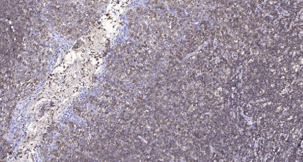
| Sample | Paraffin-embedded human tonsil tissue; fixative not specified (datasheet A05103) |
| Fixation | Image fixative and duration unreported (datasheet A05103); verify before use. |
| Sectioning | 4–5 µm sections on charged slides (standard) |
| Deparaffinisation | Xylene, graded ethanol series to water (standard) |
| Antigen retrieval | Heat retrieval: Tris-EDTA pH 9.0 (datasheet A05103); 20 min, 95–100 °C (standard) |
| Peroxidase block | 3% H2O2, 10 min, room temperature (standard) |
| Blocking | 10% normal serum of the secondary host, 30 min, room temperature (standard) |
| Primary antibody | Rabbit anti-PRKG2, 1:100-1:300 (datasheet A05103) |
| Primary incubation | Overnight at 4 °C (standard) |
| Detection | HRP-polymer secondary, DAB chromogen 5–10 min (standard) |
| Counterstain | Hematoxylin, blue, dehydrate and mount (standard) |
| Expected result | PRKG2-positive staining in glandular cells of appendix (HPA tissue IHC: High). HPA tissue profile: General cytoplasmic expression. No signal in the no-primary control. |
PRKG2 is annotated at the apical cell membrane without a transmembrane segment (UniProt Q13237 topology). In paraffin sections, expect staining in glandular cells of appendix, gallbladder and small intestine, and respiratory epithelial cells of bronchus (HPA: High). HPA also describes a general cytoplasmic pattern (HPA: tissue IHC). Interpret the pattern cautiously: HPA rates its IHC data Approved but reports low agreement with RNA expression and pending external verification (HPA: reliability).
| Epithelial cytoplasmic staining, sometimes concentrated toward the apical surface, in the listed positive tissues. | This fits HPA's general cytoplasmic IHC profile and UniProt's apical membrane annotation (HPA: tissue IHC; UniProt Q13237 localisation). Score the observed compartment and cell type separately; an apical accent is plausible, but HPA does not establish it as a required feature of every positive gland. |
| Predominantly nuclear staining, with little epithelial cytoplasmic signal. | That compartment is discordant with the supplied localisation evidence (HPA: general cytoplasmic expression; UniProt Q13237: apical cell membrane). Treat an isolated nuclear pattern as suspect, then compare it with a positive tissue and appropriate detection controls before calling it PRKG2 (general IHC practice). |
| Strong staining mainly in fibroblasts or splenic red pulp cells. | Those cells are reported at Low staining, whereas several glandular and respiratory epithelial populations are High (HPA: tissue IHC). A dominant signal in these cells warrants a specificity check; cross-reactivity or endogenous detection activity are possibilities, not conclusions from appearance alone (general IHC practice). |
| Broad, nearly uniform colour across cells and tissue spaces. | A uniform haze obscures the cell and compartment pattern needed for interpretation (general IHC practice). Compare with a section lacking primary antibody and inspect blocking, detection and wash steps; those checks distinguish background from a tissue pattern without presuming a PRKG2-specific cause (general IHC practice). |
| No convincing staining in appendix, bronchus, gallbladder or small intestine. | Each has a High-staining epithelial population in the supplied HPA IHC record (HPA: tissue IHC). An absent signal therefore makes the run or specimen worth checking, although HPA's low RNA–staining consistency means one negative section cannot, by itself, settle antibody specificity (HPA: reliability). |
| Compartment and topology (UniProt Q13237) | UniProt places PRKG2 at the apical cell membrane and reports no transmembrane segment; HPA describes general cytoplasmic IHC staining (UniProt Q13237 topology; HPA: tissue IHC). Evaluate both descriptions against the visible epithelial pattern. Neither source supplies a rule that every positive paraffin section must show a sharp membrane outline. |
| Choice of comparison tissue (HPA: tissue IHC) | Appendix, bronchus, gallbladder and small intestine contain specified High-staining epithelial populations; fibroblasts in soft tissue and splenic red pulp cells are Low (HPA: tissue IHC). Compare like cell types when judging a run. No tissue in the supplied HPA list is designated negative (HPA: negative list empty). |
| Strength of validation (HPA: antibodies and reliability) | HPA lists two rabbit polyclonal antibodies, HPA007386 and CAB018739, as IHC Approved; it reports low agreement between antibody staining and RNA expression and says external verification is pending (HPA: antibodies; HPA: reliability). These data support cautious pattern comparison, not an assertion of independently reproduced or definitive specificity. |
| Isoforms and antigen features (UniProt Q13237) | UniProt records two PRKG2 isoforms and several phosphorylated residues, including Ser110, Ser117, Ser431 and Thr609 (UniProt Q13237). The supplied record gives no antibody epitope or isoform selectivity, so these facts cannot predict which isoform a stain detects or whether phosphorylation changes its IHC signal. |
| Can this pattern be carried over to IF/ICC? (HPA: subcellular and antibodies) | The supplied HPA ICC-IF record has no main location or cell-line images, and neither listed antibody has an ICC validation entry (HPA: subcellular; HPA: antibodies). Use the tissue IHC pattern as context only; the supplied evidence does not establish an IF/ICC staining pattern or support an IF/ICC protocol choice. |
| Situation | Likely cause | Next action |
|---|---|---|
| No signal in a listed High-staining tissue. | The run may have a specimen or detection problem; a single blank section does not identify the cause (general IHC practice; HPA: tissue IHC). | Confirm the expected epithelial cells are present, review the catalog antibody's IHC-P instructions, and check the run's positive and detection controls (general IHC practice). |
| Signal appears chiefly nuclear. | The pattern conflicts with the supplied cytoplasmic and apical annotations (HPA: tissue IHC; UniProt Q13237 localisation). | Recheck compartment assignment against the counterstain and a positive tissue; assess primary-omission background before interpreting the nuclear signal (general IHC practice). |
| Fibroblasts or red pulp cells dominate the staining. | Those populations are Low in the supplied HPA record; specificity or detection background may need investigation (HPA: tissue IHC; general IHC practice). | Compare epithelial and low-staining cells on matched sections, then inspect primary-omission and detection controls (HPA: tissue IHC; general IHC practice). |
| Diffuse chromogen obscures cell borders. | Nonspecific detection or inadequate washing can produce background in chromogenic IHC (general IHC practice). | Inspect a primary-omission control and review blocking, wash and detection steps before scoring compartment or intensity (general IHC practice). |
| Cytoplasmic signal is present but lacks a crisp apical rim. | HPA reports general cytoplasmic expression, while UniProt annotates apical membrane localisation; the records do not require an apical rim in every section (HPA: tissue IHC; UniProt Q13237). | Judge cell identity and background first, record the observed distribution, and avoid rejecting an epithelial cytoplasmic pattern solely for this feature (HPA: tissue IHC; general IHC practice). |
| Staining varies between sections or tissues. | Cell composition and assay performance can affect IHC comparisons; HPA also flags low agreement between staining and RNA data (general IHC practice; HPA: reliability). | Compare the same annotated cell populations, use run controls, and report variation without converting the supplied Low entries into validated negatives (HPA: tissue IHC; general IHC practice). |
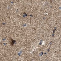
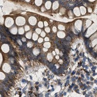


Comprehensive Human Protein Atlas IHC scoring per tissue (reliability: Approved — Low consistency between antibody staining and RNA expression data. Pending external verification.). Rows are taken directly from the HPA tissue chart — click any row's HPA link to view the source.
| Tissue | Cell type | Level | Evidence | Source |
|---|---|---|---|---|
| Appendix | Glandular cells | High | Protein (IHC) | HPA → |
| Bronchus | Respiratory epithelial cells | High | Protein (IHC) | HPA → |
| Gallbladder | Glandular cells | High | Protein (IHC) | HPA → |
| Small intestine | Glandular cells | High | Protein (IHC) | HPA → |
| Adipose tissue | Adipocytes | Medium | Protein (IHC) | HPA → |
| Tissue | Cell type | Level | Evidence | Source |
|---|---|---|---|---|
| None in HPA: PRKG2 is detected in all 45 scored tissues. Use a no-primary (secondary-only) and an isotype control instead. | ||||
Troubleshoot PRKG2 staining in paraffin sections by checking retrieval, cellular distribution, controls, and scoring before interpreting chromogenic signal.
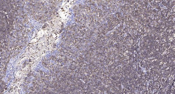
Two anti-PRKG2 antibodies have human tonsil paraffin-section IHC images (catalog: A05103, A30481); both list IF and Human, Mouse, Rat reactivity (catalog: applications/reactivity).
A05103 will render with a paraffin-section human tonsil IHC image using primary antibody at 1:200 and Tris-EDTA retrieval at pH 9.0 (A05103 image caption). A30481 will render with its own paraffin-section human tonsil IHC image using the same reported dilution and retrieval conditions (A30481 image caption).
Which to pick: For tissue IHC, either SKU has its own paraffin-section image; the fixative is unreported for both (A05103 and A30481 image captions). For IF/ICC, choose A30481 because it lists both applications, while A05103 lists IF only; neither has an IF image in the payload (catalog: applications/images). For Human, Mouse, or Rat work, both list reactivity and are rabbit polyclonals, but their pictured IHC evidence is human tonsil only (catalog: reactivity/host/clone; A05103 and A30481 image captions).