This website uses cookies to ensure you get the best experience on our website.


- Table of Contents
Plan PTCH1 chromogenic IHC in paraffin sections using the observed cytoplasmic tissue pattern, while considering its membrane and ciliary localisation (HPA tissue IHC; UniProt). The IHC-validated antibody has a stated dilution range of 1:50–1:200; interpret staining cautiously because tissue evidence has uncertain concordance with RNA expression (datasheet; HPA tissue IHC).

Expected localisation, validated protocols, controls and antibodies — the at-a-glance facts below, then the full design guide.
| Expected localisation | Cytoplasmic tissue staining (HPA tissue IHC); membrane and ciliary localisation (UniProt) | |
| Staining pattern | Cytoplasmic staining in most tissues (HPA tissue IHC) | |
| Antigen retrieval | Citrate pH 6.0 HIER, 95–98 °C, 20 min (rule: cytoplasmic / membrane antigen) | |
| Positive control | Adrenal gland+4 more · see all | |
| Negative control | None in HPA (detected in all 45 tissues); use no-primary + isotype controls |
| Fixation | Keep fixation consistent across paraffin sections (standard IHC practice; not target-specific); Matched tissue-IHC evidence does not establish the fixation claim. Validate the specimen-specific method before use. (selected-SKU IHC image A00441) | |
| Caveat | Staining has uncertain concordance with RNA expression (HPA tissue IHC) | |
| Regulation | Hedgehog may alter ciliary PTCH1 (UniProt) | |
| Isoform / epitope | 4 isoforms; check whether the epitope is extracellular or cytoplasmic (UniProt) |
The catalog antibody’s IHC-P protocol is accompanied by published PTCH1 staining protocols for breast carcinoma (PMC2633285), rat cartilage (PMC4377216), and pancreatic tumor tissue (PMC4085644).
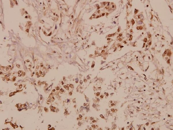
| Sample | Paraffin-embedded human breast carcinoma tissue; fixative not specified (datasheet A00441) |
| Fixation | Image fixative and duration unreported (datasheet A00441); verify before use. |
| Sectioning | 4–5 µm sections on charged slides (standard) |
| Deparaffinisation | Xylene, graded ethanol series to water (standard) |
| Antigen retrieval | Heat-induced epitope retrieval in citrate buffer, pH 6.0, 20 min at 95–98 °C (standard rule: cytoplasmic / membrane antigen) |
| Peroxidase block | 3% H2O2, 10 min, room temperature (standard) |
| Blocking | 10% normal serum of the secondary host, 30 min, room temperature (standard) |
| Primary antibody | Rabbit anti-PTCH1, 1:50-1:200 (datasheet A00441) |
| Primary incubation | Overnight at 4 °C (standard) |
| Detection | HRP-polymer secondary, DAB chromogen 5–10 min (standard) |
| Counterstain | Hematoxylin, blue, dehydrate and mount (standard) |
| Expected result | PTCH1-positive staining in glandular cells of adrenal gland (HPA tissue IHC: High). HPA tissue profile: Cytoplasmic expression in most tissues. No signal in the no-primary control. |
PTCH1 is a 12-pass membrane protein at the cell membrane and primary cilium (UniProt Q13635 topology; UniProt Q13635 subcellular location). In tissue IHC, HPA describes predominantly cytoplasmic staining and reports high staining in selected glandular, hematopoietic, neuronal and endothelial cells (HPA tissue IHC). Treat that distribution as a reference pattern, not definitive validation: HPA rates its tissue staining Uncertain because antibody staining and RNA expression show low consistency (HPA tissue IHC).
| Distinct staining in glandular cells of adrenal gland, appendix or breast, or hematopoietic cells in bone marrow (HPA tissue IHC). | These are reported high-staining cell populations (HPA tissue IHC). Score the relevant cells and their compartment, rather than calling an entire section positive from isolated color (general IHC practice). HPA's Uncertain reliability limits how strongly a match confirms PTCH1 (HPA tissue IHC). |
| Cytoplasmic staining in an expected cell population, with or without a discernible membrane rim (HPA tissue IHC; UniProt Q13635 subcellular location). | Cytoplasmic staining matches HPA's tissue IHC profile, while a membrane-associated component is compatible with UniProt localization (HPA tissue IHC; UniProt Q13635 subcellular location). Routine tissue IHC cannot establish primary-cilium localization from broad cytoplasmic color alone (general IHC practice). |
| Predominantly nuclear staining, with no convincing cytoplasmic or membrane-associated pattern (HPA tissue IHC; UniProt Q13635 subcellular location). | Neither source places PTCH1 in the nucleus (HPA tissue IHC; UniProt Q13635 subcellular location). Investigate a compartment mismatch as possible nonspecific staining or detection artefact before interpreting it as PTCH1 (general IHC practice). |
| Strong color in a cell population outside the reported pattern, especially when expected cells remain unstained (HPA tissue IHC). | The mismatch raises possible cross-reactivity or endogenous detection activity; it does not identify which occurred (general IHC practice). HPA lists low staining in smooth muscle cells and soft-tissue fibroblasts, but supplies no negative-tissue list (HPA tissue IHC). |
| Diffuse color across cells and surrounding tissue, or no staining in an HPA high-staining reference population (HPA tissue IHC). | Diffuse color weakens compartment and cell-type interpretation; absent reference staining may reflect an assay failure or a biological/sample difference (general IHC practice). Neither result alone resolves specificity, particularly with HPA's Uncertain tissue reliability (HPA tissue IHC). |
| Tissue evidence quality (HPA tissue IHC) | HPA calls tissue staining Uncertain because antibody staining and RNA expression have low consistency; its high-staining examples guide comparison but do not prove specificity in a new specimen (HPA tissue IHC). |
| Membrane topology and antibody access (UniProt Q13635 topology) | PTCH1 has 12 transmembrane segments plus extracellular and cytoplasmic regions (UniProt Q13635 topology). Without the antibody epitope, topology alone cannot specify a retrieval or permeabilisation setting (general IHC practice). |
| Isoform coverage (UniProt Q13635 isoforms) | UniProt lists 4 isoforms: L, L′, M and S (UniProt Q13635 isoforms). Their presence does not show which isoforms an unspecified antibody recognizes; interpret tissue differences without assuming universal isoform detection (general IHC practice). |
| Glycosylation and processing (UniProt Q13635 features) | Six glycosylation sites are annotated, and the listed protein chain spans residues 1–1447 with no signal peptide or propeptide (UniProt Q13635 features). These annotations alone do not establish epitope masking, shedding or fixation sensitivity (general IHC practice). |
| Primary-cilium context (UniProt Q13635 subcellular location) | PTCH1 localizes to the primary cilium and delocalizes after hedgehog binding, according to UniProt (UniProt Q13635 subcellular location). Therefore, absence of a visible ciliary punctum in paraffin IHC should not override an otherwise interpretable cell pattern (general IHC practice). |
| IF/ICC Q&A (HPA subcellular) | Q: What pattern is reported for IF/ICC? A: Mainly Golgi apparatus, with additional primary-cilium localization; HPA marks both locations Approved (HPA subcellular). This is an IF/ICC observation, not a paraffin-IHC protocol or required slide pattern (HPA subcellular; general IHC practice). |
| Situation | Likely cause | Next action |
|---|---|---|
| Expected cells show only faint or absent staining despite an HPA high-staining reference choice (HPA tissue IHC). | An ineffective assay step or sample-specific expression difference is possible; HPA's high designation does not guarantee every specimen will stain (HPA tissue IHC; general IHC practice). | Check the validated IHC-P instructions for retrieval, antibody dilution and detection, and review the run's control section before changing interpretation (general IHC practice). |
| A uniform brown haze obscures cell borders and compartments (general IHC practice). | Excess chromogen development, insufficient blocking or overly concentrated antibody can elevate background (general IHC practice). | Compare with a reagent-omission control; review blocking, washes, dilution and development time under the chosen chromogenic workflow (general IHC practice). |
| Brown signal follows blood-rich or unrelated cells when HRP detection is used (general IHC practice). | Endogenous peroxidase activity can contribute color in an HRP-based assay, so the signal may not reflect antibody binding (general IHC practice). | Inspect an antibody-omission control and verify that the protocol's peroxidase-blocking step was performed effectively (general IHC practice). |
| Nuclear color dominates while expected cytoplasmic staining is unclear (HPA tissue IHC). | This compartment conflicts with HPA tissue IHC and UniProt localization; nonspecific staining or detection artefact is plausible (HPA tissue IHC; UniProt Q13635 subcellular location; general IHC practice). | Review controls and antibody concentration, then require a reproducible expected-cell pattern before scoring PTCH1 positivity (general IHC practice). |
| Smooth muscle cells or soft-tissue fibroblasts stain more strongly than the chosen reference population (HPA tissue IHC). | HPA reports low staining in those cells; cross-reactivity, endogenous detection activity or specimen variation could explain a reversal (HPA tissue IHC; general IHC practice). | Compare matched controls and the reported cell distribution, then record the discrepancy rather than treating those cells as a validated positive reference (HPA tissue IHC; general IHC practice). |
| No discrete ciliary signal is visible in a paraffin section (general IHC practice). | A cilium may be hard to resolve in routine chromogenic sections, and PTCH1 can leave the cilium after hedgehog binding (UniProt Q13635 subcellular location; general IHC practice). | Assess the overall cell and compartment pattern for IHC; use the separate IF/ICC guide if resolving Golgi or ciliary localization is the experimental goal (HPA tissue IHC; HPA subcellular; general IHC practice). |


Comprehensive Human Protein Atlas IHC scoring per tissue (reliability: Uncertain — Low consistency between antibody staining and RNA expression data. Pending external verification.). Rows are taken directly from the HPA tissue chart — click any row's HPA link to view the source.
| Tissue | Cell type | Level | Evidence | Source |
|---|---|---|---|---|
| Adrenal gland | Glandular cells | High | Protein (IHC) | HPA → |
| Appendix | Glandular cells | High | Protein (IHC) | HPA → |
| Bone marrow | Hematopoietic cells | High | Protein (IHC) | HPA → |
| Breast | Glandular cells | High | Protein (IHC) | HPA → |
| Caudate | Neuronal cells | High | Protein (IHC) | HPA → |
| Tissue | Cell type | Level | Evidence | Source |
|---|---|---|---|---|
| None in HPA: PTCH1 is detected in all 45 scored tissues. Use a no-primary (secondary-only) and an isotype control instead. | ||||
Troubleshoot PTCH1 staining in paraffin sections by checking retrieval, compartment, controls and scoring before interpreting signal.
The catalog antibody has pictured PTCH1 IHC in human paraffin sections (catalog IHC image caption). It lists Human, Mouse and Rat reactivity, with no IF image (catalog reactivity; catalog IF image alts).
A00441 will render with an IHC image of paraffin-embedded human breast carcinoma tissue at 1:50 (catalog IHC image caption). Its listed applications are IHC and WB, and its listed reactivity is Human, Mouse and Rat (catalog applications; catalog reactivity).
Which to pick: Choose A00441 for paraffin-section IHC: its own caption shows human breast carcinoma tissue at 1:50, but does not report the fixative (catalog IHC image caption). No IF/ICC choice is supported by this payload: A00441 has no listed IF application or IF image (catalog applications; catalog IF image alts). For cross-species work, A00441 lists Human, Mouse and Rat reactivity, although the pictured IHC result is human only (catalog reactivity; catalog IHC image caption).