This website uses cookies to ensure you get the best experience on our website.


- Table of Contents
Plan CD45 IHC on paraffin sections using the catalog antibody's 1:50–1:200 dilution range (datasheet A00555-1) and assess selective cytoplasmic staining in lymphoid tissue and immune cells (HPA tissue IHC). Interpret that observed pattern alongside CD45's membrane topology (UniProt).

Expected localisation, validated protocols, controls and antibodies — the at-a-glance facts below, then the full design guide.
| Expected localisation | Cytoplasmic tissue staining (HPA tissue IHC); membrane expected (UniProt) | |
| Staining pattern | Selective cytoplasmic signal in lymphoid tissue and immune cells (HPA tissue IHC) | |
| Antigen retrieval | Citrate pH 6.0 HIER, heat-mediated (datasheet A00555-1) | |
| Positive control | Appendix+4 more · see all | |
| Negative control | Adipose tissue+4 more · see all |
| Fixation | Keep fixation consistent across sections (standard IHC practice; not target-specific); Matched tissue-IHC evidence does not establish the fixation claim. Validate the specimen-specific method before use. (selected-SKU IHC image A00555-1) | |
| Caveat | Immune-cell infiltrates can dominate the tissue signal (HPA tissue IHC) | |
| Regulation | Expression regulation not specified (UniProt) | |
| Isoform / epitope | 8 isoforms; map extracellular vs cytoplasmic epitopes (UniProt) |
The catalog antibody uses citrate pH 6.0 retrieval (datasheet A00555-1). One published salivary gland IHC protocol provides an alternative (PMC10545362).
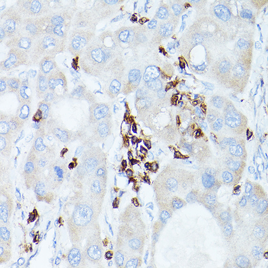
| Sample | Paraffin-embedded human liver cancer tissue; fixative not specified (datasheet A00555-1) |
| Fixation | Image fixative and duration unreported (datasheet A00555-1); verify before use. |
| Sectioning | 4–5 µm sections on charged slides (standard) |
| Deparaffinisation | Xylene, graded ethanol series to water (standard) |
| Antigen retrieval | Heat retrieval: Citrate pH 6.0 (datasheet A00555-1); 20 min, 95–100 °C (standard) |
| Peroxidase block | 3% H2O2, 10 min, room temperature (standard) |
| Blocking | 10% normal serum of the secondary host, 30 min, room temperature (standard) |
| Primary antibody | Rabbit anti-PTPRC, 1:50-1:200 (datasheet A00555-1) |
| Primary incubation | Overnight at 4 °C (standard) |
| Detection | HRP-polymer secondary, DAB chromogen 5–10 min (standard) |
| Counterstain | Hematoxylin, blue, dehydrate and mount (standard) |
| Expected result | PTPRC-positive staining in germinal center cells of appendix (HPA tissue IHC: High). HPA tissue profile: Selective cytoplasmic expression in lymphoid tissue and immune cells. No signal in the no-primary control. |
PTPRC (CD45) is a cell-membrane protein with an extracellular region and cytoplasmic phosphatase domains (UniProt P08575 topology). In paraffin-section IHC, expect staining in immune and lymphoid cells, including germinal center cells and bone-marrow hematopoietic cells (HPA tissue IHC: High). HPA describes its observed tissue pattern as selective cytoplasmic expression and rates the IHC evidence Enhanced, with medium consistency between staining and RNA data (HPA tissue IHC).
| Distinct staining in germinal center cells of tonsil, lymph node, or appendix, with surrounding cells easier to distinguish. | This matches HPA's High staining in those annotated cell populations (HPA tissue IHC). Assess the stained cells and their location on the section; a dark region alone does not establish that the expected cells are positive (standard IHC practice). |
| Staining outlines immune-cell membranes, or appears partly cytoplasmic in the expected cells. | Membrane staining fits PTPRC topology (UniProt P08575 topology). HPA reports selective cytoplasmic staining in tissue IHC (HPA tissue IHC), so cytoplasmic signal should be judged alongside cell identity and controls rather than rejected solely for lacking a crisp membrane rim. |
| Predominantly nuclear staining in paraffin-section IHC, without convincing staining of expected immune cells. | A nuclear-dominant IHC pattern does not match UniProt's membrane assignment or HPA's tissue-IHC profile (UniProt P08575 subcellular location; HPA tissue IHC). HPA's uncertain nucleoplasm finding comes from ICC-IF and does not validate a nuclear IHC interpretation (HPA subcellular ICC-IF). |
| Strong staining in adipocytes or respiratory epithelial cells, especially when expected immune cells show little signal. | HPA lists adipocytes in adipose tissue and respiratory epithelial cells in bronchus as Not detected (HPA tissue IHC). Consider nonspecific antibody binding or endogenous detection activity; review controls before interpreting those cells as PTPRC-positive (standard IHC practice). |
| Diffuse color across tissue, or no signal in expected cells of a known-positive section. | Diffuse color obscures cell-specific interpretation (standard IHC practice). Absence in a section containing HPA-positive germinal center or bone-marrow hematopoietic cells calls for a control and workflow check before concluding that the specimen lacks PTPRC (HPA tissue IHC; standard IHC practice). |
| Compartment and epitope | PTPRC spans the membrane at residues 578–598, with extracellular residues 26–577 and cytoplasmic residues 599–1306 (UniProt P08575 topology). The supplied evidence does not identify the catalog antibody's epitope; do not infer whether it recognizes an extracellular or cytoplasmic region. |
| Observed IHC pattern and confidence | HPA calls tissue-IHC expression selective cytoplasmic and rates reliability Enhanced, while reporting medium consistency with RNA and staining in some unannotated cells or structures (HPA tissue IHC). Interpret localisation and unexpected cells with those limits in view. |
| Isoform coverage | UniProt lists 8 isoforms; isoforms 1, 2, 3, 5, and 7 are detected in thymocytes, whereas 4, 6, and 8 are not (UniProt P08575 isoforms and tissue specificity). Antibody recognition across isoforms is not established by this record. |
| Extracellular glycosylation | UniProt lists 17 glycosylation sites in the extracellular region (UniProt P08575 topology and glycosylation). Their presence alone does not establish epitope accessibility, retrieval requirements, or fixation sensitivity for this antibody. |
| IF/ICC Q&A: Should nuclear or vesicular IF signal define the expected IHC result? | No. HPA labels nucleoplasm and vesicle localisation uncertain in ICC-IF, and the listed antibody HPA000440 has ICC status Uncertain (HPA subcellular ICC-IF; HPA antibodies). Use the tissue-IHC evidence to interpret this paraffin-section guide (HPA tissue IHC). |
| Situation | Likely cause | Next action |
|---|---|---|
| No staining in tonsil or lymph-node germinal center cells. | Those cells are High in HPA tissue IHC, so a negative result may reflect a failed IHC run or an absent suitable positive population (HPA tissue IHC; standard IHC practice). | Confirm the expected cells are present; compare an established positive section and reagent controls, then review retrieval and detection steps (standard IHC practice). |
| Broad brown signal covers the section and obscures individual cells. | Diffuse background can arise from nonspecific binding, insufficient washing, or endogenous detection activity (standard chromogenic IHC practice). | Inspect the negative control, blocking and wash steps, and the detection system's endogenous-activity control (standard chromogenic IHC practice). |
| Adipocytes or respiratory epithelial cells stain strongly. | These annotated cell types are Not detected in HPA tissue IHC; the signal may be nonspecific or due to endogenous detection activity (HPA tissue IHC; standard IHC practice). | Compare expected immune cells with the negative control and reassess antibody dilution and detection background (HPA tissue IHC; standard IHC practice). |
| Only nuclei appear strongly positive. | This is discordant with the UniProt membrane assignment and HPA's selective cytoplasmic tissue-IHC profile (UniProt P08575 subcellular location; HPA tissue IHC). | Check the counterstain and negative control, then verify that any proposed positive signal is cell-associated in expected immune cells (standard IHC practice; HPA tissue IHC). |
| Signal is faint in expected cells but background remains low. | Weak signal alone cannot distinguish limited assay sensitivity from lower staining in this particular section; HPA's High calls describe specified annotated populations, not every section (HPA tissue IHC; standard IHC practice). | Compare a positive control processed in the same run; review antibody dilution, antigen retrieval, incubation, and detection according to the antibody's IHC instructions (standard IHC practice). |
| Membrane rims are hard to see, but expected cells show cytoplasmic color. | HPA reports selective cytoplasmic tissue-IHC staining despite UniProt's membrane assignment (HPA tissue IHC; UniProt P08575 subcellular location). | Score cell identity and controlled contrast first; record the observed compartment without claiming that cytoplasmic staining proves a cytosolic PTPRC pool (HPA tissue IHC; standard IHC practice). |
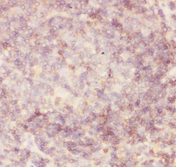
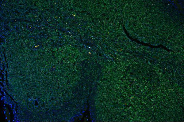


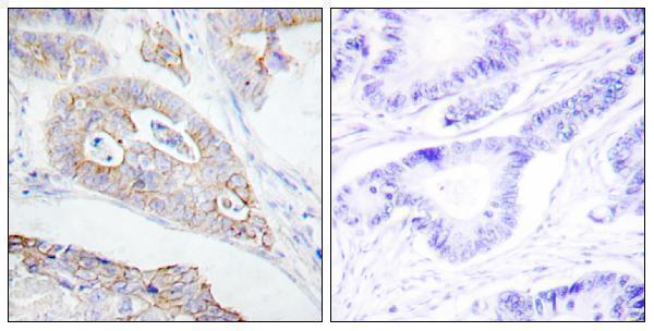
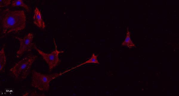
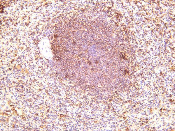
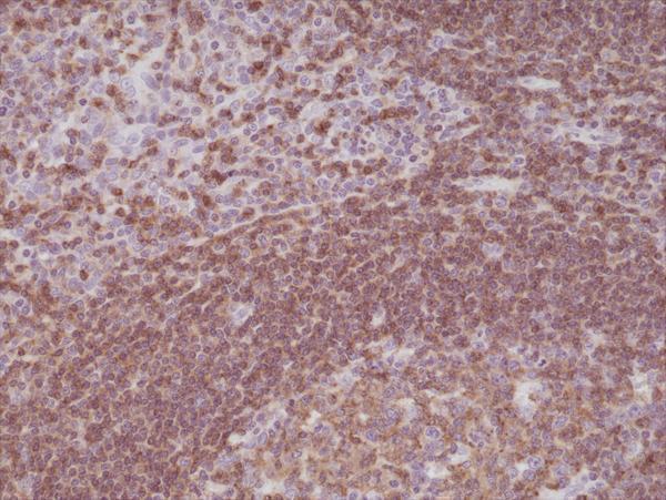
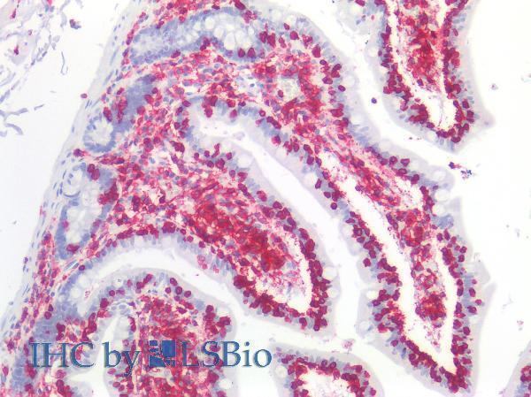
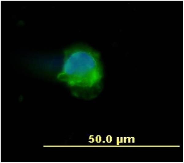


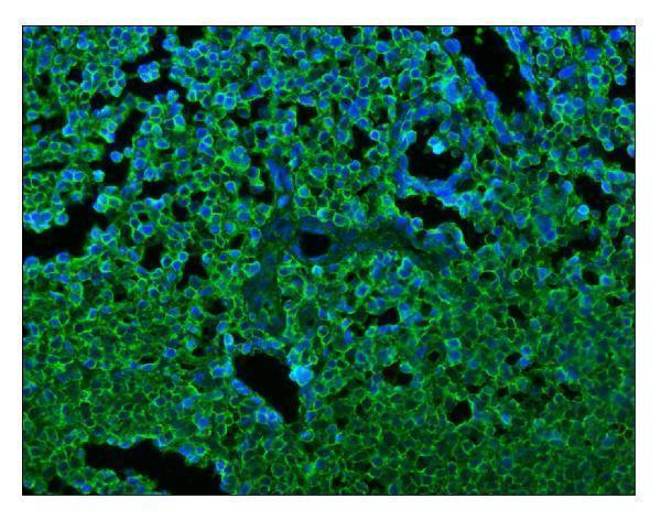
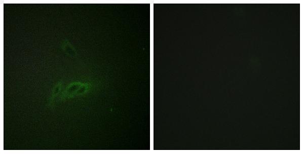
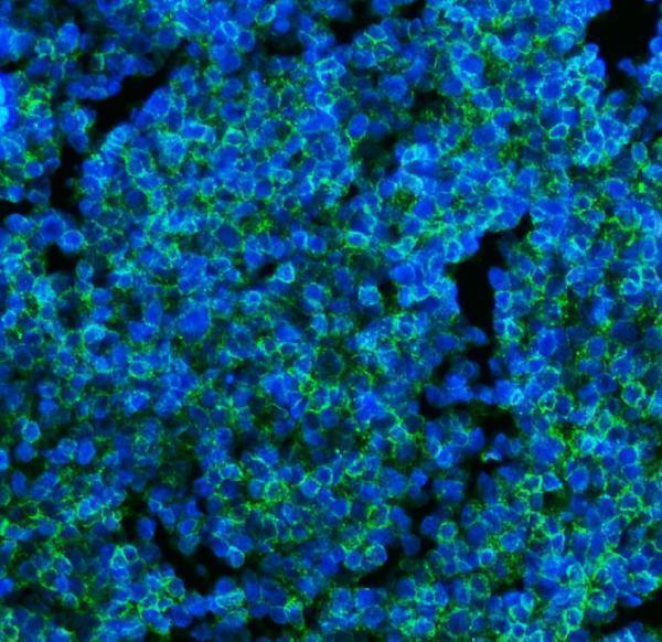
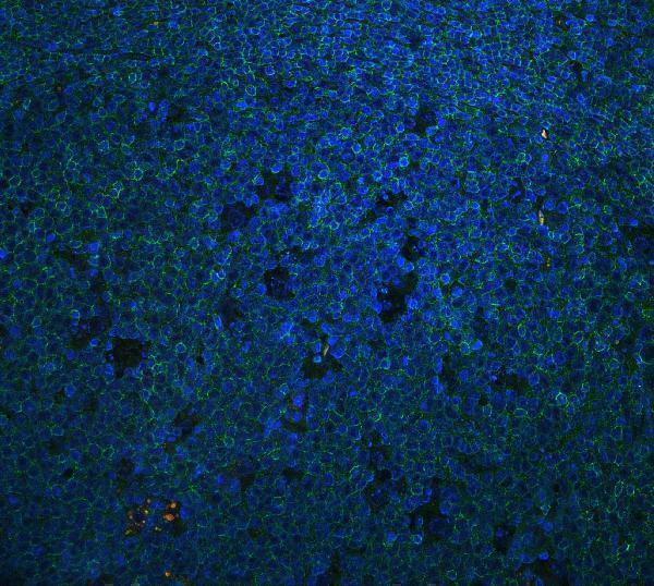
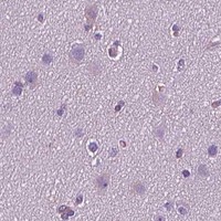


Comprehensive Human Protein Atlas IHC scoring per tissue (reliability: Enhanced — Medium consistency between antibody staining and RNA expression data. Antibody staining in cells/structures not annotated, view images.). Rows are taken directly from the HPA tissue chart — click any row's HPA link to view the source.
| Tissue | Cell type | Level | Evidence | Source |
|---|---|---|---|---|
| Appendix | Germinal center cells | High | Protein (IHC) | HPA → |
| Bone marrow | Hematopoietic cells | High | Protein (IHC) | HPA → |
| Lymph node | Germinal center cells | High | Protein (IHC) | HPA → |
| Skin | Fibrohistiocytic cells | High | Protein (IHC) | HPA → |
| Spleen | Cells in red pulp | High | Protein (IHC) | HPA → |
| Tissue | Cell type | Level | Evidence | Source |
|---|---|---|---|---|
| Adipose tissue | Adipocytes | Not detected | Protein (IHC) | HPA → |
| Adrenal gland | Glandular cells | Not detected | Protein (IHC) | HPA → |
| Breast | Adipocytes | Not detected | Protein (IHC) | HPA → |
| Bronchus | Respiratory epithelial cells | Not detected | Protein (IHC) | HPA → |
| Caudate | Glial cells | Not detected | Protein (IHC) | HPA → |
Troubleshoot PTPRC/CD45 staining in paraffin section chromogenic IHC using the catalog image, tissue expression profile, and membrane topology as reference points.
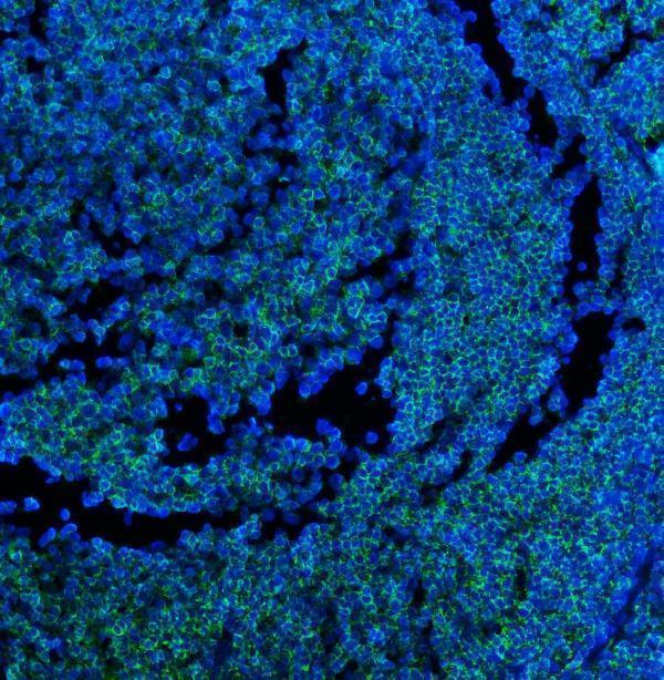
Anti-PTPRC antibodies have IHC images from human tissue, including paraffin sections, and IF images from cells and tissue (catalog IHC and IF captions). Catalog reactivity also includes mouse and rat (A00555-1 reactivity).
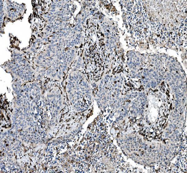
A00555-1 shows human liver cancer paraffin sections; M00555-5 shows human lung cancer paraffin sections; PB9096 and PB9785 each show human tonsil paraffin sections (respective IHC captions). A00555 shows human colon carcinoma paraffin sections, while M00555-10 shows human spleen staining without a reported preparation method (respective IHC captions).
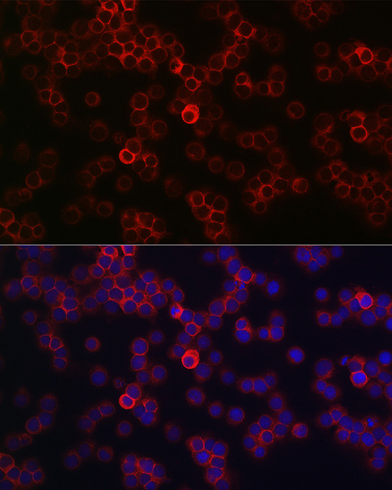
Which to pick: For tissue IHC, PB9096 provides a documented human tonsil paraffin example with EDTA pH 8.0 retrieval and 1 μg/ml primary antibody; its caption does not report the fixative (PB9096 IHC caption). For IF/ICC, A00555-1 lists both applications and shows Jurkat cell IF at 1:100 (A00555-1 applications; A00555-1 IF caption). For work across species, A00555 lists human, mouse and rat reactivity, although its own IHC image documents only human colon carcinoma paraffin sections and does not report the fixative (A00555 reactivity; A00555 IHC caption).