This website uses cookies to ensure you get the best experience on our website.


- Table of Contents
Plan chromogenic PTPRO IHC in paraffin sections using selective cytoplasmic glomerular staining as a reference (HPA tissue IHC). Start within the catalog antibody’s 1:50–1:200 IHC range (datasheet) and include a no-primary control (standard IHC practice).

Expected localisation, validated protocols, controls and antibodies — the at-a-glance facts below, then the full design guide.
| Expected localisation | Glomerular cytoplasm (HPA tissue IHC); membrane topology (UniProt) | |
| Staining pattern | Selective cytoplasmic staining in glomerular cells (HPA tissue IHC) | |
| Antigen retrieval | Citrate pH 6.0 HIER, 95–98 °C, 20 min (rule: cytoplasmic / membrane antigen) | |
| Positive control | Kidney+2 more · see all | |
| Negative control | Adipose tissue+4 more · see all |
| Fixation | Keep fixation consistent across paraffin sections. (standard IHC practice; not target-specific) | |
| Caveat | Splice/transcript discrepancy may affect staining concordance (HPA tissue IHC) | |
| Regulation | Stimulus-dependent regulation unreported (UniProt) | |
| Isoform / epitope | 5 isoforms; extracellular vs cytoplasmic epitope matters (UniProt) |
The catalog antibody’s IHC-P protocol is accompanied by one published PTPRO immunohistochemistry protocol (PMC4496227 methods).
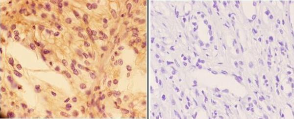
| Sample | Paraffin-embedded human kidney carcinoma tissue; fixative not specified (datasheet A04781) |
| Fixation | Image fixative and duration unreported (datasheet A04781); verify before use. |
| Sectioning | 4–5 µm sections on charged slides (standard) |
| Deparaffinisation | Xylene, graded ethanol series to water (standard) |
| Antigen retrieval | Heat-induced epitope retrieval in citrate buffer, pH 6.0, 20 min at 95–98 °C (standard rule: cytoplasmic / membrane antigen) |
| Peroxidase block | 3% H2O2, 10 min, room temperature (standard) |
| Blocking | 10% normal serum of the secondary host, 30 min, room temperature (standard) |
| Primary antibody | Rabbit anti-PTPRO, 1:50-1:200 (datasheet A04781) |
| Primary incubation | Overnight at 4 °C (standard) |
| Detection | HRP-polymer secondary, DAB chromogen 5–10 min (standard) |
| Counterstain | Hematoxylin, blue, dehydrate and mount (standard) |
| Expected result | PTPRO-positive staining in cells in glomeruli of kidney (HPA tissue IHC: High). HPA tissue profile: Selective cytoplasmic expression of glomeruli. No signal in the no-primary control. |
PTPRO is a membrane protein with an extracellular region, one transmembrane segment, and a cytoplasmic region (UniProt Q16827 topology). In tissue IHC, expect selective staining of kidney glomerular cells, reported as cytoplasmic and high (HPA tissue IHC). HPA rates the tissue staining evidence Enhanced while noting medium agreement with RNA data and a possible splice or transcript discrepancy (HPA tissue IHC).
| High staining is concentrated in cells of kidney glomeruli (HPA tissue IHC). | This matches the strongest listed tissue IHC signal and the selective glomerular profile (HPA tissue IHC). Interpret the visible cytoplasmic pattern alongside PTPRO's membrane topology; HPA's tissue image description does not resolve which side of the membrane contains the detected epitope (HPA tissue IHC; UniProt Q16827 topology). |
| Medium staining appears in Paneth cells of duodenum or small intestine (HPA tissue IHC). | This is a second reported positive cell population, so intestinal staining is not automatically off target (HPA tissue IHC). Judge its cell specificity and intensity against the listed Paneth cell pattern; the kidney glomerulus remains the stronger reference signal (HPA tissue IHC). |
| Nuclear-only staining, with no selective glomerular pattern, dominates the section (HPA tissue IHC; UniProt Q16827 topology). | Treat this as suspect because the reported tissue pattern is cytoplasmic and PTPRO is annotated at the membrane (HPA tissue IHC; UniProt Q16827 subcellular location). Check controls and detection before calling it PTPRO; nuclear color alone does not establish target localization (general IHC practice). |
| Strong color appears in adipocytes or adrenal glandular cells (HPA tissue IHC: not detected). | These listed cell populations lack detectable HPA tissue IHC staining (HPA tissue IHC). Widespread signal there raises concern for cross-reactivity or endogenous detection activity; compare a no-primary control and review blocking and detection reagents (general IHC practice). |
| Diffuse color covers glomeruli and surrounding tissue without a clear cell pattern (HPA tissue IHC: selective glomerular expression). | Diffuse background obscures the selective result needed for interpretation (HPA tissue IHC). If a known-positive kidney section has no discernible glomerular signal, assess the run's positive control before scoring the sample negative (general IHC practice). |
| Tissue and cell choice (HPA tissue IHC) | Kidney glomerular cells offer the listed high positive reference; Paneth cells in duodenum and small intestine are medium references (HPA tissue IHC). Adipocytes and adrenal glandular cells are listed as not detected, so use them to examine unwanted staining within an appropriately controlled run (HPA tissue IHC; general IHC practice). |
| Compartment assignment (UniProt Q16827 topology; HPA tissue IHC) | PTPRO has extracellular residues 30–822, a transmembrane segment at 823–843, and cytoplasmic residues 844–1216 (UniProt Q16827 topology). HPA describes tissue staining as cytoplasmic and its subcellular summary as membrane; neither record identifies the catalog antibody's epitope (HPA tissue IHC; HPA subcellular). |
| Isoforms and evidence limits (UniProt Q16827; HPA tissue IHC) | UniProt lists 5 isoforms (UniProt Q16827 isoforms). HPA flags a splice or transcript discrepancy and medium consistency between antibody staining and RNA expression (HPA tissue IHC). These facts support cautious interpretation of discordant results but do not identify which isoform an IHC signal represents. |
| Antibody evidence (HPA antibodies; HPA tissue IHC) | HPA034525 is listed as a rabbit polyclonal antibody with Enhanced IHC validation (HPA antibodies). The tissue profile is also rated Enhanced, with the stated RNA agreement caveat (HPA tissue IHC). Validation supports the reported pattern; it does not make staining in an unlisted cell type a confirmed PTPRO result. |
| IF/ICC applicability? (HPA subcellular; HPA antibodies) | HPA's subcellular summary says membrane, but it provides no ICC-IF cell-line images, and the listed antibody has no ICC validation status (HPA subcellular; HPA antibodies). Use the separate IF/ICC guide for that application; the tissue IHC pattern alone cannot establish an expected fluorescent cell image. |
| Situation | Likely cause | Next action |
|---|---|---|
| No glomerular signal in a kidney positive control (HPA tissue IHC: high in glomerular cells). | The run may have failed at retrieval, primary incubation, or chromogenic detection (general IHC practice); the supplied sources give no PTPRO-specific retrieval condition. | Review the control and each assay step, then optimize retrieval and antibody use under the laboratory's IHC workflow (general IHC practice). Do not score an unknown section negative until the positive control works. |
| Glomeruli stain, but surrounding tissue is also diffusely colored (HPA tissue IHC: selective glomerular expression). | Excess background may come from nonspecific binding or detection chemistry (general IHC practice); HPA does not attribute this pattern to PTPRO. | Compare a no-primary control, inspect blocking and washes, and adjust assay conditions using the positive reference section (general IHC practice). Preserve a readable glomerular pattern when judging the result (HPA tissue IHC). |
| Adipocytes or adrenal glandular cells show strong staining (HPA tissue IHC: not detected). | The mismatch raises concern for cross-reactivity or endogenous detection activity (general IHC practice); it does not prove either cause. | Check a no-primary control and the assay's endogenous-activity block, then reassess cell-specific staining with the kidney reference (general IHC practice; HPA tissue IHC). |
| Only nuclei appear positive in kidney tissue (HPA tissue IHC: cytoplasmic glomerular profile). | Nuclear-only color conflicts with HPA's observed profile and UniProt's membrane annotation (HPA tissue IHC; UniProt Q16827 subcellular location). | Inspect counterstain and detection controls, then repeat interpretation against a section showing selective glomerular staining (general IHC practice; HPA tissue IHC). Do not assign nuclear localization from this result alone. |
| Intestinal Paneth cells stain less strongly than glomerular cells (HPA tissue IHC). | HPA reports medium Paneth cell staining and high glomerular cell staining (HPA tissue IHC). | Compare each cell population with its own reported level and keep the kidney glomerulus as the stronger positive reference (HPA tissue IHC). A weaker intestinal result alone does not show assay failure. |
| IHC and an IF/ICC image appear to disagree (HPA tissue IHC; HPA subcellular). | HPA reports a tissue IHC pattern but supplies no ICC-IF cell-line images or ICC validation for the listed antibody (HPA tissue IHC; HPA subcellular; HPA antibodies). | Evaluate the IHC section against tissue-specific controls and the separate IF/ICC evidence for that application (general IHC practice). Do not use the unvalidated IF/ICC appearance to overturn the reported glomerular IHC profile (HPA tissue IHC; HPA antibodies). |
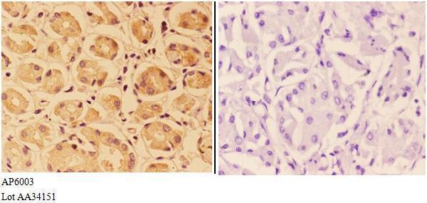
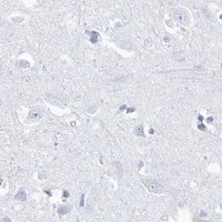


Comprehensive Human Protein Atlas IHC scoring per tissue (reliability: Enhanced — Medium consistency between antibody staining and RNA expression data. Caution, Splice and/or transcript discrepancy exists.). Rows are taken directly from the HPA tissue chart — click any row's HPA link to view the source.
| Tissue | Cell type | Level | Evidence | Source |
|---|---|---|---|---|
| Kidney | Cells in glomeruli | High | Protein (IHC) | HPA → |
| Duodenum | Paneth cells | Medium | Protein (IHC) | HPA → |
| Small intestine | Paneth cells | Medium | Protein (IHC) | HPA → |
| Tissue | Cell type | Level | Evidence | Source |
|---|---|---|---|---|
| Adipose tissue | Adipocytes | Not detected | Protein (IHC) | HPA → |
| Adrenal gland | Glandular cells | Not detected | Protein (IHC) | HPA → |
| Appendix | Endocrine cells | Not detected | Protein (IHC) | HPA → |
| Bone marrow | Hematopoietic cells | Not detected | Protein (IHC) | HPA → |
| Breast | Adipocytes | Not detected | Protein (IHC) | HPA → |
These questions address paraffin-section chromogenic IHC for PTPRO, with one IF-specific question for planning a companion experiment (page application setting).
A04781 has real IHC data from a human kidney carcinoma paraffin section (A04781 figure caption); its listed reactivity covers human, mouse and rat (catalog: A04781). No IF data are supplied (catalog: A04781).
A04781 is the only SKU that will render (catalog: A04781). Its IHC figure shows staining in a human kidney carcinoma paraffin section at 1:50 (A04781 figure caption); the catalog lists human, mouse and rat reactivity (catalog: A04781).
Which to pick: Choose A04781 for paraffin-section IHC: it is a rabbit polyclonal antibody (catalog: A04781; A04781 figure caption) with an IHC range of 1:50–1:200 (datasheet: A04781), and its own figure shows human kidney carcinoma staining (A04781 figure caption). The fixative is unreported (A04781 figure caption). For mouse or rat IHC, A04781 has listed reactivity but no supplied species-specific IHC figure (catalog: A04781; A04781 figure caption); for IF/ICC, no SKU in this payload lists that application or supplies IF data (catalog: A04781).