This website uses cookies to ensure you get the best experience on our website.


- Table of Contents
Plan PTPRZ1 chromogenic IHC in paraffin sections using brain staining reported in glial and neuronal subsets as a reference (HPA tissue IHC). Consider the membrane-bound and shed forms when interpreting staining, and check which region the antibody recognizes (UniProt).

Expected localisation, validated protocols, controls and antibodies — the at-a-glance facts below, then the full design guide.
| Expected localisation | Cerebellar molecular layer cells: cytoplasm/membrane (HPA tissue IHC) | |
| Staining pattern | Subsets of glia and neurons; cerebellar cytoplasm/membrane (HPA tissue IHC) | |
| Antigen retrieval | Tris-EDTA pH 9.0 HIER, heat-mediated (datasheet A03510) | |
| Positive control | Cerebral cortex+3 more · see all | |
| Negative control | Adipose tissue+4 more · see all |
| Fixation | Keep fixation consistent across sections. (standard IHC practice; not target-specific) | |
| Caveat | A shed extracellular form may shift the staining pattern (UniProt) | |
| Regulation | Developmentally regulated in brain (UniProt) | |
| Isoform / epitope | 3 isoforms; check extracellular vs cytoplasmic epitope (UniProt) |
The catalog antibody protocol specifies heat-mediated Tris-EDTA pH 9.0 retrieval (datasheet A03510). Published IHC methods add conditions for tumor sections and glioma tissue microarrays (PMC12667478; PMC13523290).
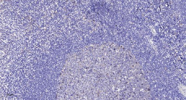
| Sample | Paraffin-embedded human tonsil tissue; fixative not specified (datasheet A03510) |
| Fixation | Image fixative and duration unreported (datasheet A03510); verify before use. |
| Sectioning | 4–5 µm sections on charged slides (standard) |
| Deparaffinisation | Xylene, graded ethanol series to water (standard) |
| Antigen retrieval | Heat retrieval: Tris-EDTA pH 9.0 (datasheet A03510); 20 min, 95–100 °C (standard) |
| Peroxidase block | 3% H2O2, 10 min, room temperature (standard) |
| Blocking | 10% normal serum of the secondary host, 30 min, room temperature (standard) |
| Primary antibody | Rabbit anti-PTPRZ1, 1:50 recommended; image 1:200 (datasheet A03510) |
| Primary incubation | Overnight at 4 °C (standard) |
| Detection | HRP-polymer secondary, DAB chromogen 5–10 min (standard) |
| Counterstain | Hematoxylin, blue, dehydrate and mount (standard) |
| Expected result | PTPRZ1-positive staining in glial cells of cerebral cortex (HPA tissue IHC: Medium). HPA tissue profile: Distinct expression in brain, both in subsets of neurons and a subset of glial cells. No signal in the no-primary control. |
PTPRZ1 is a cell-membrane protein with a secreted form generated by extracellular-domain shedding (UniProt P23471). In paraffin-section IHC, expect staining chiefly in subsets of brain neurons and glial cells (HPA tissue IHC: Enhanced); HPA reports medium staining in the listed positive cells and medium consistency between antibody staining and RNA expression. Cerebellar molecular-layer cells show cytoplasmic/membranous staining (HPA tissue IHC).
| Medium staining in cerebral-cortex or caudate glial cells, or hippocampal neurons (HPA tissue IHC). | These are documented positive cell populations (HPA tissue IHC). Score staining by cell type and region; an unstained neighboring cell does not negate a subset-specific pattern (HPA tissue IHC: subsets of neurons and glia). |
| Medium cytoplasmic/membranous staining in cerebellar molecular-layer cells (HPA tissue IHC). | This matches the reported cerebellar appearance (HPA tissue IHC) and is compatible with membrane-associated PTPRZ1 (UniProt P23471 topology). Cytoplasmic appearance alone is not grounds for rejection: HPA also reports cytosol and vesicles in ICC-IF (HPA subcellular). |
| Predominantly nuclear staining, without the documented brain-cell pattern. | A nuclear location is not reported in the supplied UniProt or HPA localization records (UniProt P23471; HPA tissue IHC; HPA subcellular). Treat the result as suspect and review controls and staining conditions before assigning it to PTPRZ1. |
| Strong staining in cells reported as not detected, such as adipocytes or colon endothelial cells (HPA tissue IHC). | The cell-type mismatch raises concern for nonspecific binding, cross-reactivity, or endogenous chromogenic activity; it does not independently identify the cause. Compare with a no-primary control and a documented brain-positive section before interpreting it as target staining. |
| Diffuse staining across the section, or no signal in documented positive brain cells (HPA tissue IHC). | Diffuse signal obscures the reported cell-restricted pattern; absent signal prevents its assessment. Check the no-primary and positive-tissue controls, detection reagents, background blocking, and primary-antibody conditions before drawing a biological conclusion. |
| Membrane topology and shedding | PTPRZ1 has an extracellular region at residues 25–1636, one transmembrane segment at 1637–1662, and a cytoplasmic region at 1663–2315 (UniProt P23471 topology). Extracellular-domain shedding can produce a secreted form (UniProt P23471); do not require every signal to trace a cell boundary. |
| Antibody epitope and isoforms | UniProt lists 3 isoforms and a shed extracellular form (UniProt P23471). The supplied record does not locate this antibody’s epitope, so it cannot establish which forms the stain detects. Interpret compartment differences with that limit in mind. |
| Evidence strength | The tissue profile is rated Enhanced, but its description reports medium consistency between staining and RNA data and notes that secretion complicates their correspondence (HPA tissue IHC). HPA015103 is IHC Enhanced and ICC Uncertain (HPA antibodies); these labels should not be interchanged. |
| IF/ICC: what pattern is reported? | HPA reports mainly cytosolic signal with additional vesicular signal in ICC-IF (HPA subcellular: approved locations). That observation belongs to a different application; the listed antibody has ICC Uncertain status (HPA antibodies), so do not turn it into an IHC compartment requirement. |
| Situation | Likely cause | Next action |
|---|---|---|
| No staining in a cerebral-cortex or caudate glial-cell area reported as positive (HPA tissue IHC). | The section may lack an evaluable positive population, or the IHC workflow may have failed; the supplied sources do not establish PTPRZ1-specific fixation sensitivity. | Confirm the relevant cell type is present, then review the positive control, primary-antibody conditions, antigen-retrieval step, and detection reagents as general IHC checks. |
| Diffuse chromogen obscures individual cells. | Background from antibody or detection reagents can hide the subset-specific brain pattern (HPA tissue IHC: neuronal and glial subsets). | Compare a no-primary control; review blocking, washes, primary concentration, and chromogen development. Reassess whether staining resolves into the documented positive cells (HPA tissue IHC). |
| Adipocytes or colon endothelial cells stain strongly despite being reported as not detected (HPA tissue IHC). | Nonspecific binding, cross-reactivity, or endogenous detection activity are possible explanations; the slide alone cannot distinguish them. | Run a no-primary control and a documented brain-positive section; review endogenous-activity blocking where applicable to the chromogenic system, then compare cell-specific signal. |
| Signal appears nuclear while documented cerebellar cytoplasmic/membranous staining is absent (HPA tissue IHC). | The observed compartment does not match the supplied IHC reference pattern or UniProt membrane topology (HPA tissue IHC; UniProt P23471). | Check counterstain interpretation and no-primary background, then reassess antibody and detection conditions using a documented positive brain section. |
| Cerebellar staining looks cytoplasmic rather than sharply membrane-bound. | Cytoplasmic/membranous staining is itself reported in molecular-layer cells (HPA tissue IHC); secreted protein may also complicate a simple membrane-only expectation (UniProt P23471). | Score the documented cell population and distribution first. Avoid rejecting the section solely because cell borders are indistinct; compare with controls if the staining is diffuse. |
| An ICC-IF image appears cytosolic or vesicular while the IHC interpretation emphasizes brain-cell distribution. | HPA reports cytosol and vesicles for ICC-IF, while its tissue IHC profile describes neuronal and glial subsets (HPA subcellular; HPA tissue IHC). | Interpret each application against its own HPA record. For this paraffin-section IHC result, prioritize the documented tissue, cell type, and cerebellar cytoplasmic/membranous appearance (HPA tissue IHC). |


Comprehensive Human Protein Atlas IHC scoring per tissue (reliability: Enhanced — Medium consistency between antibody staining and RNA expression data. At least one protein variant secreted, tissue location of RNA and protein might differ and correlation is complex.). Rows are taken directly from the HPA tissue chart — click any row's HPA link to view the source.
| Tissue | Cell type | Level | Evidence | Source |
|---|---|---|---|---|
| Cerebral cortex | Glial cells | Medium | Protein (IHC) | HPA → |
| Caudate | Glial cells | Medium | Protein (IHC) | HPA → |
| Cerebellum | Molecular layer cells - cytoplasm/membrane | Medium | Protein (IHC) | HPA → |
| Hippocampus | Neuronal cells | Medium | Protein (IHC) | HPA → |
| Tissue | Cell type | Level | Evidence | Source |
|---|---|---|---|---|
| Adipose tissue | Adipocytes | Not detected | Protein (IHC) | HPA → |
| Adrenal gland | Glandular cells | Not detected | Protein (IHC) | HPA → |
| Appendix | Glandular cells | Not detected | Protein (IHC) | HPA → |
| Bone marrow | Hematopoietic cells | Not detected | Protein (IHC) | HPA → |
| Breast | Adipocytes | Not detected | Protein (IHC) | HPA → |
Troubleshoot PTPRZ1 chromogenic IHC in paraffin sections using the catalog image conditions, tissue distribution, and known membrane and secreted forms (datasheet A03510; HPA tissue IHC; UniProt P23471).
The catalog lists one anti-PTPRZ1 antibody for IHC in human, mouse and rat; its image shows paraffin-embedded human tonsil, with no IF image provided (catalog applications/reactivity; IHC image caption).
A03510 is listed for IHC and shows staining of paraffin-embedded human tonsil (catalog applications; IHC image caption). Human, mouse and rat reactivity is listed, but the supplied image documents only human tissue (catalog reactivity; IHC image caption).
Which to pick: Choose A03510 for tissue IHC: its own caption documents paraffin-embedded human tonsil, while the fixative is unreported (IHC image caption). For cross-species work, A03510 lists human, mouse and rat reactivity, but the supplied IHC image shows only human tissue (catalog reactivity; IHC image caption). No IF/ICC application or image is supplied for A03510, so this payload does not support an IF/ICC recommendation (catalog applications; IF image list).