This website uses cookies to ensure you get the best experience on our website.


- Table of Contents
Plan RAB11FIP3 paraffin-section IHC around cytoplasmic staining in kidney tubular cells (HPA tissue IHC). This guide covers consistent fixation, the catalog antibody’s 1:100–1:300 IHC dilution range (datasheet), and controls for presumed off-target binding (HPA tissue IHC).

Expected localisation, validated protocols, controls and antibodies — the at-a-glance facts below, then the full design guide.
| Expected localisation | Cytoplasmic staining (HPA tissue IHC); endosomal and Golgi localization (UniProt) | |
| Staining pattern | Tubular and GI glandular cells show cytoplasmic staining (HPA tissue IHC) | |
| Antigen retrieval | Citrate pH 6.0 HIER, 95–98 °C, 20 min (rule: cytoplasmic / membrane antigen) | |
| Positive control | Kidney+4 more · see all | |
| Negative control | Adipose tissue+4 more · see all |
| Fixation | Keep fixation conditions consistent across sections. (standard IHC practice; not target-specific) | |
| Caveat | Presumed off-target binding may confound staining (HPA tissue IHC) | |
| Regulation | Expression regulation is unannotated (UniProt) | |
| Isoform / epitope | Three isoforms; epitope coverage is unspecified (UniProt) |
Compare the catalog antibody’s IHC-P protocol with a published IHC method for human papillary thyroid carcinoma sections (PMC6126170).
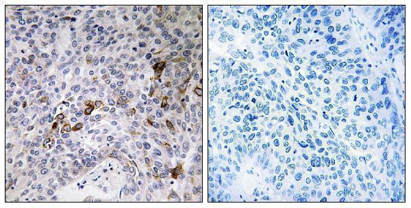
| Sample | Paraffin-embedded human lung carcinoma tissue; fixative not specified (datasheet A09469-1) |
| Fixation | Image fixative and duration unreported (datasheet A09469-1); verify before use. |
| Sectioning | 4–5 µm sections on charged slides (standard) |
| Deparaffinisation | Xylene, graded ethanol series to water (standard) |
| Antigen retrieval | Heat-induced epitope retrieval in citrate buffer, pH 6.0, 20 min at 95–98 °C (standard rule: cytoplasmic / membrane antigen) |
| Peroxidase block | 3% H2O2, 10 min, room temperature (standard) |
| Blocking | 10% normal serum of the secondary host, 30 min, room temperature (standard) |
| Primary antibody | Rabbit anti-RAB11FIP3, 1:100 - 1:300 (datasheet A09469-1) |
| Primary incubation | Overnight at 4 °C (standard) |
| Detection | HRP-polymer secondary, DAB chromogen 5–10 min (standard) |
| Counterstain | Hematoxylin, blue, dehydrate and mount (standard) |
| Expected result | RAB11FIP3-positive staining in cells in tubules of kidney (HPA tissue IHC: High). HPA tissue profile: Cytoplasmic expression in several tissues including cells in tubules and glandular cells in the gastrointestinal tract. No signal in the no-primary control. |
RAB11FIP3 is an intracellular protein associated with recycling endosomes, the centrosome, Golgi membranes and structures involved in cell division (UniProt O75154 subcellular location; UniProt O75154 topology: no transmembrane segment). In tissue IHC, expect cytoplasmic staining, most prominently in kidney tubular cells; several glandular and epithelial populations show medium staining (HPA tissue IHC). HPA rates the tissue pattern Enhanced, while noting medium agreement with RNA data and disregarded presumed off-target binding (HPA tissue IHC).
| Kidney tubular cells show clear cytoplasmic chromogen, with weaker or absent staining in nearby cells. | This matches the strongest listed tissue result: High in kidney tubular cells (HPA tissue IHC). Compare the stain within identifiable tubules rather than treating every cell on the section as an equally strong positive; HPA reports staining by cell population (HPA tissue IHC). |
| Glandular or epithelial cells show moderate cytoplasmic staining outside the kidney. | Medium staining is reported in adrenal, colon, duodenum, fallopian tube and gallbladder glandular cells, bronchial respiratory epithelium and hepatocytes (HPA tissue IHC). A moderate result in these populations can fit the reported pattern; intensity alone does not establish antibody specificity (HPA tissue IHC: presumed off-target binding disregarded). |
| Strong, predominantly nuclear staining appears, with little cytoplasmic signal. | A nuclear-dominant result conflicts with HPA’s cytoplasmic tissue profile and UniProt’s intracellular membrane and cytoskeletal locations (HPA tissue IHC; UniProt O75154 subcellular location). Treat it as suspect and check the control section and detection background before assigning it to RAB11FIP3 (general IHC practice). |
| Chromogen appears in a cell population reported as undetected, or broadly across unlike cell types. | For example, HPA reports no detection in adipocytes or esophageal squamous epithelial cells (HPA tissue IHC). Unexpected staining warrants a cross-reactivity or endogenous detection-activity check; an HPA ‘Not detected’ result is a comparison point, not proof that every specimen of that cell type must be negative (HPA tissue IHC; general IHC practice). |
| A diffuse haze covers cells and extracellular areas, obscuring cell boundaries. | A field-wide haze cannot be scored as the cell-associated cytoplasmic pattern reported by HPA (HPA tissue IHC). Compare a no-primary control, inspect wash and blocking steps, and assess whether excessive chromogen development or counterstain is hiding the distribution (general IHC practice). |
| Tissue and cell selection | Kidney tubular cells provide the clearest reported positive population, at High staining (HPA tissue IHC). The listed Medium populations offer a less intense comparison, while HPA’s ‘Not detected’ populations can help reveal unexpected colour; these are cell-specific observations, not whole-organ guarantees (HPA tissue IHC). |
| Strength of tissue evidence | The HPA tissue IHC profile is rated Enhanced, but its description reports medium consistency with RNA expression and presumed off-target binding that was disregarded (HPA tissue IHC). Interpret borderline or unexpected staining with controls and cell morphology rather than using the rating as a guarantee for an individual slide (general IHC practice). |
| Intracellular distribution | UniProt places RAB11FIP3 at recycling endosomes, centrosomes, Golgi-associated membranes and, during cytokinesis, the furrow and midbody (UniProt O75154 subcellular location). Routine chromogenic tissue IHC supports a cytoplasmic assessment (HPA tissue IHC); a tiny punctum need not be resolved for the section to be interpretable (general IHC practice). |
| Cell-cycle context | UniProt describes changing distribution across mitosis, with vesicles moving toward the cleavage furrow and midbody late in division (UniProt O75154 subcellular location). A rare dividing cell can therefore differ from a typical interphase cell, but the reported tissue-level expectation remains cytoplasmic staining in the named cell populations (HPA tissue IHC). |
| Isoforms and epitope coverage | Three isoforms are listed (UniProt O75154 isoforms). No antibody epitope or isoform coverage is supplied here, so neither a missing signal nor an unusual distribution can be assigned to isoform recognition from these records alone (UniProt O75154 isoforms; supplied antibody-validation summary). |
| IF/ICC Q&A: what distribution should fluorescence show? | HPA reports mainly vesicles, centriolar satellites and the basal body, with additional cytokinetic-bridge and primary-cilium localisation (HPA subcellular ICC-IF). Those are fluorescence observations; they do not set a required punctate appearance for chromogenic tissue IHC (HPA subcellular ICC-IF; HPA tissue IHC). |
| Situation | Likely cause | Next action |
|---|---|---|
| No stain in the kidney tubular cells selected as the positive control. | The result conflicts with HPA’s High tubular-cell staining, but the source does not identify a target-specific fixation or retrieval failure (HPA tissue IHC). A run-level staining failure remains possible (general IHC practice). | Confirm tubules are present, inspect the control run, and verify primary-antibody, retrieval and detection steps against the assay instructions (general IHC practice). Do not infer that RAB11FIP3 is absent from this negative slide alone. |
| Only weak colour appears in a tissue expected to stain. | The comparison may be a Medium or Low HPA cell population rather than the High kidney tubular population (HPA tissue IHC). Weak colour may also reflect the staining run (general IHC practice). | Identify and score the relevant cell type first, then compare it with a kidney tubular control stained in the same run (HPA tissue IHC; general IHC practice). Review retrieval and detection conditions as general assay parameters, without assuming RAB11FIP3-specific fixation sensitivity. |
| Nuclei dominate the signal. | That distribution conflicts with the reported cytoplasmic tissue pattern and UniProt locations (HPA tissue IHC; UniProt O75154 subcellular location). Background or cross-reactivity is a possible explanation, not a demonstrated diagnosis (general IHC practice). | Check the no-primary control, distinguish chromogen from the nuclear counterstain, and compare cytoplasmic staining in the positive-control tubules (general IHC practice; HPA tissue IHC). Withhold a positive call if the expected cell-associated pattern is missing. |
| Unexpected colour appears in adipocytes or another HPA-undetected population. | Adipocytes are reported as Not detected (HPA tissue IHC). Cross-reactivity or endogenous enzyme activity could contribute to a chromogenic signal (general IHC practice). | Inspect no-primary and appropriate detection controls, review endogenous-activity blocking, and compare the distribution with a same-run positive tissue (general IHC practice). Record the cell type and discrepancy rather than labelling an entire tissue positive. |
| Brown haze appears over tissue and blank areas. | Widespread background can arise from nonspecific reagent binding, inadequate washing or excessive detection development (general IHC practice); it does not reproduce HPA’s cell-associated cytoplasmic profile (HPA tissue IHC). | Compare blank areas and a no-primary section, then review blocking, washes and development time (general IHC practice). Score RAB11FIP3 only where cellular boundaries and the expected compartment remain distinguishable (HPA tissue IHC; general IHC practice). |
| A small dot or bridge-like signal appears in an occasional dividing cell. | Centrosomal and cytokinetic locations are described by UniProt, while bridge localisation is also reported in ICC-IF (UniProt O75154 subcellular location; HPA subcellular ICC-IF). Chromogenic section morphology alone may not identify those structures securely (general IHC practice). | Assess the cell’s morphology and surrounding cytoplasmic pattern, then retain a compartment-specific claim only if the structure is distinguishable (general IHC practice). Use the HPA tissue cell populations to judge the IHC result overall (HPA tissue IHC). |


Comprehensive Human Protein Atlas IHC scoring per tissue (reliability: Enhanced — Medium consistency between antibody staining and RNA expression data. Presumed off target binding observed and disregarded.). Rows are taken directly from the HPA tissue chart — click any row's HPA link to view the source.
| Tissue | Cell type | Level | Evidence | Source |
|---|---|---|---|---|
| Kidney | Cells in tubules | High | Protein (IHC) | HPA → |
| Adrenal gland | Glandular cells | Medium | Protein (IHC) | HPA → |
| Bronchus | Respiratory epithelial cells | Medium | Protein (IHC) | HPA → |
| Colon | Glandular cells | Medium | Protein (IHC) | HPA → |
| Duodenum | Glandular cells | Medium | Protein (IHC) | HPA → |
| Tissue | Cell type | Level | Evidence | Source |
|---|---|---|---|---|
| Adipose tissue | Adipocytes | Not detected | Protein (IHC) | HPA → |
| Caudate | Glial cells | Not detected | Protein (IHC) | HPA → |
| Cerebellum | Cells in granular layer | Not detected | Protein (IHC) | HPA → |
| Cerebral cortex | Endothelial cells | Not detected | Protein (IHC) | HPA → |
| Esophagus | Squamous epithelial cells | Not detected | Protein (IHC) | HPA → |
Troubleshoot chromogenic IHC for RAB11FIP3 in paraffin sections using the catalog image, tissue staining patterns, and known intracellular localisation (catalog caption A09469-1; HPA tissue IHC; UniProt O75154).
A09469-1 is listed for human, mouse and rat IHC, IF and ICC (catalog: applications/reactivity); its pictured evidence shows paraffin-embedded human lung carcinoma IHC (catalog: IHC image caption).
A09469-1 is a rabbit polyclonal antibody listed for IHC, IF and ICC in human, mouse and rat (catalog: host/clonality, applications/reactivity). Its own IHC image shows paraffin-embedded human lung carcinoma with synthesized-peptide blocking; no IF image is supplied (catalog: IHC/IF image captions).
Which to pick: Choose A09469-1 for paraffin-section tissue IHC: its own image documents human lung carcinoma, and the listed dilution is 1:100–1:300 (catalog: IHC image caption; datasheet: IHC dilution). For IF/ICC or mouse and rat samples, A09469-1 is listed for those applications and species, with IF at 1:50; its pictured evidence remains human tissue IHC (catalog: applications/reactivity, IF dilution, IHC/IF image captions). The IHC caption specifies paraffin embedding but does not report the fixative (catalog: IHC image caption).