This website uses cookies to ensure you get the best experience on our website.


- Table of Contents
Plan RNF125 IHC on paraffin sections using the catalog antibody at 1:100–1:300 (datasheet A08878). Assess cytoplasmic and nuclear staining while accounting for uncertain agreement between antibody staining and RNA expression (HPA tissue IHC).

Expected localisation, validated protocols, controls and antibodies — the at-a-glance facts below, then the full design guide.
| Expected localisation | Cytoplasmic and nuclear staining in tissue (HPA tissue IHC) | |
| Staining pattern | Cytoplasmic and nuclear signal in several tissues (HPA tissue IHC) | |
| Antigen retrieval | Tris-EDTA pH 9.0 HIER, heat-mediated (datasheet A08878) | |
| Positive control | Adrenal gland+4 more · see all | |
| Negative control | Adipose tissue+3 more · see all |
| Fixation | Keep fixation consistent across sections (standard IHC practice; not target-specific) | |
| Caveat | Low agreement between antibody staining and RNA expression (HPA tissue IHC) | |
| Regulation | Preferential expression in CD4+ and CD8+ T cells (UniProt) | |
| Isoform / epitope | No isoforms annotated; chain spans aa 2–232 (UniProt) |
The catalog antibody protocol uses heat-mediated Tris-EDTA pH 9.0 retrieval (datasheet A08878). These tables summarize two published RNF125 IHC protocols (PMC12075997; PMC5564816).
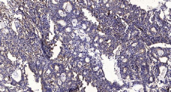
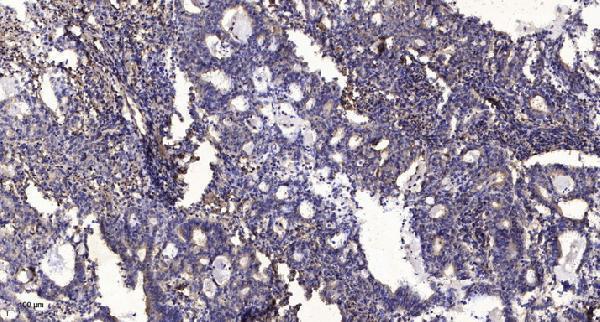
| Sample | Paraffin-embedded human Gastric adenocarcinoma tissue; fixative not specified (datasheet A08878) |
| Fixation | Image fixative and duration unreported (datasheet A08878); verify before use. |
| Sectioning | 4–5 µm sections on charged slides (standard) |
| Deparaffinisation | Xylene, graded ethanol series to water (standard) |
| Antigen retrieval | Heat retrieval: Tris-EDTA pH 9.0 (datasheet A08878); 20 min, 95–100 °C (standard) |
| Peroxidase block | 3% H2O2, 10 min, room temperature (standard) |
| Blocking | 10% normal serum of the secondary host, 30 min, room temperature (standard) |
| Primary antibody | Rabbit anti-RNF125, 1:100-1:300 (datasheet A08878) |
| Primary incubation | Overnight at 4 °C (standard) |
| Detection | HRP-polymer secondary, DAB chromogen 5–10 min (standard) |
| Counterstain | Hematoxylin, blue, dehydrate and mount (standard) |
| Expected result | RNF125-positive staining in glandular cells of adrenal gland (HPA tissue IHC: Medium). HPA tissue profile: Cytoplasmic and nuclear expression in several tissues. No signal in the no-primary control. |
RNF125 is mainly associated with the Golgi apparatus and can show a reticular intracellular pattern; UniProt reports no transmembrane segment and no plasma membrane detection (UniProt Q96EQ8). Expression is predominant in CD4(+) and CD8(+) T cells (UniProt Q96EQ8). HPA reports cytoplasmic and nuclear tissue staining, but rates its IHC pattern Uncertain because antibody staining has low consistency with RNA expression (HPA tissue IHC).
| Intracellular cytoplasmic staining, especially around the Golgi region, in a lymphoid cell population. | This fits the reported Golgi and reticular localization and the preference for T cells (UniProt Q96EQ8). Treat morphology as supportive, not confirmation: HPA rates its tissue IHC pattern Uncertain (HPA tissue IHC). |
| Strong staining confined to cell borders, with little intracellular signal. | A surface-only pattern conflicts with the report that RNF125 is not detected at the plasma membrane (UniProt Q96EQ8). Reassess the chromogenic signal and antibody specificity before scoring these cells as positive; compartment alone cannot establish the cause (standard IHC practice). |
| Prominent staining in a cell type outside the expected lymphoid population. | UniProt describes preferential expression in T cells, while HPA reports Medium staining in several nonlymphoid cells, including colon glandular cells (UniProt Q96EQ8; HPA tissue IHC). Unexpected staining warrants checks for cross-reactivity or endogenous detection activity; it is not automatically false (standard IHC practice). |
| Even chromogen across tissue, stroma, and areas lacking recognizable cellular localization. | A diffuse deposit is difficult to reconcile with the reported intracellular distribution (UniProt Q96EQ8). Consider nonspecific background or endogenous detection activity and compare with a negative reagent control before assigning RNF125 positivity (standard IHC practice). |
| No staining in a section selected because HPA reports Medium signal there. | For example, HPA lists adrenal gland glandular cells and appendix glandular cells at Medium intensity (HPA tissue IHC). Absence prompts review of tissue identity, controls, and assay performance; HPA’s Uncertain IHC reliability means either tissue is a provisional reference, not a guaranteed positive control (HPA tissue IHC; standard IHC practice). |
| Cell population used for interpretation | UniProt reports predominant expression in lymphoid tissues, including bone marrow, spleen, and thymus, and a preference for CD4(+) and CD8(+) T cells (UniProt Q96EQ8). HPA reports tissue-enhanced RNA expression in bone marrow but does not provide a dependable IHC-positive T-cell control (HPA tissue IHC). |
| Reported compartment versus tissue staining | Golgi localization is supported in HPA ICC-IF, whereas additional nucleolar localization is Uncertain (HPA subcellular). HPA tissue IHC describes cytoplasmic and nuclear expression but assigns the overall pattern Uncertain reliability (HPA tissue IHC). Interpret nuclear IHC cautiously rather than treating it as independently confirmed localization. |
| Antibody evidence | For HPA041514, IHC is rated Uncertain and ICC is rated Approved (HPA antibodies). Approval in ICC does not resolve the stated low consistency between tissue antibody staining and RNA expression in IHC (HPA antibodies; HPA tissue IHC). |
| IF/ICC Q: What localization should be expected? | A: Mainly Golgi-associated signal is supported; nucleolar signal is an additional Uncertain localization (HPA subcellular). HPA lists ICC-IF images from A-431, SK-MEL-30, and U2OS, but those images do not establish a paraffin IHC protocol or validate tissue IHC staining (HPA subcellular; HPA antibodies). |
| Situation | Likely cause | Next action |
|---|---|---|
| The entire section has weak or absent signal. | The staining run may have failed, or the chosen tissue may have little detectable signal; UniProt reports weaker expression outside predominantly lymphoid tissues (UniProt Q96EQ8; standard IHC practice). | Check the run’s reagent and detection controls, then compare a documented HPA Medium staining site such as adrenal gland glandular cells; interpret a negative result cautiously because HPA tissue IHC reliability is Uncertain (HPA tissue IHC; standard IHC practice). |
| Signal appears mainly at the plasma membrane. | This distribution conflicts with UniProt’s report of intracellular membranes and no detected plasma membrane localization; the image alone does not identify the artefact (UniProt Q96EQ8). | Inspect adjacent morphology and negative reagent controls for edge deposits or nonspecific staining, and require convincing intracellular signal before calling the result consistent with RNF125 localization (UniProt Q96EQ8; standard IHC practice). |
| Nuclear staining dominates while cytoplasmic staining is scarce. | HPA reports nuclear as well as cytoplasmic tissue IHC, but its tissue pattern is Uncertain; HPA’s additional nucleolar ICC-IF location is also Uncertain (HPA tissue IHC; HPA subcellular). | Record nuclear and cytoplasmic scores separately and compare with controls; avoid presenting dominant nuclear IHC as confirmed RNF125 localization on these sources alone (HPA tissue IHC; HPA subcellular; standard IHC practice). |
| Brown signal covers stroma and multiple cell types without clear intracellular detail. | Diffuse staining can arise from background or endogenous detection activity in chromogenic IHC; it does not show the reported Golgi-associated intracellular distribution (standard IHC practice; UniProt Q96EQ8). | Review the negative reagent control, blocking and wash performance, and tissue morphology before scoring; interpret only localized cellular staining above control background (standard IHC practice). |
| Unexpected nonlymphoid cells stain strongly. | T-cell preference does not imply exclusive expression: HPA reports Medium staining in several nonlymphoid cell types, although its IHC reliability is Uncertain (UniProt Q96EQ8; HPA tissue IHC). | Document the exact cell type and compartment, compare the negative reagent control, and seek independent antibody or orthogonal evidence before attributing a surprising pattern to RNF125 (HPA tissue IHC; standard IHC practice). |
| A tissue expected to be negative shows staining. | HPA lists cardiomyocytes in heart muscle and cholangiocytes in liver as Not detected, but its tissue IHC pattern has Uncertain reliability (HPA tissue IHC). | Confirm the stained cell type and compare controls; report the observation without using a single HPA negative entry to declare cross-reactivity or RNF125 expression (HPA tissue IHC; standard IHC practice). |


Comprehensive Human Protein Atlas IHC scoring per tissue (reliability: Uncertain — Low consistency between antibody staining and RNA expression data.). Rows are taken directly from the HPA tissue chart — click any row's HPA link to view the source.
| Tissue | Cell type | Level | Evidence | Source |
|---|---|---|---|---|
| Adrenal gland | Glandular cells | Medium | Protein (IHC) | HPA → |
| Appendix | Glandular cells | Medium | Protein (IHC) | HPA → |
| Breast | Adipocytes | Medium | Protein (IHC) | HPA → |
| Caudate | Neuronal cells | Medium | Protein (IHC) | HPA → |
| Cerebellum | Cells in granular layer | Medium | Protein (IHC) | HPA → |
| Tissue | Cell type | Level | Evidence | Source |
|---|---|---|---|---|
| Adipose tissue | Adipocytes | Not detected | Protein (IHC) | HPA → |
| Heart muscle | Cardiomyocytes | Not detected | Protein (IHC) | HPA → |
| Liver | Cholangiocytes | Not detected | Protein (IHC) | HPA → |
| Parathyroid gland | Glandular cells | Not detected | Protein (IHC) | HPA → |
Troubleshoot RNF125 staining in paraffin sections by checking retrieval, cellular context and compartment before comparing chromogenic signal across samples.
Both anti-RNF125 antibodies have paraffin-section IHC images and list IF and human, mouse, and rat reactivity (catalog: A08878 and A30664 applications, reactivity, IHC image captions).
A08878 lists IHC and IF for human, mouse, and rat; its IHC image shows paraffin-embedded human gastric adenocarcinoma (catalog: A08878 applications, reactivity, IHC image caption). A30664 lists IHC, IF, and ICC for the same species; its IHC image also shows paraffin-embedded human gastric adenocarcinoma (catalog: A30664 applications, reactivity, IHC image caption).
Which to pick: For paraffin-section tissue IHC, either SKU has its own image using 1:200 primary antibody and Tris-EDTA retrieval at pH 9.0; neither caption reports the fixative (catalog: A08878 and A30664 IHC image captions). For IF/ICC, choose A30664 if ICC is required because it lists both applications and is described as polyclonal; A08878 lists IF but does not list ICC (catalog: A30664 applications and dilution_raw; A08878 applications). For cross-species work, both list human, mouse, and rat reactivity, while their supplied IHC images show human tissue (catalog: A08878 and A30664 reactivity, IHC image captions).