RRS1 / Ribosome biogenesis regulatory protein homolog · IHC design guide

Design Immunohistochemistry for RRS1

Plan RRS1 chromogenic IHC in paraffin sections using the cytoplasmic and nucleolar tissue pattern (HPA tissue IHC). Select controls using high staining in neuronal or Leydig cells and compare antibody concentrations within 2–5 μg/ml (HPA tissue IHC; datasheet A04418-3).

Evidence assembled Oct 2026 · For research use; verify linked source records and product datasheet before use
Immunohistochemistry protocol sheet for RRS1 (IHC for RRS1): expected localisation Cytoplasmic and nucleolar tissue staining (HPA tissue IHC), antibody A04418-3, validated IHC image, and IHC protocol steps
Printable RRS1 IHC protocol sheet — expected localisation Cytoplasmic and nucleolar tissue staining (HPA tissue IHC), antibody A04418-3, controls and protocol steps. Open the full RRS1 IHC guide →

RRS1 Immunohistochemistry Experimental Design Guide

Expected localisation, validated protocols, controls and antibodies — the at-a-glance facts below, then the full design guide.

Must know before staining
Expected localisation Cytoplasmic and nucleolar tissue staining (HPA tissue IHC)
Staining pattern Most tissues: cytoplasmic and nucleolar cell staining (HPA tissue IHC)
Antigen retrieval EDTA pH 8.0 HIER, heat-mediated (datasheet A04418-3)
Positive control ⓘ Cerebral cortex+4 more · see all
Negative control ⓘ Adipose tissue+4 more · see all
Important caveats
Reasons your staining may differ from the expected pattern.
Fixation Keep fixation consistent across sections (standard IHC practice; not target-specific)
Caveat Staining has low consistency with RNA expression (HPA tissue IHC)
Regulation No expression regulator annotated (UniProt)
Isoform / epitope No annotated isoforms; one 1–365 chain (UniProt)
Section 1

Recommended RRS1 IHC & IF Protocols

The catalog antibody uses heat-mediated EDTA pH 8.0 retrieval (datasheet A04418-3). Published RRS1 IHC examples cover retinoblastoma, colorectal cancer, and breast cancer tissues (PMC7652605; PMC5669942; PMC6237566).

Recommended immunohistochemistry (IHC-P) protocol parameters
SampleParaffin-embedded rat testis tissue; fixative not specified (datasheet A04418-3)
FixationImage fixative and duration unreported (datasheet A04418-3); verify before use.
Sectioning4–5 µm sections on charged slides (standard)
DeparaffinisationXylene, graded ethanol series to water (standard)
Antigen retrievalHeat retrieval: EDTA pH 8.0 (datasheet A04418-3); 20 min, 95–100 °C (standard)
Peroxidase block3% H2O2, 10 min, room temperature (standard)
Blocking10% goat serum (datasheet A04418-3)
Primary antibodyRabbit anti-RRS1, 2-5 μg/ml (datasheet A04418-3)
Primary incubationOvernight at 4 °C (datasheet A04418-3)
DetectionHRP-conjugated secondary, DAB chromogen (datasheet A04418-3)
CounterstainHematoxylin, blue, dehydrate and mount (standard)
Expected resultRRS1-positive staining in neuronal cells of cerebral cortex (HPA tissue IHC: High). HPA tissue profile: Cytoplasmic and nucleolar expression in most tissues. No signal in the no-primary control.
💡Decision noteStart with EDTA pH 8.0 retrieval for the catalog antibody (datasheet A04418-3); citrate pH 6 was reported for retinoblastoma tissue (PMC7652605).
Section 2

What Is the Expected RRS1 Staining Pattern?

RRS1 is a nucleolar protein with no transmembrane segment (UniProt Q15050). In paraffin-section IHC, HPA reports cytoplasmic and nucleolar staining in most tissues, with high staining in cerebral-cortex and hippocampal neuronal cells and testicular Leydig cells (HPA tissue IHC). Treat this as an observed pattern: HPA rates its tissue IHC reliability “Approved” but reports low consistency between antibody staining and RNA expression (HPA tissue IHC).

What am I looking at on my slide?
Distinct nucleolar staining in neuronal cells of cerebral cortex or hippocampus, or in testicular Leydig cells.This fits the strongest listed tissue IHC examples, each rated High (HPA tissue IHC). A nucleolar component also agrees with UniProt localization and HPA ICC-IF localization (UniProt Q15050; HPA subcellular).
Cytoplasmic staining accompanies nucleolar staining in an otherwise expected tissue and cell type.This can fit HPA’s tissue IHC profile of cytoplasmic and nucleolar expression in most tissues (HPA tissue IHC). Score the compartments separately; cytoplasmic color alone gives less support for the nucleolar localization reported by UniProt and HPA ICC-IF (UniProt Q15050; HPA subcellular).
Strong membrane-dominant or exclusively extranuclear staining, without a discernible nucleolar component.The compartment disagrees with nucleolar localization and the absence of a transmembrane segment (UniProt Q15050; HPA subcellular). Check tissue morphology and detection controls before assigning that color to RRS1 (general IHC practice).
Strong staining in a listed ‘Not detected’ cell population, such as adipocytes or lymph-node germinal-center cells.That result conflicts with the listed HPA tissue IHC observations (HPA tissue IHC). Cross-reactivity or endogenous detection activity is possible; a negative control can help distinguish assay color from specific staining (general IHC practice).
Diffuse color obscures cell borders and nuclei, or a known-positive tissue has no detectable staining.Diffuse color limits compartment scoring (general IHC practice). A blank cerebral-cortex neuronal or testicular Leydig-cell result conflicts with HPA’s High observations, but does not alone identify the failed assay step (HPA tissue IHC).
💡Expected RRS1 appearanceCall a result supportive when nucleoli are discernible in the listed High neuronal or Leydig-cell examples, allowing accompanying cytoplasmic color; strong membrane-dominant color or staining confined to listed ‘Not detected’ cells warrants investigation (HPA tissue IHC; UniProt Q15050).
How each factor affects the staining
Compartment assignmentUniProt places RRS1 in the nucleus and nucleolus, while HPA tissue IHC reports cytoplasmic and nucleolar expression (UniProt Q15050; HPA tissue IHC). Record nucleolar and cytoplasmic staining independently so the observed IHC profile is clear.
Tissue and cell selectionCerebral-cortex and hippocampal neuronal cells and testicular Leydig cells are High examples; skin keratinocytes are Medium (HPA tissue IHC). Adipocytes in adipose tissue and breast are listed as Not detected (HPA tissue IHC). Compare the stated cell populations rather than whole sections.
Strength of tissue evidenceHPA calls the tissue IHC reliability Approved while noting low consistency with RNA expression (HPA tissue IHC). Its antibody entries list IHC as Approved for HPA055549 and HPA060937 (HPA antibodies). These ratings support cautious interpretation of a matching pattern, not certainty about every stained cell.
Protein topology and processingRRS1 has no transmembrane segment or signal peptide, and its annotated chain spans residues 1–365 (UniProt Q15050). These annotations favor scrutiny of membrane-dominant staining; they do not establish an IHC epitope or a target-specific retrieval requirement.
Antigen retrievalNo target-specific retrieval or fixation sensitivity is established by the supplied UniProt and HPA records. If staining fails, review the assay’s recorded retrieval conditions as a general paraffin-section IHC check (general IHC practice); do not infer an RRS1-specific fixation effect from tissue staining levels.
Why is my staining missing, weak or wrong?
SituationLikely causeNext action
No signal in a cerebral-cortex neuronal or testicular Leydig-cell sample listed as High.The result disagrees with HPA’s listed IHC observations; the record does not identify whether tissue handling, retrieval, antibody application, or detection caused it (HPA tissue IHC).Check tissue morphology, the run’s positive and negative controls, antibody application, retrieval record, and chromogenic detection steps (general IHC practice). Reassess the specified cell population.
Signal is visible, but nucleoli cannot be distinguished from surrounding color.Diffuse chromogen or counterstain may prevent compartment scoring (general IHC practice). HPA tissue IHC permits cytoplasmic staining, but HPA ICC-IF identifies nucleoli as the main location (HPA tissue IHC; HPA subcellular).Inspect a less intensely stained area and compare the negative control; review counterstaining and detection intensity before calling the compartment (general IHC practice).
A listed ‘Not detected’ population stains strongly.The observation conflicts with HPA’s adipocyte, lymph-node germinal-center, parathyroid glandular-cell, or chondrocyte examples, as applicable (HPA tissue IHC). Cross-reactivity or endogenous detection activity may contribute (general IHC practice).Verify cell identity and inspect a no-primary control for detection-derived color; compare an HPA-listed High population in the same run where available (HPA tissue IHC; general IHC practice).
Only membrane-dominant staining is present.That distribution is discordant with nucleolar localization and no annotated transmembrane segment (UniProt Q15050; HPA subcellular). The supplied sources do not establish the cause.Recheck morphology and controls, then report the observed compartment explicitly instead of scoring it as a nucleolar RRS1 pattern (general IHC practice; UniProt Q15050).
Two tissue areas appear to differ in intensity.HPA reports High neuronal and Leydig-cell examples, Medium keratinocytes, and several Low or Not detected populations; its reliability note cautions against treating every difference as confirmed biology (HPA tissue IHC).Compare like cell types and compartments within the same staining run; document intensity and localization separately (general IHC practice).
IF/ICC Q: Where should RRS1 appear?HPA reports a mainly nucleolar ICC-IF location and marks nucleoli as supported; HPA055549 has Supported ICC status (HPA subcellular; HPA antibodies).A: Look for nucleolar fluorescence and assess it against the nuclear counterstain (HPA subcellular; general IF practice). This location answer does not specify an IF/ICC protocol.

Sample controls for RRS1 IHC & IF

🧪Run testis first and score Leydig cells for RRS1 staining (HPA: High in Leydig cells); use parathyroid glandular cells as the negative tissue comparison (HPA: Not detected in parathyroid glandular cells). On the positive slide, internal background areas should show counterstain without discrete nucleolar DAB; other testicular cell types are not established negatives by these HPA rows (HPA: High in Leydig cells; HPA: nucleoli supported).
Positive control tissue: Cerebral cortex (Neuronal cells, HPA High)
Negative control tissue: Adipose tissue (HPA Not detected)
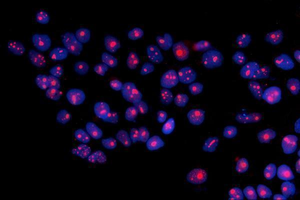
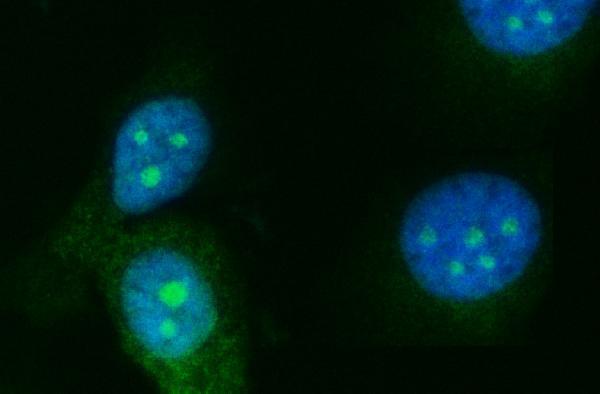
ICC-IF cell lines (HPA subcellular resource): HPA ICC-IF images show RRS1 in A-431, A-549, U2OS, NIH 3T3, with annotated localisation: Nucleoli (supported) (HPA subcellular).
Technical controls: Include no-primary (secondary-only) and concentration-matched rabbit isotype controls alongside a knockout or RRS1-depleted biological control, if available (caption: rabbit primary antibody; standard IHC specificity controls). Quench endogenous peroxidase and check background in the testis section before interpreting HRP/DAB staining (caption: HRP/DAB detection in rat testis; standard chromogenic IHC practice).
⚠️Feasibility: No target-specific fixation window or fixation effect is reported, and the selected A04418-3 paraffin-section caption does not state a fixative (caption: fixative unreported). The caption uses heat-mediated retrieval in EDTA at pH 8.0, but does not establish that retrieval is required (caption: EDTA retrieval, pH 8.0). ICC-IF images support nucleolar localisation, but the supplied evidence does not show that frozen sections or IF are easier than paraffin IHC; check testis sections for endogenous peroxidase background when scoring nucleolar DAB (HPA: nucleoli supported; caption: HRP/DAB detection in rat testis; standard chromogenic IHC practice).

HPA tissue IHC evidence for RRS1

Comprehensive Human Protein Atlas IHC scoring per tissue (reliability: Approved — Low consistency between antibody staining and RNA expression data.). Rows are taken directly from the HPA tissue chart — click any row's HPA link to view the source.

Positive expression · recommended positive controls

TissueCell typeLevelEvidenceSource
Cerebral cortex Neuronal cells High Protein (IHC) HPA →
Hippocampus Neuronal cells High Protein (IHC) HPA →
Skin Keratinocytes Medium Protein (IHC) HPA →
Testis Leydig cells High Protein (IHC) HPA →
Adrenal gland Glandular cells Medium Protein (IHC) HPA →

Undetected expression · recommended negative controls

TissueCell typeLevelEvidenceSource
Adipose tissue Adipocytes Not detected Protein (IHC) HPA →
Breast Adipocytes Not detected Protein (IHC) HPA →
Lymph node Germinal center cells Not detected Protein (IHC) HPA →
Parathyroid gland Glandular cells Not detected Protein (IHC) HPA →
Soft tissue Chondrocytes Not detected Protein (IHC) HPA →
Section 3

Advanced RRS1 IHC Tips

Troubleshoot RRS1 staining in paraffin sections by checking retrieval, intracellular access, compartment-specific signal and cell-level controls before scoring.

What retrieval should I try first if RRS1 staining is weak in paraffin sections?
Start with heat-mediated antigen retrieval in EDTA at pH 8.0 for paraffin sections (datasheet A04418-3). The selected antibody's rat testis IHC image used this retrieval, followed by 2 μg/ml primary antibody overnight at 4°C (datasheet A04418-3). If staining remains weak, check retrieval temperature and section adhesion before comparing a fallback buffer on matched sections (standard IHC practice). Keep antibody concentration, detection chemistry and development conditions constant during that comparison (standard IHC practice). Judge improvement by clearer nucleolar staining, the supported subcellular location, while monitoring any rise in diffuse background (HPA subcellular; standard IHC practice).
How should I troubleshoot fixation-related loss of RRS1 staining?
The selected rat testis caption describes a paraffin section but does not state its fixative, so RRS1-specific fixation sensitivity is unknown (datasheet A04418-3). Record the fixative and fixation duration for each specimen, then compare sections processed together under the same EDTA pH 8.0 retrieval conditions (standard IHC practice; datasheet A04418-3). If signal varies across a block, inspect morphology and compare central and peripheral regions before changing antibody concentration (standard IHC practice). Include a concurrently processed positive control to distinguish a specimen-processing problem from a staining-run problem (standard IHC practice). Neither the reported tissue pattern nor the protein's topology establishes a fixation requirement (HPA tissue IHC; UniProt Q15050).
Where should convincing RRS1 staining appear in chromogenic IHC?
Prioritize nucleolar staining when assessing RRS1 because UniProt places it in the nucleus and nucleolus, and HPA supports nucleoli as its main location (UniProt Q15050; HPA subcellular). HPA tissue IHC also reports cytoplasmic and nucleolar expression in most tissues, so record those compartments separately (HPA tissue IHC). Inspect high magnification fields with a nuclear counterstain to distinguish discrete intranuclear nucleoli from stain deposited over the entire nucleus (standard IHC practice). RRS1 has no annotated transmembrane segment, so a crisp plasma-membrane outline alone does not match its annotated topology (UniProt Q15050). Compare each compartment with a negative reagent control and consistent tissue morphology (standard IHC practice).
Could an isoform or modified epitope explain inconsistent RRS1 IHC?
UniProt lists one 1–365 chain and no annotated isoforms for RRS1, so the supplied record offers no isoform-specific explanation for differing IHC patterns (UniProt Q15050). It lists modified residues including N-acetylmethionine at 1, phosphoserine at 5 and citrulline at 273 (UniProt Q15050). The payload does not map the catalog antibody's epitope, so do not attribute staining differences to any of those modifications without epitope evidence (datasheet A04418-3; UniProt Q15050). Compare matched sections across retrieval conditions while holding detection and exposure to chromogen constant (standard IHC practice). If available, use independent epitope validation to investigate a persistent discrepancy (standard IHC practice).
How can IF help assess ambiguous RRS1 IHC localisation?
Use IF/ICC as a separate localisation check: HPA supports mainly nucleolar RRS1 localisation in its subcellular images (HPA subcellular). For multiplexing, pair RRS1 with a marker identifying the expected cell type, such as Leydig cells in testis, where HPA reports high staining (HPA tissue IHC; standard IF practice). Choose fluorophores after inspecting tissue autofluorescence and place the weaker signal in a cleaner spectral channel (standard IF practice). Because RRS1 lacks a transmembrane segment and is nucleolar, provide permeabilisation that permits antibody access to intracellular and nuclear epitopes, then check morphology (UniProt Q15050; standard IF practice). Keep IF controls separate from the chromogenic IHC assessment (standard IF/IHC practice).
What should I change when RRS1 DAB staining is diffuse?
First compare a no-primary control with the stained section to identify signal arising from the detection system or tissue (standard IHC practice). The selected IHC example used 10% goat serum block, 2 μg/ml primary antibody and DAB development, providing a documented starting point for this antibody (datasheet A04418-3). Check endogenous peroxidase blocking and shorten chromogen development if the no-primary control is brown; these are general chromogenic IHC steps (standard IHC practice). If background appears only with primary antibody, titrate it and reassess washing and blocking on matched sections (standard IHC practice). Preserve discrete nucleolar signal while reducing diffuse nuclear or cytoplasmic deposit (HPA subcellular; standard IHC practice).
How should I score RRS1 IHC across samples? ⚠ ANSWER MARKED FOR VERIFICATION
Define the compartment and cell population before scoring; HPA supports nucleoli as the main location and reports cytoplasmic staining in tissue IHC (HPA subcellular; HPA tissue IHC). For each annotated cell population, record the percentage of positive cells and an intensity-based H-score, keeping nucleolar and cytoplasmic scores separate (standard IHC practice). Alternatively, report positive-cell density per mm² of viable tissue when cell counts differ substantially (standard IHC practice). Normalize counts to the number of eligible cells or measured viable area, and apply the same positivity threshold and imaging settings across samples (standard IHC practice). Document exclusions for folds, damaged edges and necrosis before comparison (standard IHC practice).
How can I distinguish true RRS1 signal from artefact?
Convincing RRS1 staining should include the expected nucleolar pattern within intact cells, consistent with its supported main location (HPA subcellular; standard IHC practice). In testis, assess whether signal maps to Leydig cells, which HPA lists as highly stained, while recognizing that the selected rat testis caption does not assign staining to a cell type (HPA tissue IHC; datasheet A04418-3). Treat isolated membrane outlines, strong section-edge deposits and staining confined to necrosis as reasons to investigate artefact (UniProt Q15050; standard IHC practice). A brown no-primary control points to detection-system or endogenous-enzyme signal rather than specific primary-antibody binding (standard IHC practice). Interpret discrepancies cautiously because HPA reports low consistency between antibody staining and RNA expression data (HPA tissue IHC).
Boster reagents

Best RRS1 / Ribosome biogenesis regulatory protein homolog IHC Antibodies

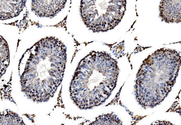
Two anti-RRS1 antibodies have paraffin-section IHC and IF/ICC images (catalog image captions); the catalog lists human, mouse and rat reactivity for A04418-3 and human reactivity for A04418-4 (catalog reactivity).

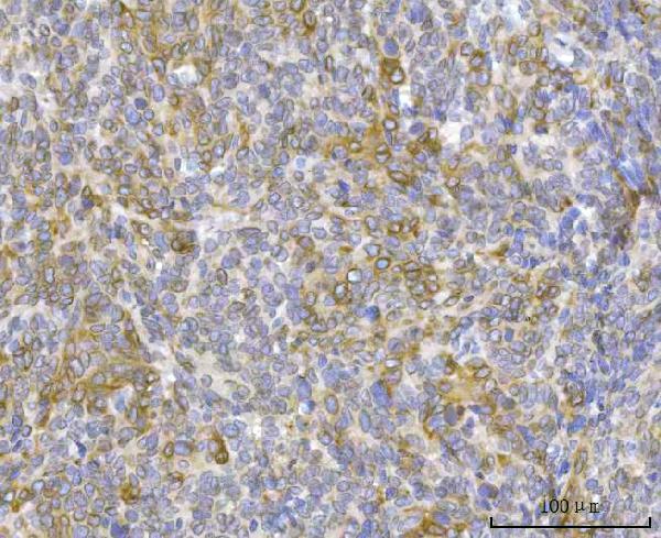
Real IHC data IHC analysis of RRS1 using anti-RRS1 antibody (A04418-3). RRS1 was detected in a paraffin-embedded section of rat testis tissue. Heat mediated antigen retrieval was performed in EDTA buffer (pH 8.0, epitope retrieval solution). The tissue section was blocked with 10% goat serum. The tissue section was then incubated with 2 μg/ml rabbit anti-RRS1 Antibody (A04418-3) overnight at 4°C. Peroxidase Conjugated Goat Anti-rabbit IgG was used as secondary antibody and incubated for 30 minutes at 37°C. The tissue section was developed using HRP Conjugated Rabbit IgG Super Vision Assay Kit (Catalog # SV0002) with DAB as the chromogen.
Anti-RRS1 Antibody ®
Cat # A04418-3
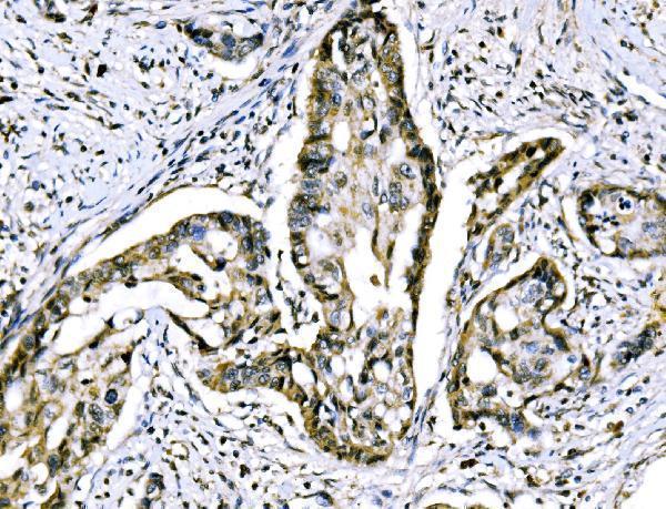
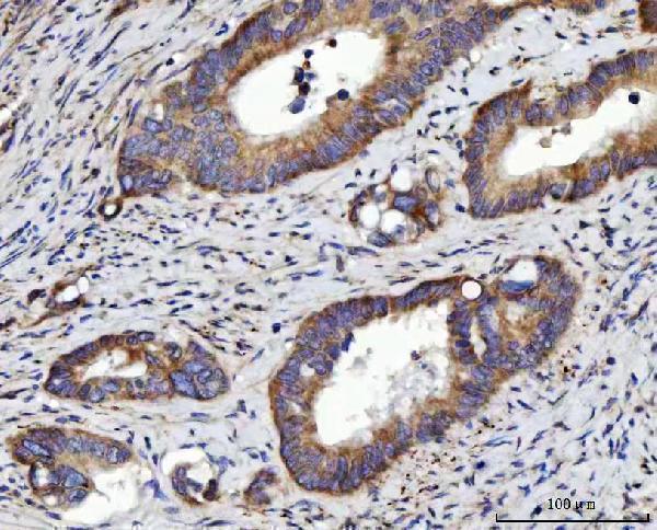
Real IHC data IHC analysis of RRS1 using anti-RRS1 antibody (A04418-4). RRS1 was detected in a paraffin-embedded section of human colorectal adenocarcinoma tissue. Heat mediated antigen retrieval was performed in EDTA buffer (pH 8.0, epitope retrieval solution). The tissue section was blocked with 10% goat serum. The tissue section was then incubated with 2 μg/ml rabbit anti-RRS1 Antibody (A04418-4) overnight at 4°C. Peroxidase Conjugated Goat Anti-rabbit IgG was used as secondary antibody and incubated for 30 minutes at 37°C. The tissue section was developed using HRP Conjugated Rabbit IgG Super Vision Assay Kit (Catalog # SV0002) with DAB as the chromogen.
Anti-RRS1 Antibody ®
Cat # A04418-4

A04418-3 has paraffin-section IHC images from rat testis and human tissues, plus IF/ICC imaging in A431 cells (A04418-3 image captions). A04418-4 has paraffin-section IHC images from human cancers, plus IF images from PC-3 cells and a paraffin-embedded human colon cancer section (A04418-4 image captions).

Which to pick: For tissue IHC, choose A04418-3 when rat or human image evidence is useful, or A04418-4 for human cancer image evidence; each SKU’s IHC captions describe paraffin sections, and neither reports the fixative (A04418-3 and A04418-4 IHC image captions). For IF/ICC, both have cell images; A04418-4 also has a paraffin-section IF image (A04418-3 and A04418-4 IF image captions). For cross-species work, A04418-3 lists human, mouse and rat reactivity, while A04418-4 lists human only; the supplied images do not establish mouse IHC performance (catalog reactivity; A04418-3 and A04418-4 IHC image captions).

Each figure is that product's own IHC / IF validation image from its datasheet.

References

  1. UniProt Consortium. UniProt entry Q15050 (RRS1_HUMAN, Ribosome biogenesis regulatory protein homolog).
  2. Human Protein Atlas. RRS1 tissue IHC expression (reliability: Approved).
  3. Human Protein Atlas. RRS1 subcellular location (ICC-IF): Mainly localized to the nucleoli..
  4. Human Protein Atlas. RRS1 antibody validation summary (2 antibodies).
  5. RRS1 Promotes Retinoblastoma Cell Proliferation and Invasion via Activating the AKT/mTOR Signaling Pathway. BioMed research international 2020 — PMC7652605.
  6. RRS1 silencing suppresses colorectal cancer cell proliferation and tumorigenesis by inhibiting G2/M progression and angiogenesis. Oncotarget 2017 — PMC5669942.
  7. Functional role of RRS1 in breast cancer cell proliferation. Journal of cellular and molecular medicine 2018 — PMC6237566.
  8. Genomic gain/methylation modification/hsa-miR-132-3p increases RRS1 overexpression in liver hepatocellular carcinoma. Cancer science 2023 — PMC10637089.
  9. PubMed PMID:7788527 — UniProt-cited evidence.
  10. PubMed PMID:15489334 — UniProt-cited evidence.
  11. PubMed PMID:12429849 — UniProt-cited evidence.