This website uses cookies to ensure you get the best experience on our website.


- Table of Contents
Plan chromogenic IHC for SCGB2A1 in paraffin sections using cytoplasmic gland staining as a reference (HPA tissue IHC). This guide covers fixation, controls, and interpretation when tissue RNA and protein locations differ for a secreted protein (HPA tissue IHC).

Expected localisation, validated protocols, controls and antibodies — the at-a-glance facts below, then the full design guide.
| Expected localisation | Cytoplasmic tissue staining (HPA tissue IHC) | |
| Staining pattern | Cytoplasmic staining in uterine and epididymal glands (HPA tissue IHC) | |
| Antigen retrieval | Citrate pH 6.0 HIER, 95–98 °C, 20 min (rule: cytoplasmic / membrane antigen) | |
| Positive control | Cervix+4 more · see all | |
| Negative control | Adipose tissue+4 more · see all |
| Fixation | Keep fixation consistent across paraffin sections. (standard IHC practice; not target-specific) | |
| Caveat | RNA and protein locations may differ for this secreted protein (HPA tissue IHC) | |
| Regulation | May be regulated by steroid hormones (UniProt) | |
| Isoform / epitope | No annotated isoforms; mature chain spans residues 19–95 (UniProt) |
The catalog antibody’s IHC-P protocol (datasheet) is accompanied by a published gastric cancer tissue microarray protocol (PMC6995300).
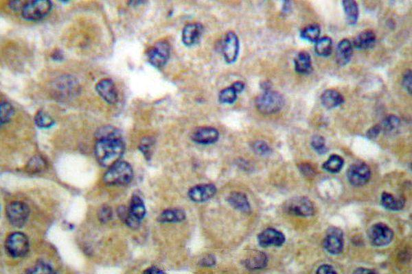
| Sample | Paraffin-embedded human breast carcinoma tissue; fixative not specified (datasheet A07927) |
| Fixation | Image fixative and duration unreported (datasheet A07927); verify before use. |
| Sectioning | 4–5 µm sections on charged slides (standard) |
| Deparaffinisation | Xylene, graded ethanol series to water (standard) |
| Antigen retrieval | Heat-induced epitope retrieval in citrate buffer, pH 6.0, 20 min at 95–98 °C (standard rule: cytoplasmic / membrane antigen) |
| Peroxidase block | 3% H2O2, 10 min, room temperature (standard) |
| Blocking | 10% normal serum of the secondary host, 30 min, room temperature (standard) |
| Primary antibody | Rabbit anti-SCGB2A1, 1:50-1:200 (datasheet A07927) |
| Primary incubation | Overnight at 4 °C (standard) |
| Detection | HRP-polymer secondary, DAB chromogen 5–10 min (standard) |
| Counterstain | Hematoxylin, blue, dehydrate and mount (standard) |
| Expected result | SCGB2A1-positive staining in glandular cells of cervix (HPA tissue IHC: High). HPA tissue profile: Cytoplasmic expression mainly in uterine and epididymal glands. No signal in the no-primary control. |
SCGB2A1 is secreted and has no transmembrane segment (UniProt O75556: subcellular location and topology). In paraffin-section IHC, expect predominantly cytoplasmic staining in uterine and epididymal glands (HPA: tissue IHC profile). HPA rates the tissue pattern Enhanced for agreement with RNA data, while noting that secretion can separate protein location from RNA location and that external verification is pending (HPA: reliability description).
| Cytoplasmic staining in uterine or epididymal glandular epithelium, with relatively little staining in surrounding cells. | This matches the reported tissue pattern (HPA: cytoplasmic expression mainly in uterine and epididymal glands). Compare cells within the same section; secretion means extracellular staining may also need context (UniProt O75556: secreted). |
| Strong, sharply nuclear or membrane-restricted staining, with little cytoplasmic signal in the expected glands. | That distribution does not match the reported IHC profile (HPA: cytoplasmic glandular expression) or a membrane-spanning topology (UniProt O75556: no transmembrane segment). Review controls and staining conditions before calling it specific (general IHC practice). |
| Prominent staining in cells outside the reported positive populations, especially when expected glands are unstained. | Treat this as uncertain specificity; cross-reactivity or endogenous detection activity can mimic a positive result (general IHC practice). HPA reports selected cell populations and staining levels, not proof that every other cell must be negative (HPA: tissue IHC). |
| A broad haze covers glands, stroma, and blank areas with little distinction between cells. | This is difficult to score as localized antigen staining (general IHC practice). Compare a no-primary control and assess blocking, washing, and chromogen development before interpreting the glandular signal (general IHC practice). |
| No convincing signal in an adequately preserved cervix, endometrium, or epididymis section. | HPA reports high staining in cervix glandular cells, endometrial ciliated epithelial cells, and epididymal glandular cells (HPA: tissue IHC). Confirm the relevant cells are present, then check the IHC workflow; a negative run alone does not establish absent SCGB2A1 (general IHC practice). |
| Tissue and cell selection | Cervix glandular cells, endometrial ciliated epithelial cells, and epididymal glandular cells are reported high; nasopharyngeal ciliated cells and salivary glandular cells are medium (HPA: tissue IHC). Evaluate the named cells, since an entire section has no single expected intensity (general IHC practice). |
| Secreted protein and topology | SCGB2A1 is secreted and lacks a transmembrane segment (UniProt O75556: location and topology). The HPA cytoplasmic IHC pattern describes observed cells; extracellular material should be interpreted cautiously because protein and RNA locations can differ (HPA: reliability description). |
| Protein processing | UniProt annotates a signal peptide at residues 1–18 and a mature chain at 19–95 (UniProt O75556: processing). The supplied evidence does not locate the catalog antibody epitope, so it cannot predict which form the stain detects. |
| Antibody evidence | The listed rabbit polyclonal antibody HPA034584 has Enhanced IHC status (HPA: antibody validation). This supports the reported pattern but does not certify every positive cell or replace tissue and detection controls (general IHC practice). |
| IF/ICC question: should vesicular signal appear in IHC? | Vesicles are the approved ICC-IF location (HPA: subcellular). That observation may fit secretory trafficking (UniProt O75556: secreted), but it does not define a required vesicular pattern in paraffin-section IHC; judge IHC against the tissue profile (HPA: tissue IHC). |
| Situation | Likely cause | Next action |
|---|---|---|
| The positive tissue has no staining. | The sampled section may lack the reported positive cells, or the staining run may have failed (HPA: tissue IHC; general IHC practice). | Check for cervix or epididymis glandular cells or endometrial ciliated epithelial cells (HPA: tissue IHC). Review antibody dilution, retrieval, detection reagents, and run controls using standard IHC practice; no SCGB2A1-specific retrieval condition is supplied. |
| The whole section shows brown haze. | Excess background from detection chemistry, incomplete blocking, or insufficient washing is possible (general IHC practice). | Compare a no-primary control, review blocking and wash steps, and shorten chromogen development if needed (general IHC practice). Score only cellular staining distinguishable from background. |
| Nuclear or sharply membrane-bound signal dominates. | This conflicts with the reported cytoplasmic IHC profile (HPA: tissue IHC) and with the lack of a transmembrane segment (UniProt O75556: topology). | Inspect the counterstain and negative control, then reassess detection background and antibody concentration before assigning SCGB2A1 localization (general IHC practice). |
| Unexpected cells stain more strongly than the expected glands. | Cross-reactivity or endogenous detection activity is possible (general IHC practice); HPA's profile describes particular tissues and cells, not universal absence elsewhere (HPA: tissue IHC). | Compare the unexpected cells with a no-primary control and a positive tissue; record cell type and compartment before scoring (general IHC practice). |
| A low-staining tissue appears negative. | HPA reports low staining in breast glandular cells, fallopian-tube ciliated cells, and several other specified populations (HPA: tissue IHC). | Use an HPA high-staining cell population to assess whether the run worked, then describe the low-staining sample as below detection under the tested conditions (HPA: tissue IHC; general IHC practice). |
| An ICC-IF image looks punctate, but the IHC section looks broadly cytoplasmic. | HPA approves a vesicular ICC-IF location and separately reports cytoplasmic tissue IHC expression (HPA: subcellular; HPA: tissue IHC). | Interpret each image in its assay context; for paraffin IHC, assess the documented cell type and cytoplasmic pattern, with controls for background (HPA: tissue IHC; general IHC practice). |


Comprehensive Human Protein Atlas IHC scoring per tissue (reliability: Enhanced — High consistency between antibody staining and RNA expression data. Secreted protein, tissue location of RNA and protein is expected to differ. Pending external verification.). Rows are taken directly from the HPA tissue chart — click any row's HPA link to view the source.
| Tissue | Cell type | Level | Evidence | Source |
|---|---|---|---|---|
| Cervix | Glandular cells | High | Protein (IHC) | HPA → |
| Endometrium | Ciliated epithelial cells | High | Protein (IHC) | HPA → |
| Epididymis | Glandular cells | High | Protein (IHC) | HPA → |
| Nasopharynx | Ciliated cells (cell body) | Medium | Protein (IHC) | HPA → |
| Salivary gland | Glandular cells | Medium | Protein (IHC) | HPA → |
| Tissue | Cell type | Level | Evidence | Source |
|---|---|---|---|---|
| Adipose tissue | Adipocytes | Not detected | Protein (IHC) | HPA → |
| Adrenal gland | Glandular cells | Not detected | Protein (IHC) | HPA → |
| Appendix | Glandular cells | Not detected | Protein (IHC) | HPA → |
| Bone marrow | Hematopoietic cells | Not detected | Protein (IHC) | HPA → |
| Caudate | Glial cells | Not detected | Protein (IHC) | HPA → |
Troubleshoot SCGB2A1 staining in paraffin sections by checking retrieval, controls, cellular location and scoring before interpreting chromogenic signal.
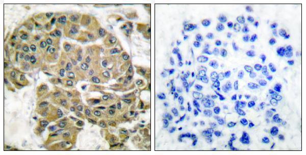
Two anti-SCGB2A1 antibodies have paraffin-section human breast carcinoma IHC images; both list IF, but neither provides an IF image (catalog image captions and applications).
A07927 has an IHC image from paraffin-embedded human breast carcinoma and lists IHC/IF reactivity with human, mouse and rat (A07927 catalog). A07927-1 has a paraffin-embedded human breast carcinoma IHC image with a peptide-blocked comparison and lists IHC/IF reactivity with human and rat (A07927-1 catalog).
Which to pick: For human tissue IHC, consider A07927-1 because its own paraffin-section image includes a peptide-blocked comparison (A07927-1 image caption). For IF, both list the application, but neither provides an IF image; ICC validation is unreported (catalog applications and image captions). For mouse samples, choose A07927 because only it lists mouse reactivity; both antibodies are rabbit-hosted, their clonality is unreported, and neither IHC caption reports the fixative (catalog reactivity, host and clone fields; IHC image captions).