SDC4 / Syndecan-4 · IHC design guide

Design Immunohistochemistry for SDC4

Plan SDC4 paraffin-section IHC with the catalog antibody at 0.5–1 μg/mL (datasheet) and a breast or colon glandular-cell control (HPA tissue IHC). Assess cytoplasmic staining by cell type while considering SDC4’s membrane location and shed ectodomain (HPA tissue IHC; UniProt).

Evidence assembled Oct 2026 · For research use; verify linked source records and product datasheet before use
Immunohistochemistry protocol sheet for SDC4 (IHC for SDC4): expected localisation Cytoplasmic tissue staining (HPA tissue IHC); membrane and soluble forms (UniProt), antibody PB9388, validated IHC image, and IHC protocol steps
Printable SDC4 IHC protocol sheet — expected localisation Cytoplasmic tissue staining (HPA tissue IHC); membrane and soluble forms (UniProt), antibody PB9388, controls and protocol steps. Open the full SDC4 IHC guide →

SDC4 Immunohistochemistry Experimental Design Guide

Expected localisation, validated protocols, controls and antibodies — the at-a-glance facts below, then the full design guide.

Must know before staining
Expected localisation Cytoplasmic tissue staining (HPA tissue IHC); membrane and soluble forms (UniProt)
Staining pattern Cytoplasmic staining in most tissues; high in glandular cells (HPA tissue IHC)
Antigen retrieval Citrate pH 6.0 HIER, 95–98 °C, 20 min (rule: cytoplasmic / membrane antigen)
Positive control ⓘ Adrenal gland+4 more · see all
Negative control ⓘ None in HPA (detected in all 45 tissues); use no-primary + isotype controls
Important caveats
Reasons your staining may differ from the expected pattern.
Fixation Keep fixation consistent across sections. (standard IHC practice; not target-specific)
Caveat Ectodomain shedding can alter the observed distribution (UniProt)
Regulation Stimulus-linked regulation unreported (UniProt)
Isoform / epitope 2 isoforms; extracellular epitopes may detect shed protein (UniProt)
Section 1

Recommended SDC4 IHC & IF Protocols

The catalog antibody protocol is paired with published SDC4 IHC methods for human placenta, biliary atresia liver tissue, and papillary thyroid carcinoma tissue (PMC6629623; PMC9932896; PMC6182223).

Recommended immunohistochemistry (IHC-P) protocol parameters
SampleTissue sections; selected-image fixative not specified (standard IHC workflow)
FixationImage fixative and duration unreported (datasheet PB9388); verify before use.
Sectioning4–5 µm sections on charged slides (standard)
DeparaffinisationXylene, graded ethanol series to water (standard)
Antigen retrievalHeat-induced epitope retrieval in citrate buffer, pH 6.0, 20 min at 95–98 °C (standard rule: cytoplasmic / membrane antigen)
Peroxidase block3% H2O2, 10 min, room temperature (standard)
Blocking10% normal serum of the secondary host, 30 min, room temperature (standard)
Primary antibodyRabbit anti-SDC4, 0.5-1μg/ml (datasheet PB9388)
Primary incubationOvernight at 4 °C (standard)
DetectionHRP-polymer secondary, DAB chromogen 5–10 min (standard)
CounterstainHematoxylin, blue, dehydrate and mount (standard)
Expected resultSDC4-positive staining in glandular cells of adrenal gland (HPA tissue IHC: High). HPA tissue profile: Cytoplasmic expression in most tissues. No signal in the no-primary control.
💡Decision noteStart with citrate pH 6.0 retrieval at 95–98 °C for 20 min (page antigen-retrieval rule); compare the paper-specific methods when adapting to each specimen.
Section 2

What Is the Expected SDC4 Staining Pattern?

SDC4 is a membrane protein with an extracellular domain at residues 19–145 and a transmembrane segment at 146–170; its ectodomain can be shed (UniProt P31431 topology). In tissue IHC, expect chiefly cytoplasmic staining in many tissues, including strong staining in several glandular and epithelial cell populations (HPA: tissue IHC). Interpret that pattern with HPA’s Enhanced tissue reliability, which reflects medium agreement between staining and RNA data (HPA: tissue IHC).

What am I looking at on my slide?
Strong cytoplasmic staining in bronchial respiratory epithelium or colon glandular cells.This matches reported high staining in those cell populations (HPA: High in bronchial respiratory epithelial cells; High in colon glandular cells). Record the stained cell population and compartment when scoring; a tissue label alone cannot distinguish the expected pattern from staining elsewhere on the section (general IHC practice).
Predominantly nuclear staining, with little staining in the expected cytoplasmic or membrane-associated locations.A dominant nuclear pattern conflicts with the reported tissue profile and protein topology (HPA: cytoplasmic expression in most tissues; UniProt P31431 topology). Treat it as suspect, then compare antibody controls and the known-positive tissue before assigning it to SDC4 (general IHC practice).
Strong staining concentrated in smooth muscle cells or splenic red-pulp cells.Both populations are reported at low staining levels, so strong signal there needs scrutiny; they are not established negative controls (HPA: Low in smooth muscle cells; Low in splenic red-pulp cells). Cross-reactivity or endogenous detection activity is possible. Check the stained cell identity and a no-primary control (general IHC practice).
Diffuse chromogen covers cells and surrounding tissue without a clear cellular pattern.This cannot be read as a specific SDC4 distribution simply because SDC4 has a shed ectodomain (UniProt P31431: ectodomain shedding). Review background in a no-primary control and assess blocking, washing and detection conditions before scoring (general IHC practice).
No detectable stain in bronchial respiratory epithelial cells or colon glandular cells.An absent signal conflicts with the reported high staining in these populations, but does not by itself identify the failed step (HPA: High in bronchial respiratory epithelial cells; High in colon glandular cells). Check section quality, antibody use and detection controls before calling the tissue negative (general IHC practice).
💡Expected SDC4 appearanceCall a result consistent when identifiable glandular or epithelial cells show clear, strong cytoplasmic staining, as reported for colon glands and bronchial epithelium (HPA: High in colon glandular cells; High in bronchial respiratory epithelial cells; cytoplasmic expression in most tissues); diffuse slide-wide color or dominant nuclear stain is suspect (general IHC practice; UniProt P31431 topology).
How each factor affects the staining
Antibody validationHPA lists CAB013240 as IHC Enhanced and HPA005716 as IHC Supported; these are antibody-level statuses, while the tissue profile has Enhanced reliability (HPA: antibody validation; tissue IHC). Apply the status to the antibody actually used, and interpret staining against its controls (general IHC practice).
Epitope location and sheddingThe extracellular region spans residues 19–145, and shedding can release the ectodomain (UniProt P31431 topology; shedding). The supplied record does not locate either antibody’s epitope, so it cannot predict whether an antibody detects retained protein, released material, or both.
Isoforms and processingTwo isoforms are listed, with a signal peptide at residues 1–18 and a mature chain at 19–198 (UniProt P31431: isoforms; processing). Because antibody epitope coverage is unspecified, the record cannot establish equal detection of both isoforms or which molecular form contributes to a tissue stain.
Antigen retrievalNo SDC4-specific retrieval condition or fixation-sensitivity result is supplied. If retrieval needs adjustment, compare conditions on a known-positive section using matched detection and controls (general IHC practice); neither HPA staining levels nor UniProt topology establishes a target-specific retrieval effect.
What should IF/ICC show?HPA reports a mainly Golgi location with an additional plasma-membrane location in ICC-IF (HPA: approved Golgi apparatus; approved plasma membrane). This is a separate cell-imaging observation; interpret paraffin-section chromogenic IHC against the tissue profile (HPA: cytoplasmic expression in most tissues).
Why is my staining missing, weak or wrong?
SituationLikely causeNext action
Known-positive bronchial or colon epithelium is blank.The result disagrees with reported high staining, but the payload does not identify a specific failed reagent or retrieval condition (HPA: High in bronchial respiratory epithelial cells; High in colon glandular cells).Verify the cell population and section quality, then check antibody dilution, detection reagents and a positive control in the same run (general IHC practice).
Color appears across tissue and on the no-primary control.Signal without primary antibody points to background from the detection workflow, not an interpretable SDC4 distribution (general IHC practice).Review blocking, washing and the chromogenic detection steps; repeat with the same no-primary control before scoring (general IHC practice).
Nuclei dominate the stain.This location conflicts with the tissue profile and membrane topology (HPA: cytoplasmic expression in most tissues; UniProt P31431 topology).Compare a known-positive tissue and controls, and check whether the pattern follows cell boundaries or counterstained nuclei (general IHC practice).
Smooth muscle or splenic red pulp looks strongly positive.Those cells have low reported staining, so strong signal may reflect cross-reactivity or endogenous activity; low does not mean absent (HPA: Low in smooth muscle cells; Low in splenic red-pulp cells).Confirm cell identity and compare a no-primary control with expected high-staining cells on the run (general IHC practice; HPA: High in colon glandular cells).
Stain is difficult to assign to cells near extracellular material.SDC4 has a shed ectodomain, but the supplied evidence does not establish where shed protein accumulates in a section (UniProt P31431: ectodomain shedding).Score clearly identifiable cells separately from ambiguous extracellular color; document the latter without calling it a specific location (general IHC practice).
Two antibodies give different patterns.Their IHC validation statuses differ, and their epitopes are not supplied (HPA: CAB013240 IHC Enhanced; HPA005716 IHC Supported). The record cannot establish whether the difference reflects epitope recognition.Keep antibody-specific results separate, compare staining in the same reported high-staining cell population, and review matched controls (HPA: High in bronchial respiratory epithelial cells; general IHC practice).

Sample controls for SDC4 IHC & IF

🧪Run breast first: glandular cells should stain (HPA: High in breast glandular cells; selected PB9388 IHC-P caption: human mammary cancer tissue). HPA detects SDC4 in all 45 scored tissues, so there is no supported negative tissue; use no-primary and isotype controls, and treat cells without visible staining on the positive slide as internal background references rather than confirmed SDC4-negative cells (HPA: no negative rows).
Positive control tissue: Adrenal gland (Glandular cells, HPA High)
Negative control tissue: None in HPA: SDC4 is detected in all 45 scored tissues. Use a no-primary (secondary-only) and an isotype control instead.
ICC-IF cell lines (HPA subcellular resource): HPA ICC-IF images show SDC4 in A-431, U-251MG, U2OS, with annotated localisation: Golgi apparatus (approved) (HPA subcellular).
Technical controls: Include a no-primary, secondary-only control; a host-species- and isotype-matched control for a monoclonal primary, or matched normal IgG for a polyclonal primary; and SDC4 knockout material or a validated peptide-block control as a biological specificity check (standard IHC practice). For chromogenic breast IHC, block endogenous peroxidase and check background staining against the controls; assess endogenous biotin if using biotin-based detection (standard IHC practice).
⚠️Feasibility: A target-specific fixation window, fixation effect, and antigen-retrieval dependency are unreported in the supplied evidence; the selected PB9388 tissue-IHC caption also does not report a fixative (selected PB9388 IHC-P caption). The evidence does not establish whether frozen sections or IF/ICC are easier than paraffin IHC; HPA ICC-IF images show mainly Golgi and additional plasma-membrane localization (HPA subcellular). In breast sections, assess extracellular or luminal signal cautiously because the SDC4 ectodomain can be shed (UniProt P31431 processing and localization).

HPA tissue IHC evidence for SDC4

Comprehensive Human Protein Atlas IHC scoring per tissue (reliability: Enhanced — Medium consistency between antibody staining and RNA expression data. At least one protein variant secreted, tissue location of RNA and protein might differ and correlation is complex.). Rows are taken directly from the HPA tissue chart — click any row's HPA link to view the source.

Positive expression · recommended positive controls

TissueCell typeLevelEvidenceSource
Adrenal gland Glandular cells High Protein (IHC) HPA →
Appendix Glandular cells High Protein (IHC) HPA →
Breast Glandular cells High Protein (IHC) HPA →
Bronchus Respiratory epithelial cells High Protein (IHC) HPA →
Caudate Neuronal cells High Protein (IHC) HPA →

Undetected expression · recommended negative controls

TissueCell typeLevelEvidenceSource
None in HPA: SDC4 is detected in all 45 scored tissues. Use a no-primary (secondary-only) and an isotype control instead.
Section 3

Advanced SDC4 IHC Tips

Troubleshoot SDC4 chromogenic IHC in paraffin sections using its membrane topology, shedding, and reported tissue and subcellular staining patterns.

What retrieval conditions should I start with for SDC4 paraffin IHC?
Use citrate buffer at pH 6.0 for heat-induced epitope retrieval at 95–98 °C for 20 min before chromogenic IHC (page retrieval rule). Keep section thickness, heating time, and cooling conditions consistent across the comparison set so a change in staining can be assigned to the variable under test (standard IHC practice). SDC4 has an extracellular region at residues 19–145 and a cytoplasmic region at 171–198, so record which region the antibody recognizes when assessing retrieval (UniProt P31431 topology). If signal remains weak, compare a small retrieval time series alongside a known positive section, while checking whether stronger heating also increases background or damages morphology (standard IHC practice).
How should I assess whether fixation is masking the SDC4 epitope?
Target-specific fixation sensitivity is unknown from the supplied evidence; the PB9388 tissue caption identifies an IHC(P) image but does not state its fixative (PB9388 tissue-IHC caption). Record the fixative, fixation duration, processing history, and section age for each specimen before comparing staining intensities (standard IHC practice). When fixation varies, stain a consistently processed positive control in the same run and inspect morphology as well as signal (standard IHC practice). If a fixation effect is suspected, compare otherwise matched specimens under the same pH 6.0, 95–98 °C, 20 min retrieval condition; differences then remain an observation to validate, not an established SDC4-specific fixation response (page retrieval rule; standard IHC practice).
Should SDC4 appear at the cell surface or in the cytoplasm?
Assess both cell-associated cytoplasmic and membrane staining: HPA reports cytoplasmic expression in most tissues, while its subcellular data place SDC4 mainly at the Golgi apparatus and additionally at the plasma membrane (HPA tissue IHC; HPA subcellular). A surface pattern is biologically plausible because residues 146–170 form the transmembrane segment (UniProt P31431 topology). Extracellular staining requires care because shedding produces a soluble ectodomain, which can separate protein location from the producing cell (UniProt P31431 subcellular; HPA tissue IHC reliability description). Score membrane, cytoplasmic, and extracellular signal separately rather than treating every brown deposit as equivalent cellular expression (standard IHC practice).
How do epitope location and SDC4 isoforms affect staining interpretation?
First establish whether the IHC-validated antibody recognizes an extracellular or cytoplasmic epitope; the supplied material does not identify its binding sequence (supplied antibody evidence; UniProt P31431 topology). The extracellular segment spans residues 19–145, whereas the cytoplasmic tail spans 171–198, so an antibody against one side need not report material retained on the other after ectodomain shedding (UniProt P31431 topology and subcellular annotation). SDC4 has 2 annotated isoforms, but their epitope coverage is not specified here (UniProt P31431 isoforms; supplied antibody evidence). Its extracellular glycosylation sites at residues 39, 61, 63, and 95 are relevant context when assessing an extracellular epitope, without establishing that they alter this antibody’s IHC staining (UniProt P31431 glycosylation).
How can I check an SDC4 IHC pattern with multiplex IF?
Use IF as a separate cross-check of the chromogenic IHC pattern, and pair SDC4 with a marker that identifies the cell population being assessed (standard IF practice). For example, interpret overlap within epithelial or fibroblast populations only after validating the chosen cell marker, since SDC4 is reported in epithelial cells and fibroblasts (UniProt P31431 tissue specificity; standard IF practice). Choose a fluorophore whose emission is distinguishable from the specimen’s autofluorescence, and include single-label and no-primary controls when evaluating overlap (standard IF practice). Select permeabilisation according to the documented antibody epitope: access to the cytoplasmic tail at 171–198 generally requires permeabilisation, while an extracellular epitope at 19–145 may be assessed without it (UniProt P31431 topology; standard IF practice).
What should I change when SDC4 DAB staining is diffuse?
Inspect a no-primary control and the tissue edges before adjusting antibody concentration; diffuse DAB can reflect background, edge effects, or endogenous peroxidase activity (standard chromogenic IHC practice). Apply a peroxidase block before the enzyme-based detection step, then keep blocking, antibody incubation, and DAB development consistent across comparison sections (standard chromogenic IHC practice). SDC4 shedding makes extracellular signal biologically possible, so classify extracellular deposits by their distribution and control staining before calling them specific (UniProt P31431 subcellular annotation; standard IHC practice). HPA lists low staining in smooth muscle cells and spleen red-pulp cells, but supplies no negative tissue here; use these as contextual comparators rather than guaranteed blank controls (HPA tissue IHC).
How should I quantify SDC4 staining across paraffin sections? ⚠ ANSWER MARKED FOR VERIFICATION
Define the cell population and compartment before scoring, since SDC4 can be membrane-associated and HPA also reports cytoplasmic tissue staining (UniProt P31431 topology; HPA tissue IHC). For a defined population, record the % of positive cells and an H-score based on staining intensity; for spatially restricted populations, cell density per mm² can supplement those measures (standard IHC scoring practice). Normalise counts to the viable tissue area or the number of eligible cells, and apply the same positivity threshold to every slide in a comparison (standard IHC scoring practice). Keep membrane, cytoplasmic, and extracellular measurements separate so shed material does not inflate cell-associated expression estimates (UniProt P31431 subcellular annotation; standard IHC scoring practice).
How can I distinguish convincing SDC4 staining from artefact?
A convincing result follows identifiable cells and compartments and is reproducible in a same-run positive control; HPA reports high glandular-cell staining in breast, colon, and several other tissues (standard IHC practice; HPA tissue IHC). Membrane or cytoplasmic signal can fit SDC4 biology, while extracellular signal needs separate assessment because its ectodomain can be shed (UniProt P31431 topology and subcellular annotation; HPA tissue IHC). Treat staining confined to cut edges, necrotic areas, or the no-primary control as suspect, and check the peroxidase block when DAB appears independently of primary antibody (standard chromogenic IHC practice). A signal in an unexpected cell type warrants confirmation with morphology and a cell-identity marker before assigning it to SDC4-positive cells (standard IHC practice).
Boster reagents

Best SDC4 / Syndecan-4 IHC Antibodies

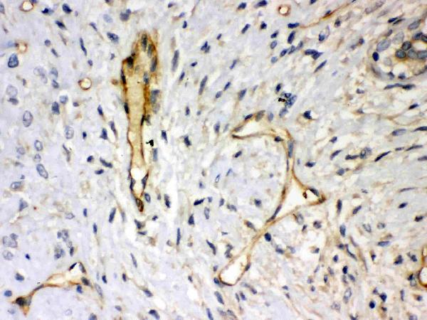
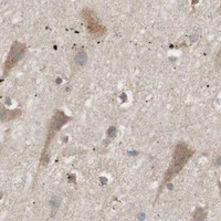
PB9388 has IHC images from human mammary cancer tissue and rat and mouse intestine (PB9388 IHC image captions). IF/ICC data are unreported (PB9388 applications and IF image alts).

Real IHC data Anti-Syndecan 4 vendor antibody, PB9388, IHC(P) IHC(P): Human Mammary Cancer Tissue
Anti-Syndecan 4/SDC4 Antibody ®
Cat # PB9388

PB9388 has paraffin-section IHC images from human mammary cancer tissue and rat and mouse intestine (PB9388 IHC image captions). Its catalog lists IHC reactivity with human, mouse, and rat (PB9388 applications and reactivity).

Which to pick: For tissue IHC, choose PB9388 for paraffin sections supported by its own images; the fixative is unreported (PB9388 IHC image captions). For cross-species IHC, PB9388 lists human, mouse, and rat reactivity and has an IHC image for each; its host is rabbit and clonality is unreported (PB9388 catalog reactivity, host, clone and image alts). No listed SKU has IF/ICC validation (PB9388 applications and IF image alts).

Each figure is that product's own IHC / IF validation image from its datasheet.

References

  1. UniProt Consortium. UniProt entry P31431 (SDC4_HUMAN, Syndecan-4).
  2. Human Protein Atlas. SDC4 tissue IHC expression (reliability: Enhanced).
  3. Human Protein Atlas. SDC4 subcellular location (ICC-IF): Mainly localized to the Golgi apparatus. In addition localized to the plasma membrane..
  4. Human Protein Atlas. SDC4 antibody validation summary (2 antibodies).
  5. Syndecan-4 regulates extravillous trophoblast migration by coordinating protein kinase C activation. Scientific reports 2019 — PMC6629623.
  6. Bioinformatics analysis identifies heparan sulfate proteoglycans acting as different progress subtypes of biliary atresia. Frontiers in pediatrics 2023 — PMC9932896.
  7. Role of Syndecan-4 in the Inhibition of Articular Cartilage Degeneration in Osteoarthritis. Biomedicines 2023 — PMC10452293.
  8. SDC4 Gene Silencing Favors Human Papillary Thyroid Carcinoma Cell Apoptosis and Inhibits Epithelial Mesenchymal Transition via Wnt/β-Catenin Pathway. Molecules and cells 2018 — PMC6182223.
  9. PubMed PMID:1500433 — UniProt-cited evidence.
  10. PubMed PMID:7916598 — UniProt-cited evidence.
  11. PubMed PMID:8797100 — UniProt-cited evidence.