SERINC2 / Serine incorporator 2 · IHC design guide

Design Immunohistochemistry for SERINC2

Plan chromogenic IHC on paraffin sections using the IHC-validated antibody at 1:100–1:300 (datasheet A30553). Evaluate the reported cytoplasmic tissue pattern (HPA tissue IHC) alongside the annotated cell-membrane location (UniProt).

Evidence assembled Oct 2026 · For research use; verify linked source records and product datasheet before use
Immunohistochemistry protocol sheet for SERINC2 (IHC for SERINC2): expected localisation General cytoplasmic tissue staining (HPA tissue IHC), antibody A30553, validated IHC image, and IHC protocol steps
Printable SERINC2 IHC protocol sheet — expected localisation General cytoplasmic tissue staining (HPA tissue IHC), antibody A30553, controls and protocol steps. Open the full SERINC2 IHC guide →

SERINC2 Immunohistochemistry Experimental Design Guide

Expected localisation, validated protocols, controls and antibodies — the at-a-glance facts below, then the full design guide.

Must know before staining
Expected localisation General cytoplasmic tissue staining (HPA tissue IHC)
Staining pattern Cytoplasmic staining in glandular and other positive cells (HPA tissue IHC)
Antigen retrieval Tris-EDTA pH 9.0 HIER, heat-mediated (datasheet A30553)
Positive control ⓘ Adrenal gland+4 more · see all
Negative control ⓘ None in HPA (detected in all 45 tissues); use no-primary + isotype controls
Important caveats
Reasons your staining may differ from the expected pattern.
Fixation Keep fixation consistent across paraffin sections. (standard IHC practice; not target-specific)
Caveat Medium staining–RNA concordance; external verification pending (HPA tissue IHC)
Regulation Liver-enhanced RNA; regulation unreported (HPA tissue RNA)
Isoform / epitope Four isoforms, 11 membrane spans; epitope side needs mapping (UniProt)
Section 1

Recommended SERINC2 IHC & IF Protocols

The catalog antibody’s IHC-P protocol (datasheet A30553) is accompanied by one published SERINC2 IHC method (PMC9260995).

Recommended immunohistochemistry (IHC-P) protocol parameters
SampleParaffin-embedded human colon cancer tissue; fixative not specified (datasheet A30553)
FixationImage fixative and duration unreported (datasheet A30553); verify before use.
Sectioning4–5 µm sections on charged slides (standard)
DeparaffinisationXylene, graded ethanol series to water (standard)
Antigen retrievalHeat retrieval: Tris-EDTA pH 9.0 (datasheet A30553); 20 min, 95–100 °C (standard)
Peroxidase block3% H2O2, 10 min, room temperature (standard)
Blocking10% normal serum of the secondary host, 30 min, room temperature (standard)
Primary antibodyRabbit anti-SERINC2, 1:100 - 1:300 (datasheet A30553)
Primary incubationOvernight at 4 °C (standard)
DetectionHRP-polymer secondary, DAB chromogen 5–10 min (standard)
CounterstainHematoxylin, blue, dehydrate and mount (standard)
Expected resultSERINC2-positive staining in glandular cells of adrenal gland (HPA tissue IHC: High). HPA tissue profile: General cytoplasmic expression. No signal in the no-primary control.
💡Decision noteStart with Tris-EDTA pH 9.0 heat retrieval (datasheet A30553); the published method uses high temperature and pressure without specifying a buffer (PMC9260995).
Section 2

What Is the Expected SERINC2 Staining Pattern?

SERINC2 is a cell-membrane protein with 11 transmembrane segments (UniProt Q96SA4 topology). In paraffin-section IHC, HPA describes general cytoplasmic staining and reports high staining in several glandular and other cell populations (HPA tissue IHC). Treat that pattern as a guide, not definitive validation: HPA rates its IHC evidence Enhanced but notes medium agreement with RNA data and pending external verification (HPA tissue IHC).

What am I looking at on my slide?
Cytoplasmic staining in colon or duodenal glandular cells, or bronchial respiratory epithelial cells.Consistent with HPA's general cytoplasmic IHC profile and High staining in those cells (HPA tissue IHC). Compare the stained cells with tissue morphology; a positive field alone cannot establish antibody specificity.
Membrane-associated staining is visible alongside cytoplasmic staining.Biologically plausible for an 11-pass cell-membrane protein (UniProt Q96SA4 topology and subcellular location). HPA describes tissue IHC as generally cytoplasmic, so a sharp membrane outline is not required to call the reported IHC pattern (HPA tissue IHC).
Nuclear-only staining dominates the paraffin section.Discordant with HPA's general cytoplasmic tissue-IHC profile (HPA tissue IHC). Recheck morphology and controls before calling it SERINC2: HPA also reports an additional nucleoplasmic location by ICC-IF, which does not establish nuclear-only tissue IHC (HPA subcellular ICC-IF).
Strong staining appears chiefly in cardiomyocytes, chondrocytes or the listed low-staining brain cells.These are Low populations in HPA tissue IHC (HPA tissue IHC). Strong signal there, especially without the expected positive-cell pattern, raises cross-reactivity or detection-background concerns; it does not prove either cause.
Uniform haze covers cells and stroma, or a known high-staining population shows no signal.A field-wide haze is difficult to assign to SERINC2-positive cells; absent signal in HPA high-staining cells weakens a negative call (HPA tissue IHC; general IHC practice). Assess both against matched detection controls and tissue morphology.
💡Expected SERINC2 appearanceCall a positive IHC result when glandular or other HPA-listed high-staining cells show clear, predominantly cytoplasmic signal, with possible membrane association; treat diffuse haze or staining confined to an unexpected cell population as suspect (HPA tissue IHC; UniProt Q96SA4 subcellular location; general IHC practice).
How each factor affects the staining
Which cells provide an interpretable positive reference?HPA reports High staining in colon and duodenal glandular cells, bronchial respiratory epithelial cells, cerebellar Purkinje cells and other listed populations (HPA tissue IHC). Select a listed cell population that is identifiable on the same type of section.
How should membrane topology affect interpretation?SERINC2 has 11 transmembrane segments and is assigned to the cell membrane (UniProt Q96SA4 topology and subcellular location). This supports membrane association, while HPA's observed paraffin-section profile remains generally cytoplasmic (HPA tissue IHC).
Can an unstained section distinguish isoforms?UniProt lists 4 SERINC2 isoforms (UniProt Q96SA4 isoforms). The supplied evidence gives no antibody epitope or isoform-specific IHC pattern, so staining intensity and location cannot identify an isoform.
Does processing explain missing or extracellular staining?UniProt annotates a 1–455 chain, with no signal peptide or propeptide, and lists 0 glycosylation sites (UniProt Q96SA4 processing and glycosylation). The supplied record does not establish shedding or a processing-dependent tissue pattern.
What does IF/ICC add?HPA reports plasma membrane as the main approved ICC-IF location and nucleoplasm as an additional approved location, with images in HaCaT, RT-4 and U2OS (HPA subcellular ICC-IF). Use that as a localisation comparison; this section supplies no IF/ICC protocol.
How strong is the tissue-pattern evidence?HPA marks IHC Enhanced for antibody HPA005974, but describes medium staining–RNA consistency and pending external verification (HPA tissue IHC; HPA antibody validation). Interpret a matching pattern with controls rather than treating the rating as proof in every specimen.
Why is my staining missing, weak or wrong?
SituationLikely causeNext action
No staining in a section expected to contain colon or duodenal glandular cells.The run may have failed, or those cells may be absent from the examined area; HPA reports High staining in these populations (HPA tissue IHC).Confirm the cell population on the counterstain, then check primary-antibody application, retrieval, detection reagents and run controls (general IHC practice). A negative field is inconclusive until the positive control performs.
Diffuse brown haze obscures cell boundaries.Background can arise from insufficient blocking, excess detection signal or endogenous peroxidase activity in chromogenic IHC (general IHC practice).Inspect a no-primary control, verify the peroxidase-block step if applicable, and adjust antibody or detection conditions according to the validated assay (general IHC practice). Score only signal that can be assigned to cells.
Only nuclei stain strongly.This differs from the general cytoplasmic tissue-IHC profile (HPA tissue IHC), although an additional nucleoplasmic location is approved in ICC-IF (HPA subcellular ICC-IF).Check no-primary and tissue controls, section morphology and detection background (general IHC practice). Do not transfer the ICC-IF nuclear observation directly into a nuclear-only paraffin-IHC scoring rule.
Unexpectedly strong signal appears in HPA low-staining cells.Strong staining in cardiomyocytes, chondrocytes or specified low-staining neural cells is discordant with the reported levels (HPA tissue IHC); cross-reactivity or endogenous detection activity is possible.Compare the same run with a listed high-staining population and a no-primary control (HPA tissue IHC; general IHC practice). Record the discordance before assigning it to SERINC2.
Membrane outlines are weak, but cytoplasmic staining is clear in expected cells.HPA calls the tissue-IHC profile generally cytoplasmic, despite UniProt's cell-membrane assignment (HPA tissue IHC; UniProt Q96SA4 subcellular location).Assess the cytoplasmic pattern in identifiable HPA high-staining cells and review controls (HPA tissue IHC; general IHC practice). Do not require a crisp membrane rim as the sole positive criterion.
Staining differs between sections or runs.Target-specific fixation effects are not established by the supplied assay evidence. Verify with a matched IHC source before attributing a result to fixation.Compare like tissue regions and documented run conditions, including retrieval and detection, using the same positive control (general IHC practice). Report unresolved variability without attributing it to SERINC2 epitope masking.

Sample controls for SERINC2 IHC & IF

🧪Run bone marrow first and expect staining in hematopoietic cells (HPA: High in hematopoietic cells). HPA detects SERINC2 in all 45 scored tissues, so there is no supported negative tissue; use no-primary and isotype controls, and treat cells showing only counterstain on the positive slide as local background references rather than proven SERINC2-negative cells (HPA: no negative tissue rows).
Positive control tissue: Adrenal gland (Glandular cells, HPA High)
Negative control tissue: None in HPA: SERINC2 is detected in all 45 scored tissues. Use a no-primary (secondary-only) and an isotype control instead.
ICC-IF cell lines (HPA subcellular resource): HPA ICC-IF images show SERINC2 in HaCaT, RT-4, U2OS, with annotated localisation: Plasma membrane (approved) (HPA subcellular).
Technical controls: Include a no-primary, secondary-only control, an isotype control matched to the primary antibody’s host species and clonality, and a biological specificity control using SERINC2 knockout material or peptide blocking if a suitable immunizing peptide is available (standard IHC practice). In bone marrow, quench endogenous peroxidase and compare the controls before interpreting chromogen in hematopoietic cells (HPA: High in hematopoietic cells; standard IHC practice).
⚠️Feasibility: No target-specific fixation window or fixation effect is reported, and the selected paraffin-section caption does not state a fixative (SKU A30553 caption: fixative unreported). That caption uses Tris-EDTA at pH 9.0 for retrieval and a 1:200 primary dilution overnight at 4 °C; it does not establish that retrieval is required for all specimens (SKU A30553 caption). No supplied evidence establishes that frozen sections or IF are easier; marrow peroxidase can complicate chromogenic interpretation, while plasma-membrane staining is a plausible localization to assess (HPA: High in bone-marrow hematopoietic cells; HPA: plasma membrane approved; standard IHC practice).

HPA tissue IHC evidence for SERINC2

Comprehensive Human Protein Atlas IHC scoring per tissue (reliability: Enhanced — Medium consistency between antibody staining and RNA expression data. Pending external verification.). Rows are taken directly from the HPA tissue chart — click any row's HPA link to view the source.

Positive expression · recommended positive controls

TissueCell typeLevelEvidenceSource
Adrenal gland Glandular cells High Protein (IHC) HPA →
Appendix Glandular cells High Protein (IHC) HPA →
Bone marrow Hematopoietic cells High Protein (IHC) HPA →
Bronchus Respiratory epithelial cells High Protein (IHC) HPA →
Cerebellum Purkinje cells High Protein (IHC) HPA →

Undetected expression · recommended negative controls

TissueCell typeLevelEvidenceSource
None in HPA: SERINC2 is detected in all 45 scored tissues. Use a no-primary (secondary-only) and an isotype control instead.
Section 3

Advanced SERINC2 IHC Tips

Troubleshoot SERINC2 staining by checking retrieval, cell compartment, and controls before comparing signal across paraffin sections.

What should I change when SERINC2 staining is weak in paraffin sections?
Start with heat-mediated retrieval in Tris-EDTA at pH 9.0 for paraffin-section IHC (datasheet A30553). The catalog image used paraffin-embedded human colon cancer, a 1:200 primary antibody dilution, and incubation at 4°C overnight; its fixative was not reported (caption A30553). Keep section thickness, heating, cooling, and detection conditions matched while varying retrieval duration in a small series (standard IHC practice). Compare the same tissue region across runs and watch for lost morphology or increased background, since stronger staining alone does not establish specific SERINC2 detection (standard IHC practice).
Could fixation explain variable SERINC2 staining between paraffin blocks?
Target-specific fixation sensitivity for SERINC2 is unknown from the supplied evidence; the paraffin-section caption does not identify a fixative (caption A30553). Record each block’s fixative, fixation interval, and processing history before comparing chromogenic staining across specimens (standard IHC practice). When preparing a new series, hold those conditions constant and include a similarly processed positive-control section in every staining run (standard IHC practice). If older and newer blocks differ, compare morphology, retrieval response, and background together; neither the tissue staining profile nor membrane topology establishes a SERINC2-specific fixation effect (standard IHC practice).
How should I judge cytoplasmic staining when SERINC2 is described as membrane-localised?
Score membrane-associated and diffuse cytoplasmic chromogenic signal separately, because UniProt places SERINC2 at the cell membrane while tissue IHC reports general cytoplasmic expression (UniProt Q96SA4 localisation; HPA tissue IHC profile). The subcellular IF record identifies plasma membrane as the main location and nucleoplasm as an additional location (HPA subcellular). In colon sections, examine glandular cells, which have a high tissue IHC signal, and compare their staining with adjacent structures (HPA: High in colon glandular cells). Treat isolated nuclear or diffuse staining cautiously until omission controls and a second detection approach support it (standard IHC practice).
Can this antibody distinguish SERINC2 isoforms or reveal a masked epitope?
SERINC2 has 4 annotated isoforms and 11 transmembrane segments, so epitope position matters when interpreting accessibility (UniProt Q96SA4 isoforms and topology). The supplied caption gives no immunogen sequence or epitope map, so it does not establish which isoforms this antibody recognizes (caption A30553). Start with the documented Tris-EDTA retrieval at pH 9.0, then compare staining and morphology across matched sections before changing conditions (datasheet A30553; standard IHC practice). Do not assign a positive chromogenic signal to one isoform, or infer an extracellular epitope, without independent epitope or isoform-specific evidence (standard IHC practice).
How can IF help check an ambiguous chromogenic SERINC2 pattern?
Use IF as a separate localisation check alongside the paraffin-section IHC result, with a validated marker for the expected cell type; colon glandular cells show high tissue IHC staining (HPA: High in colon glandular cells). Choose spectrally separated fluorophores and assess unstained tissue autofluorescence before interpreting overlap, especially when a weak signal is near the background level (standard IF practice). Match permeabilisation to the antibody epitope’s membrane side: compare nonpermeabilised staining for a demonstrated external epitope with mild permeabilisation for a demonstrated internal epitope (standard IF practice). The supplied evidence does not identify that epitope’s side, so do not infer it from membrane localisation alone (UniProt Q96SA4 topology; standard IF practice).
How do I distinguish SERINC2 signal from chromogenic background?
Inspect a no-primary control alongside the test section, and block endogenous peroxidase before peroxidase-based chromogenic detection (standard IHC practice). Use matched exposure to the chromogen, washing, and counterstaining across sections so that diffuse deposit is comparable between runs (standard IHC practice). The catalog image used primary antibody at 1:200 overnight at 4°C and secondary antibody at 1:200 for 45 minutes at room temperature (caption A30553). If background persists, adjust blocking or detection conditions while checking that signal in expected glandular cells remains distinguishable from the no-primary control (HPA: High in colon glandular cells; standard IHC practice).
What is a defensible way to score SERINC2 chromogenic IHC? ⚠ ANSWER MARKED FOR VERIFICATION
Define the scored cell population and compartment before reviewing intensity, then keep the same rules for every section (standard IHC practice). For a colon series, quantify glandular cells separately from other cells because high glandular staining is reported there (HPA: High in colon glandular cells). Report the percentage of positive cells and an H-score from 0–300, calculated as 1 × percent weak plus 2 × percent moderate plus 3 × percent strong (standard IHC practice). Normalise cell counts to the number of evaluable cells, or positive-cell density to measured tissue area in mm², and exclude damaged regions consistently (standard IHC practice).
When is an apparent SERINC2-positive area likely to be artefactual?
A convincing result should recur in comparable cells and compartments across well-preserved sections, rather than appearing only at tissue edges or within necrosis (standard IHC practice). For colon, high glandular-cell staining is reported, while UniProt places SERINC2 at the cell membrane and tissue IHC describes general cytoplasmic expression (HPA: High in colon glandular cells; UniProt Q96SA4 localisation; HPA tissue IHC profile). Investigate staining confined to an unexpected cell population or compartment with adjacent sections and a no-primary control (standard IHC practice). Endogenous peroxidase activity or nonspecific chromogen deposit can mimic a positive signal, so interpret those controls before assigning biological meaning (standard IHC practice).
Boster reagents

Best SERINC2 / Serine incorporator 2 IHC Antibodies

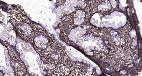
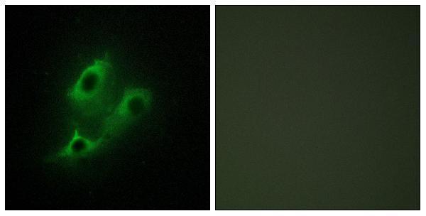
SERINC2 antibody data show paraffin-section IHC in human colon cancer (A30553 IHC image caption) and IF in U20S cells (A14175-2 IF image caption); catalog reactivity includes human, mouse, and rat (A30553 reactivity).

Real IHC data Immunohistochemical analysis of paraffin-embedded human colon cancer. 1, Tris-EDTA,pH9.0 was used for antigen retrieval. 2 Antibody was diluted at 1:200(4° overnight.3,Secondary antibody was diluted at 1:200(room temperature, 45min).
Anti-SERC2 SERINC2 Antibody
Cat # A30553
Real IF data IF analysis of SERINC2 using anti-SERINC2 antibody (A14175-2). SERINC2 was detected in an immunocytochemical section of U20S cells. Enzyme antigen retrieval was performed using IHC enzyme antigen retrieval reagent (AR0022) for 15 mins. The cells were blocked with 10% goat serum. And then incubated with 5 μg/mL rabbit anti-SERINC2 Antibody (A14175-2) overnight at 4°C. DyLight®488 Conjugated Goat Anti-Rabbit IgG (BA1127) was used as secondary antibody at 1:500 dilution and incubated for 30 minutes at 37°C. Visualize using a fluorescence microscope and filter sets appropriate for the label used.
Anti-SERINC2 Antibody ®
Cat # A14175-2

A30553 lists IHC and IF applications with human, mouse, and rat reactivity (A30553 applications/reactivity); its IHC figure shows paraffin-embedded human colon cancer (A30553 IHC image caption). A14175-2 lists human IF/ICC applications (A14175-2 applications/reactivity), with an IF figure in U20S cells (A14175-2 IF image caption).

Which to pick: Choose A30553 for tissue IHC: it is listed as polyclonal and IHC-tested (A30553 datasheet), and its own figure shows paraffin-embedded human colon cancer after Tris-EDTA retrieval at pH 9.0 (A30553 IHC image caption). The fixative is unreported (A30553 IHC image caption). Choose A14175-2 for human IF/ICC (A14175-2 applications/reactivity; A14175-2 IF image caption), or A30553 when mouse or rat reactivity is needed (A30553 reactivity).

Each figure is that product's own IHC / IF validation image from its datasheet.

References

  1. UniProt Consortium. UniProt entry Q96SA4 (SERC2_HUMAN, Serine incorporator 2).
  2. Human Protein Atlas. SERINC2 tissue IHC expression (reliability: Enhanced).
  3. Human Protein Atlas. SERINC2 subcellular location (ICC-IF): Mainly localized to the plasma membrane. In addition localized to the nucleoplasm..
  4. Human Protein Atlas. SERINC2 antibody validation summary (1 antibodies).
  5. Integrative Methylome and Transcriptome Characterization Identifies SERINC2 as a Tumor-Driven Gene for Papillary Thyroid Carcinoma. Cancers 2022 — PMC9818177.
  6. Serinc2 Drives the Progression of Cervical Cancer Through Regulating Myc Pathway. Cancer medicine 2024 — PMC11483714.
  7. SERINC2-mediated serine metabolism promotes cervical cancer progression and drives T cell exhaustion. International journal of biological sciences 2025 — PMC11781177.
  8. Serinc2 deficiency causes susceptibility to sepsis-associated acute lung injury. Journal of inflammation (London, England) 2022 — PMC9260995.
  9. PubMed PMID:12975309 — UniProt-cited evidence.
  10. PubMed PMID:14702039 — UniProt-cited evidence.
  11. PubMed PMID:16710414 — UniProt-cited evidence.