This website uses cookies to ensure you get the best experience on our website.


- Table of Contents
Plan chromogenic SFRP4 IHC in paraffin sections using endometrial stromal fibroblasts as a strong positive reference (HPA tissue IHC). Score cytoplasmic stromal and occasional glandular staining while accounting for secretion when interpreting its distribution (HPA tissue IHC; UniProt).

Expected localisation, validated protocols, controls and antibodies — the at-a-glance facts below, then the full design guide.
| Expected localisation | Cytoplasmic staining in stromal and some glandular cells (HPA tissue IHC) | |
| Staining pattern | Cytoplasmic stroma in endometrium and fallopian tube; some glands (HPA tissue IHC) | |
| Antigen retrieval | EDTA pH 8.0 HIER, heat-mediated (datasheet PB9888) | |
| Positive control | Endometrium+1 more · see all | |
| Negative control | Adipose tissue+4 more · see all |
| Fixation | Keep fixation conditions consistent across sections. (standard IHC practice; not target-specific) | |
| Caveat | Secreted SFRP4 may stain away from RNA-positive cells (HPA tissue IHC) | |
| Regulation | Ovarian tumor expression rises with stage (UniProt) | |
| Isoform / epitope | No isoforms; signal 1–18 cleaved; no membrane epitope split (UniProt) |
The catalog antibody’s IHC-P protocol (datasheet: PB9888) is followed by published SFRP4 IHC methods for prostate cancer, pancreatic cancer, and adipose tissue (PMC5660209; PMC5617497; PMC5644066).
| Sample | Paraffin-embedded human endometrial carcinoma tissue; fixative not specified (datasheet PB9888) |
| Fixation | Image fixative and duration unreported (datasheet PB9888); verify before use. |
| Sectioning | 4–5 µm sections on charged slides (standard) |
| Deparaffinisation | Xylene, graded ethanol series to water (standard) |
| Antigen retrieval | Heat retrieval: EDTA pH 8.0 (datasheet PB9888); 20 min, 95–100 °C (standard) |
| Peroxidase block | 3% H2O2, 10 min, room temperature (standard) |
| Blocking | 10% goat serum (datasheet PB9888) |
| Primary antibody | Rabbit anti-SFRP4, 0.5-1μg/ml (datasheet PB9888) |
| Primary incubation | Overnight at 4 °C (datasheet PB9888) |
| Detection | Streptavidin-biotin complex (SABC), DAB chromogen (datasheet PB9888) |
| Counterstain | Hematoxylin, blue, dehydrate and mount (standard) |
| Expected result | SFRP4-positive staining in stromal fibroblasts of endometrium (HPA tissue IHC: High). HPA tissue profile: Selective cytoplasmic expression in stromal cells in both fallopian tube and endometrium as well as a subset of endometrial glandular cells. No signal in the no-primary control. |
SFRP4 is secreted and has no transmembrane segment (UniProt Q6FHJ7: topology). In paraffin section IHC, expect selective cytoplasmic staining in endometrial and fallopian tube stromal cells, with staining in some endometrial glandular cells (HPA tissue IHC: Enhanced). Endometrial stromal fibroblasts are a strong positive reference (HPA: High). HPA reports only medium consistency between staining and RNA expression, partly because secreted protein may appear away from its site of production (HPA tissue IHC: reliability description).
| Endometrial stromal fibroblasts show strong cytoplasmic chromogen; nearby glandular cells stain variably. | This matches the clearest reference pattern: stromal fibroblasts are High, while only a subset of endometrial glandular cells stains (HPA tissue IHC). Judge the distribution by cell type as well as intensity. Secreted SFRP4 can complicate agreement between protein staining and local RNA expression (HPA tissue IHC: reliability description; UniProt Q6FHJ7: secreted). |
| Signal is predominantly nuclear or forms a sharply defined membrane rim. | That distribution conflicts with HPA's selective cytoplasmic tissue pattern and UniProt's secreted, nontransmembrane annotation (HPA tissue IHC; UniProt Q6FHJ7: topology). Treat it as suspect and compare it with the endometrial positive control and a no primary control; compartment alone cannot identify the specific artifact (general IHC practice). |
| Strong staining appears mainly in cell types reported as negative, such as adipocytes or adrenal glandular cells. | Those cells are Not detected in the supplied HPA tissue profiles (HPA tissue IHC: adipose tissue; adrenal gland). Consider antibody cross-reactivity or endogenous detection activity, especially if staining persists without primary antibody (general IHC practice). HPA's negative profile is a reference pattern, not proof that every individual specimen must be negative. |
| Brown signal spreads across many cells and tissue spaces without a selective stromal pattern. | Diffuse chromogen may reflect background from detection reagents, excess antibody, or incomplete washing (general IHC practice). Because SFRP4 is secreted, some signal beyond the producing cell remains biologically plausible (UniProt Q6FHJ7: secreted; HPA tissue IHC: reliability description). Compare morphology, control sections, and the expected stromal enrichment before calling it positive. |
| An endometrial section has no convincing stromal signal. | That is unexpected for the High stromal fibroblast reference (HPA tissue IHC: endometrium), but one blank section does not establish absent SFRP4. Check tissue identity and preservation, antibody and detection steps, and a run control (general IHC practice). Interpret glandular absence separately because HPA reports staining in only a subset of glandular cells (HPA tissue IHC). |
| Secreted topology and processing (UniProt Q6FHJ7) | A signal peptide occupies residues 1–18; the annotated secreted chain begins at residue 19, and no transmembrane segment is recorded (UniProt Q6FHJ7: processing; topology). If an antibody recognizes only the removed signal peptide, mature secreted protein may be missed; the supplied record gives no antibody epitope, so this remains a conditional interpretation. |
| Cell distribution and reference intensity (HPA tissue IHC) | Endometrial stromal fibroblasts are High, testicular pachytene spermatocytes are Medium, and the listed adipocytes and adrenal glandular cells are Not detected (HPA tissue IHC). These provide different reference patterns. Match the scored cell population to its own HPA entry; a whole tissue label alone can conceal a selective positive population. |
| Tissue IHC validation and RNA agreement (HPA tissue IHC; HPA antibodies) | HPA marks tissue staining Enhanced and records Enhanced IHC status for HPA009712 and HPA050585 (HPA tissue IHC; HPA antibodies). It also reports medium staining–RNA consistency because secreted protein and RNA can occupy different locations (HPA tissue IHC: reliability description). Use RNA agreement as context, not a requirement for each stained cell. |
| IF/ICC: does the tissue IHC pattern establish a fluorescence pattern? (HPA subcellular; HPA antibodies) | No: HPA lists SFRP4 as Secreted but provides no main subcellular location or ICC–IF image cell lines, and the supplied antibody entries have no ICC status (HPA subcellular; HPA antibodies). The tissue IHC pattern can guide interpretation, but it does not validate a particular IF/ICC compartment or intensity. |
| Situation | Likely cause | Next action |
|---|---|---|
| The positive endometrial control is blank. | A missed staining step, inactive detection reagent, unsuitable retrieval, or low primary concentration is possible (general IHC practice). | Confirm tissue identity, run controls and reagent sequence; review the antibody's IHC instructions before adjusting retrieval or concentration (general IHC practice). The expected reference is High endometrial stromal fibroblast staining (HPA tissue IHC). |
| Only glandular cells stain in endometrium. | A subset of glandular cells can stain, but absent stromal staining misses the principal HPA reference population (HPA tissue IHC). | Inspect stromal morphology and the positive control, then repeat with verified detection steps if needed (general IHC practice). Do not score glandular staining alone as a match to HPA's stromal High pattern (HPA tissue IHC). |
| Adipocytes or adrenal glandular cells stain strongly. | These cell types are Not detected in the supplied reference images; cross-reactivity or endogenous detection activity is possible (HPA tissue IHC; general IHC practice). | Compare with a no primary control and inspect whether the signal follows cells or background deposits; apply the appropriate endogenous activity block if the detection chemistry requires it (general IHC practice). |
| Chromogen is diffuse across the section. | Excess antibody, inadequate washing, or detection background can blur cell boundaries (general IHC practice); secretion can also place SFRP4 beyond its producing cell (UniProt Q6FHJ7: secreted). | Check the no primary control, washing and antibody concentration, then reassess stromal enrichment and morphology (general IHC practice; HPA tissue IHC: selective cytoplasmic profile). |
| Signal appears predominantly nuclear or as a continuous membrane outline. | That compartment differs from the observed cytoplasmic tissue pattern and the nontransmembrane annotation (HPA tissue IHC; UniProt Q6FHJ7: topology). | Review counterstain and cell boundaries, check controls, and repeat staining if the discordance persists (general IHC practice). Treat the location as unconfirmed until the expected stromal pattern is reproduced (HPA tissue IHC). |
| Fallopian tube stroma stains, but endometrial glands are negative. | HPA reports stromal staining in both tissues and staining in only a subset of endometrial glandular cells (HPA tissue IHC). | Score the stromal compartment separately from glands; confirm a functioning positive control before interpreting glandular absence (general IHC practice; HPA tissue IHC: selective cytoplasmic profile). |


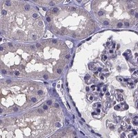
Comprehensive Human Protein Atlas IHC scoring per tissue (reliability: Enhanced — Medium consistency between antibody staining and RNA expression data. At least one protein variant secreted, tissue location of RNA and protein might differ and correlation is complex.). Rows are taken directly from the HPA tissue chart — click any row's HPA link to view the source.
| Tissue | Cell type | Level | Evidence | Source |
|---|---|---|---|---|
| Endometrium | Stromal fibroblasts | High | Protein (IHC) | HPA → |
| Testis | Pachytene spermatocytes | Medium | Protein (IHC) | HPA → |
| Tissue | Cell type | Level | Evidence | Source |
|---|---|---|---|---|
| Adipose tissue | Adipocytes | Not detected | Protein (IHC) | HPA → |
| Adrenal gland | Glandular cells | Not detected | Protein (IHC) | HPA → |
| Appendix | Glandular cells | Not detected | Protein (IHC) | HPA → |
| Bone marrow | Hematopoietic cells | Not detected | Protein (IHC) | HPA → |
| Breast | Adipocytes | Not detected | Protein (IHC) | HPA → |
Troubleshoot SFRP4 staining in paraffin section chromogenic IHC, with one entry for IF multiplexing (UniProt Q6FHJ7; HPA tissue IHC).
PB9888 has real IHC data from a human endometrial carcinoma paraffin section (catalog IHC image). M03147-1 lists IHC and ICC/IF for human, mouse and rat (catalog applications/reactivity).
The rendered card is PB9888, with IHC staining shown in a human endometrial carcinoma paraffin section (catalog IHC image). Its caption reports heat retrieval in EDTA at pH 8.0 and primary antibody at 1 μg/ml; the fixative is unreported (catalog IHC image).
Which to pick: Choose PB9888 for human paraffin-section IHC because its own caption documents staining under those conditions (catalog IHC image). Choose M03147-1 for IF/ICC or mouse/rat IHC because it lists those applications and species and is monoclonal clone 20S67 (catalog applications/reactivity/clone). No IF image is supplied for M03147-1 (catalog IF image alts).