This website uses cookies to ensure you get the best experience on our website.


- Table of Contents
Plan chromogenic IHC for SH3PXD2A in paraffin sections using the documented antibody dilution of 1:200 (datasheet A05241). Compare cytoplasmic and membranous staining with HPA tissue observations, bearing in mind their low concordance with RNA expression (HPA tissue IHC).

Expected localisation, validated protocols, controls and antibodies — the at-a-glance facts below, then the full design guide.
| Expected localisation | Cytoplasmic and membranous tissue staining (HPA tissue IHC) | |
| Staining pattern | Cytoplasmic and membranous staining in most tissues (HPA tissue IHC) | |
| Antigen retrieval | Tris-EDTA pH 9.0 HIER, heat-mediated (datasheet A05241) | |
| Positive control | Bronchus+4 more · see all | |
| Negative control | Adipose tissue+3 more · see all |
| Fixation | Keep fixation consistent across sections (standard IHC practice; not target-specific) | |
| Caveat | Low concordance between staining and RNA expression (HPA tissue IHC) | |
| Regulation | RNA has low tissue specificity (HPA tissue RNA) | |
| Isoform / epitope | 3 isoforms; check epitope coverage (UniProt) |
The catalog antibody protocol uses Tris-EDTA pH 9.0 heat retrieval (datasheet A05241). The published placental IHC protocol below uses microwave retrieval in citric acid buffer (PMC7385659).
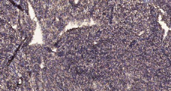
| Sample | Paraffin-embedded human uterus tissue; fixative not specified (datasheet A05241) |
| Fixation | Image fixative and duration unreported (datasheet A05241); verify before use. |
| Sectioning | 4–5 µm sections on charged slides (standard) |
| Deparaffinisation | Xylene, graded ethanol series to water (standard) |
| Antigen retrieval | Heat retrieval: Tris-EDTA pH 9.0 (datasheet A05241); 20 min, 95–100 °C (standard) |
| Peroxidase block | 3% H2O2, 10 min, room temperature (standard) |
| Blocking | 10% normal serum of the secondary host, 30 min, room temperature (standard) |
| Primary antibody | Rabbit anti-SH3PXD2A, 1:100 recommended; image 1:200 (datasheet A05241) |
| Primary incubation | Overnight at 4 °C (standard) |
| Detection | HRP-polymer secondary, DAB chromogen 5–10 min (standard) |
| Counterstain | Hematoxylin, blue, dehydrate and mount (standard) |
| Expected result | SH3PXD2A-positive staining in ciliated cells (ciliary rootlets) of bronchus (HPA tissue IHC: High). HPA tissue profile: Cytoplasmic and membranous expression in most tissues. No signal in the no-primary control. |
SH3PXD2A is cytoplasmic in normal cells and has no transmembrane segment (UniProt Q5TCZ1). In tissue IHC, expect cytoplasmic and membranous staining, with particularly strong staining at ciliary rootlets in ciliated cells of the bronchus, fallopian tube and nasopharynx (HPA: tissue IHC). Treat this as a qualified expectation: HPA rates its tissue staining Approved but reports low consistency with RNA expression (HPA: reliability).
| Strong, discrete staining at ciliary rootlets in ciliated epithelium. | This matches HPA's High staining in bronchus, fallopian tube and nasopharynx ciliated cells (HPA: tissue IHC). Evaluate the rootlet pattern against nearby cell morphology; brown color alone does not establish cell identity (general IHC practice). |
| Cytoplasmic and some membranous staining across several tissue compartments. | This fits HPA's broad tissue profile and UniProt's cytoplasmic localisation (HPA: tissue IHC; UniProt Q5TCZ1). Membranous staining by itself does not establish that SH3PXD2A spans the membrane: UniProt annotates no transmembrane segment (UniProt Q5TCZ1 topology). |
| Predominantly nuclear signal, with little cytoplasmic or ciliary-rootlet staining. | A nuclear-only pattern conflicts with the reported tissue and subcellular patterns (HPA: tissue IHC and ICC-IF; UniProt Q5TCZ1). Treat it as suspect staining and check the antibody and detection controls before assigning it to SH3PXD2A (general IHC practice). |
| Strong staining in adipocytes, duodenal glandular cells or lymph-node germinal-center cells. | These cell populations were Not detected in HPA tissue IHC (HPA: tissue IHC). Unexpected signal may reflect cross-reactivity or endogenous detection activity; compare matched controls and tissue morphology before calling it specific (general IHC practice). |
| Diffuse stain across the section, or no signal in ciliated cells expected to be positive. | Diffuse stain obscures the reported rootlet pattern; absent signal in an HPA High cell population leaves the run uninformative (HPA: tissue IHC). Examine background and positive controls before interpreting a negative specimen (general IHC practice). |
| Cell type and tissue | HPA reports High ciliary-rootlet staining in bronchus, fallopian tube and nasopharynx; Medium staining in adrenal and appendix glandular cells; and Not detected in adipocytes (HPA: tissue IHC). Score the named cell population rather than treating an entire section as uniformly positive or negative. |
| Compartment and topology | UniProt places SH3PXD2A in cytoplasm and, in SRC-transformed cells, podosomes; it annotates no transmembrane segment (UniProt Q5TCZ1). HPA describes cytoplasmic and membranous tissue staining (HPA: tissue IHC). A routine tissue section cannot establish podosome localisation from a brown punctum alone. |
| Antibody evidence | HPA rates tissue IHC Approved, while reporting low consistency between antibody staining and RNA expression (HPA: reliability). HPA037922 and HPA037923 each have Approved IHC status; the supplied antibody list gives no IHC Enhanced status (HPA: antibodies). Interpret unexpected cells conservatively. |
| IF/ICC question: where should signal appear? | HPA reports cytosol and primary cilium transition zone as approved ICC-IF locations (HPA: subcellular). That observation can inform compartment interpretation, but it is an ICC-IF result; use the tissue IHC record for intensity expectations in paraffin sections (HPA: tissue IHC). |
| Isoforms and epitope | UniProt lists 3 isoforms and a full-length chain of residues 1–1133 (UniProt Q5TCZ1). The supplied evidence gives no antibody epitope or isoform coverage, so a staining difference cannot be assigned to a particular isoform or to epitope loss. |
| Situation | Likely cause | Next action |
|---|---|---|
| No stain at ciliary rootlets in a bronchus positive-control section. | The expected High cell population is absent from the readout (HPA: bronchus); the supplied sources do not identify a target-specific fixation or retrieval cause. | Confirm ciliated cells are present, then review the run's positive control, antibody application, retrieval and detection steps as general IHC checks; avoid calling the specimen negative until the control works. |
| Whole section is diffusely brown, including areas outside recognizable cells. | Non-specific background or detection chemistry may obscure the cell-specific pattern (general IHC practice); HPA's rootlet observation cannot be assessed through uniform stain (HPA: tissue IHC). | Compare the negative reagent control and examine blocking, washes and chromogen development using standard IHC controls; then reassess ciliary rootlets and cell outlines. |
| Stain is concentrated in nuclei. | The pattern conflicts with cytoplasmic tissue staining and the approved cytosol ICC-IF location (HPA: tissue IHC and subcellular; UniProt Q5TCZ1). | Check the negative control, tissue morphology and antibody specificity before scoring it as SH3PXD2A; retain a cytoplasmic positive control in the same run (general IHC practice). |
| Adipocytes or germinal-center cells stain strongly. | Both populations are Not detected in HPA tissue IHC (HPA: adipose tissue and lymph node); cross-reactivity or endogenous detection activity is possible (general IHC practice). | Confirm the cell type on the counterstain, compare detection controls and repeat with an independently validated antibody if available; report the conflict with HPA rather than silently recoding it as expected. |
| Low staining in lung macrophages is read as a failed stain. | HPA records lung macrophages as Low, while ciliary rootlets in bronchus are High (HPA: tissue IHC); expected intensity depends on the cell population. | Use a High-staining ciliated-cell control to judge run performance, then score macrophages within their own tissue context rather than requiring rootlet-level intensity. |
| A tissue pattern differs from RNA expression or from another antibody. | HPA explicitly reports low consistency with RNA despite an Approved IHC reliability rating (HPA: reliability); the supplied antibody records show Approved IHC for HPA037922 and HPA037923 (HPA: antibodies). | Document the antibody and the specific cell populations scored, compare controls and independent staining where available, and keep any unresolved discrepancy visible in the interpretation. |
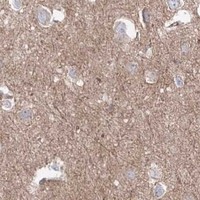


Comprehensive Human Protein Atlas IHC scoring per tissue (reliability: Approved — Low consistency between antibody staining and RNA expression data.). Rows are taken directly from the HPA tissue chart — click any row's HPA link to view the source.
| Tissue | Cell type | Level | Evidence | Source |
|---|---|---|---|---|
| Bronchus | Ciliated cells (ciliary rootlets) | High | Protein (IHC) | HPA → |
| Fallopian tube | Ciliated cells (ciliary rootlets) | High | Protein (IHC) | HPA → |
| Nasopharynx | Ciliated cells (ciliary rootlets) | High | Protein (IHC) | HPA → |
| Adrenal gland | Glandular cells | Medium | Protein (IHC) | HPA → |
| Appendix | Glandular cells | Medium | Protein (IHC) | HPA → |
| Tissue | Cell type | Level | Evidence | Source |
|---|---|---|---|---|
| Adipose tissue | Adipocytes | Not detected | Protein (IHC) | HPA → |
| Duodenum | Glandular cells | Not detected | Protein (IHC) | HPA → |
| Lymph node | Germinal center cells | Not detected | Protein (IHC) | HPA → |
| Parathyroid gland | Glandular cells | Not detected | Protein (IHC) | HPA → |
Troubleshoot SH3PXD2A staining in paraffin sections by checking retrieval, cellular compartment, controls and scoring before interpreting signal as podosome activity (UniProt Q5TCZ1).
The catalog includes an IHC image of paraffin-embedded human uterus (A05241 image caption), IF/ICC applications (catalog: application lists), and listed human, mouse, and rat reactivity (catalog: reactivity lists).
A05241 has an IHC image of paraffin-embedded human uterus and lists IHC and IF applications with human and mouse reactivity (A05241 image caption; catalog: applications and reactivity). A05241-1 lists IHC, IF, and ICC applications with human, mouse, and rat reactivity; no IHC or IF image is supplied (catalog: A05241-1 applications, reactivity, and image fields).
Which to pick: Choose A05241 for tissue IHC when a documented paraffin-section example is useful: its image shows human uterus after Tris-EDTA retrieval at pH 9.0 and primary antibody at 1:200 overnight at 4°C (A05241 IHC image caption). Choose A05241-1 for IF/ICC or rat samples because it lists both applications and rat reactivity and is described as polyclonal (catalog: A05241-1 applications, reactivity, and dilution text). The fixative is unreported for the A05241 paraffin-section image, and A05241-1 has no supplied processing image (A05241 IHC image caption; catalog: A05241-1 image fields).