This website uses cookies to ensure you get the best experience on our website.


- Table of Contents
Plan paraffin-section IHC for SLC38A2 using the reported cytoplasmic tissue pattern and high staining in kidney tubular cells (HPA tissue IHC). Interpret staining alongside its annotated plasma membrane location and insulin-dependent recruitment (UniProt).

Expected localisation, validated protocols, controls and antibodies — the at-a-glance facts below, then the full design guide.
| Expected localisation | Cytoplasmic tissue staining; plasma membrane localization is expected from protein annotation (HPA tissue IHC; UniProt) | |
| Staining pattern | Cytoplasmic staining in most tissues, including glandular cells (HPA tissue IHC) | |
| Antigen retrieval | Tris-EDTA pH 9.0 HIER, heat-mediated (datasheet A03242-1) | |
| Positive control | Duodenum+4 more · see all | |
| Negative control | Adipose tissue+4 more · see all |
| Fixation | Keep fixation consistent across sections (standard IHC practice; not target-specific) | |
| Caveat | Antibody staining has low consistency with RNA expression (HPA tissue IHC) | |
| Regulation | Insulin promotes recruitment to the plasma membrane (UniProt) | |
| Isoform / epitope | 2 isoforms; epitope coverage is unknown, and cytoplasmic versus extracellular location matters (UniProt) |
The catalog antibody’s IHC-P protocol is accompanied by two published chromogenic SLC38A2 IHC protocols (PMC7334074; PMC11876615).
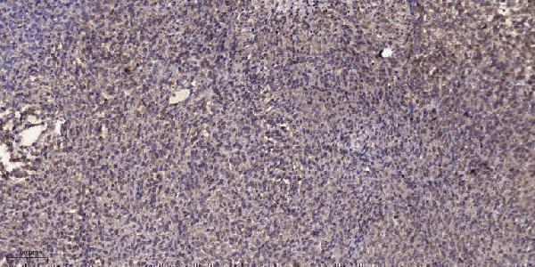
| Sample | Paraffin-embedded human Colon cancer tissue; fixative not specified (datasheet A03242-1) |
| Fixation | Image fixative and duration unreported (datasheet A03242-1); verify before use. |
| Sectioning | 4–5 µm sections on charged slides (standard) |
| Deparaffinisation | Xylene, graded ethanol series to water (standard) |
| Antigen retrieval | Heat retrieval: Tris-EDTA pH 9.0 (datasheet A03242-1); 20 min, 95–100 °C (standard) |
| Peroxidase block | 3% H2O2, 10 min, room temperature (standard) |
| Blocking | 10% normal serum of the secondary host, 30 min, room temperature (standard) |
| Primary antibody | Rabbit anti-SLC38A2, 1:50 recommended; image 1:200 (datasheet A03242-1) |
| Primary incubation | Overnight at 4 °C (standard) |
| Detection | HRP-polymer secondary, DAB chromogen 5–10 min (standard) |
| Counterstain | Hematoxylin, blue, dehydrate and mount (standard) |
| Expected result | SLC38A2-positive staining in glandular cells of duodenum (HPA tissue IHC: High). HPA tissue profile: Cytoplasmic expression in most tissues. No signal in the no-primary control. |
SLC38A2 is an 11-pass membrane transporter that can move between intracellular pools and the plasma membrane (UniProt Q96QD8 topology and subcellular location). In paraffin-section IHC, expect mainly cytoplasmic staining in many tissues, including strong staining in kidney tubule cells and duodenal glandular cells (HPA tissue IHC). HPA rates the tissue staining Approved but reports low consistency with RNA expression (HPA tissue IHC reliability).
| Strong cytoplasmic staining in kidney tubule cells or duodenal glandular cells, with possible membrane emphasis. | This fits HPA's High IHC staining in those cells and its broader cytoplasmic tissue profile (HPA tissue IHC). Membrane emphasis is biologically plausible given the cell membrane location, but HPA does not require a crisp membrane outline in paraffin sections (UniProt Q96QD8 subcellular location; HPA tissue IHC). |
| Predominantly nuclear staining, especially when cytoplasmic staining is absent. | A nuclear-only pattern conflicts with the reported cell membrane location and HPA's cytoplasmic IHC profile (UniProt Q96QD8 subcellular location; HPA tissue IHC). Treat it as suspect staining; review controls and detection conditions before assigning it to SLC38A2 (standard IHC practice). |
| Strong signal in adipocytes, marrow hematopoietic cells, or skeletal myocytes. | HPA reports SLC38A2 as not detected in these specific cell populations (HPA tissue IHC). Check whether the signal follows tissue pigment, endogenous detection activity, or another antibody target; these HPA observations do not prove absolute absence under every condition (HPA tissue IHC; standard IHC practice). |
| A uniform haze covers positive cells, negative cells, and surrounding tissue. | Diffuse staining that ignores cell boundaries is hard to reconcile with the reported cell associated distribution (HPA tissue IHC; UniProt Q96QD8 subcellular location). It can reflect excess detection background or inadequate blocking or washing; compare a no-primary control and adjust the general IHC workflow (standard IHC practice). |
| No signal in kidney tubule cells or duodenal glandular cells. | Target-specific fixation effects are not established by the supplied assay evidence. Verify with a matched IHC source before attributing a result to fixation. |
| Cellular location | UniProt places SLC38A2 at the cell membrane and notes recruitment from trans-Golgi or endosomal pools after insulin stimulation (UniProt Q96QD8 subcellular location). HPA describes predominantly cytoplasmic tissue IHC, so a cytoplasmic component is compatible with the observed slide profile; neither source sets a required membrane-to-cytoplasm ratio for paraffin IHC (HPA tissue IHC; UniProt Q96QD8). |
| Tissue and cell selection | HPA lists High staining in kidney tubule cells, duodenal and small-intestinal glandular cells, oral squamous epithelial cells, pancreatic exocrine cells, and testicular peritubular cells (HPA tissue IHC). Its not detected calls apply to specified cells, including adipocytes and skeletal myocytes, rather than every cell in those tissues (HPA tissue IHC). |
| Validation strength | The tissue IHC reliability is Approved, with low consistency between antibody staining and RNA expression (HPA tissue IHC reliability). HPA035180 is Approved for IHC, while HPA069672 is Supported for ICC; those ratings apply to different antibodies and applications (HPA antibodies). Interpret a novel cell pattern alongside controls rather than assuming it is independently confirmed (standard IHC practice). |
| Topology and epitope | The protein has 11 transmembrane segments and both cytoplasmic and extracellular regions (UniProt Q96QD8 topology). The supplied record does not locate this antibody's epitope, so topology alone cannot specify an antigen retrieval setting, predict epitope exposure, or explain a missing IHC signal (UniProt Q96QD8 topology; supplied antibody record). |
| Isoforms and processing | UniProt lists 2 isoforms, no signal peptide or propeptide, and one annotated chain spanning residues 1–506 (UniProt Q96QD8 isoforms and processing). Without an antibody epitope or isoform-specific validation in the supplied evidence, do not assign differing staining patterns to an isoform or to cleavage (UniProt Q96QD8; supplied antibody record). |
| Does IF/ICC show the same pattern? | HPA reports a supported plasma membrane location in ICC-IF, using an antibody with Supported ICC status (HPA subcellular; HPA antibodies: HPA069672). This supports membrane localization but does not replace the cytoplasmic paraffin IHC profile or establish an IF/ICC protocol here (HPA subcellular; HPA tissue IHC). |
| Situation | Likely cause | Next action |
|---|---|---|
| Positive comparator is blank. | Kidney tubule and duodenal glandular cells are rated High by HPA; a blank slide may indicate a workflow or detection failure (HPA tissue IHC; standard IHC practice). | Confirm the comparator cells are present, then review the catalog antibody's IHC-P instructions, dilution, retrieval, and detection controls (standard IHC practice). Do not infer a target-specific fixation effect from this result (HPA tissue IHC). |
| Staining is nuclear-only. | This conflicts with the reported membrane location and cytoplasmic tissue IHC profile (UniProt Q96QD8 subcellular location; HPA tissue IHC). | Compare the no-primary control and a HPA High cell population; review blocking, antibody concentration, and detection background before scoring nuclear signal as specific (HPA tissue IHC; standard IHC practice). |
| Adipocytes or skeletal myocytes stain strongly. | HPA calls these cell types not detected; cross-reactivity or endogenous detection activity is possible (HPA tissue IHC; standard IHC practice). | Check cell identity and no-primary controls, then compare staining with a HPA High population on the same run (HPA tissue IHC; standard IHC practice). |
| The whole section has a brown haze. | Uniform background can arise from detection chemistry, incomplete blocking, or insufficient washing (standard chromogenic IHC practice). | Review no-primary and detection-only controls; optimize blocking, washing, and antibody dilution using the catalog IHC-P instructions (standard IHC practice). |
| Signal is cytoplasmic without a sharp membrane rim. | HPA describes cytoplasmic expression in most tissues, while UniProt reports intracellular pools as well as cell membrane location (HPA tissue IHC; UniProt Q96QD8 subcellular location). | Score cell type and background first; do not reject an otherwise matching IHC pattern solely because the membrane edge is indistinct (HPA tissue IHC; standard IHC interpretation). |
| IHC and ICC-IF images look different. | HPA reports cytoplasmic tissue IHC and supported plasma membrane ICC-IF; its listed IHC and ICC antibodies also have different application ratings (HPA tissue IHC; HPA subcellular; HPA antibodies). | Interpret each result within its own application and antibody validation, then compare controls and cell identity before calling the patterns contradictory (HPA antibodies; standard IHC/ICC interpretation). |


![HPA ICC-IF BJ [Human fibroblast] — SLC38A2 (HPA069672)](https://images.proteinatlas.org/69672/1936_A6_1_blue_red_green.jpg)
![HPA ICC-IF BJ [Human fibroblast] — SLC38A2 (HPA069672)](https://images.proteinatlas.org/69672/1936_A6_2_blue_red_green.jpg)
Comprehensive Human Protein Atlas IHC scoring per tissue (reliability: Approved — Low consistency between antibody staining and RNA expression data.). Rows are taken directly from the HPA tissue chart — click any row's HPA link to view the source.
| Tissue | Cell type | Level | Evidence | Source |
|---|---|---|---|---|
| Duodenum | Glandular cells | High | Protein (IHC) | HPA → |
| Kidney | Cells in tubules | High | Protein (IHC) | HPA → |
| Oral mucosa | Squamous epithelial cells | High | Protein (IHC) | HPA → |
| Pancreas | Exocrine glandular cells | High | Protein (IHC) | HPA → |
| Small intestine | Glandular cells | High | Protein (IHC) | HPA → |
| Tissue | Cell type | Level | Evidence | Source |
|---|---|---|---|---|
| Adipose tissue | Adipocytes | Not detected | Protein (IHC) | HPA → |
| Bone marrow | Hematopoietic cells | Not detected | Protein (IHC) | HPA → |
| Esophagus | Squamous epithelial cells | Not detected | Protein (IHC) | HPA → |
| Ovary | Ovarian stroma cells | Not detected | Protein (IHC) | HPA → |
| Skeletal muscle | Myocytes | Not detected | Protein (IHC) | HPA → |
Troubleshoot SLC38A2 staining by checking retrieval, tissue processing, membrane localisation, controls, and scoring before interpreting chromogenic signal.
A03242-1 has an IHC image from paraffin-embedded human colon cancer (catalog image caption). Its listed reactivity covers human, mouse and rat; no IF data are supplied (catalog applications and images).
A03242-1 is listed for IHC and has an image from paraffin-embedded human colon cancer (catalog applications; catalog image caption). Human, mouse and rat are listed as reactive species, while the supplied image documents human tissue only (catalog reactivity; catalog image caption).
Which to pick: Choose A03242-1 for paraffin-section IHC: its image caption documents human colon cancer tissue, but does not report the fixative (catalog image caption). For mouse or rat tissue, A03242-1 has listed reactivity without a supplied tissue image for either species (catalog reactivity; catalog image caption). No SKU in this payload is listed for IF/ICC or has an IF image, so an IF/ICC choice is unsupported (catalog applications and images).