This website uses cookies to ensure you get the best experience on our website.


- Table of Contents
Plan SLC9A1 paraffin IHC around membranous staining, with high signal in kidney collecting ducts and several glandular tissues (HPA tissue IHC). This guide covers fixation consistency, epitope placement and cell-specific scoring (standard IHC practice; UniProt topology; HPA tissue IHC).

Expected localisation, validated protocols, controls and antibodies — the at-a-glance facts below, then the full design guide.
| Expected localisation | Membranous in tissue (HPA tissue IHC); basolateral in most epithelia (UniProt) | |
| Staining pattern | Membranous in renal tubules and glandular cells (HPA tissue IHC) | |
| Antigen retrieval | EDTA pH 8.0 HIER, heat-mediated (datasheet PB9151) | |
| Positive control | Colon+4 more · see all | |
| Negative control | Bone marrow+4 more · see all |
| Fixation | Keep fixation consistent across sections. (standard IHC practice; not target-specific) | |
| Caveat | Kidney staining is high in collecting ducts (HPA tissue IHC) | |
| Regulation | Expression regulation not specified (UniProt) | |
| Isoform / epitope | 2 isoforms; map the epitope to extracellular or cytoplasmic regions (UniProt) |
The catalog antibody’s IHC-P protocol is accompanied by one published SLC9A1 IHC protocol (PMC12605240).
| Sample | Paraffin-embedded rat kidney tissue; fixative not specified (datasheet PB9151) |
| Fixation | Image fixative and duration unreported (datasheet PB9151); verify before use. |
| Sectioning | 4–5 µm sections on charged slides (standard) |
| Deparaffinisation | Xylene, graded ethanol series to water (standard) |
| Antigen retrieval | Heat retrieval: EDTA pH 8.0 (datasheet PB9151); 20 min, 95–100 °C (standard) |
| Peroxidase block | 3% H2O2, 10 min, room temperature (standard) |
| Blocking | 10% goat serum (datasheet PB9151) |
| Primary antibody | Rabbit anti-SLC9A1, 0.5-1μg/ml recommended; image 2 μg/ml (datasheet PB9151) |
| Primary incubation | Overnight at 4 °C (datasheet PB9151) |
| Detection | Streptavidin-biotin complex (SABC), DAB chromogen (datasheet PB9151) |
| Counterstain | Hematoxylin, blue, dehydrate and mount (standard) |
| Expected result | SLC9A1-positive staining in glandular cells of colon (HPA tissue IHC: High). HPA tissue profile: Membranous expression in several tissues, highly abundant in renal tubules, intestines, gallbladder, salivary gland and seminal gland. No signal in the no-primary control. |
SLC9A1 is a 13-pass plasma-membrane protein, localized basolaterally in epithelial cells except in choroid plexus, where it is luminal (UniProt P19634 topology and subcellular location). In paraffin-section IHC, expect membranous staining in renal collecting ducts and glandular cells of the intestine, gallbladder, and salivary gland (HPA: High; membranous tissue profile). HPA rates its tissue IHC pattern Enhanced, reflecting high consistency between staining and RNA expression (HPA: reliability).
| Distinct membrane outlines in renal collecting ducts or intestinal glandular cells, with little staining away from cell borders. | This fits a positive SLC9A1 result: both cell populations stain at High levels in HPA tissue IHC, and membrane localization agrees with UniProt P19634. In epithelial cells, assess the basolateral contour where section orientation permits; a partial outline can reflect which membrane surfaces the section exposes (UniProt P19634 subcellular location; standard IHC interpretation). |
| Predominantly nuclear or diffuse cytoplasmic chromogen, without convincing membrane staining in expected positive cells. | Treat this as a compartment mismatch and investigate background or antibody specificity before scoring it positive (UniProt P19634: cell membrane; HPA: membranous tissue profile). HPA reports an uncertain additional nucleoplasmic location in ICC-IF; that observation alone does not establish a predominantly nuclear paraffin-section IHC pattern (HPA: subcellular ICC-IF). |
| Strong staining in HPA-negative cell populations, such as bone-marrow hematopoietic cells or lung alveolar cells. | That distribution conflicts with the cited HPA tissue IHC observations of Not detected in those particular cell populations (HPA: bone marrow hematopoietic cells; lung alveolar cells). Consider cross-reactivity or endogenous chromogenic detection activity, then compare with staining in a known-positive tissue and appropriate detection controls (standard IHC practice). |
| Uniform haze across cells and extracellular areas obscures membrane borders. | A field-wide deposit does not support a compartment call, even when the section contains an HPA-positive tissue (HPA: membranous tissue profile; standard IHC interpretation). Review blocking, antibody concentration, washing, and detection background with controls before interpreting intensity; haze can conceal a genuine membrane signal or mimic weak widespread staining (standard IHC practice). |
| No visible staining in renal collecting ducts or colon glandular cells. | Both are High in HPA tissue IHC, so an absent signal warrants a technical check before concluding that this preparation lacks SLC9A1 (HPA: kidney collecting ducts; colon glandular cells). Confirm tissue identity and preservation, antibody application, detection reagents, and a positive control; HPA tissue levels do not establish a target-specific fixation effect (standard IHC practice; HPA: tissue IHC). |
| Membrane polarity | UniProt places SLC9A1 basolaterally in epithelial cells, with a luminal choroid-plexus exception (UniProt P19634 subcellular location). Read membrane position in the context of tissue orientation; an apparently incomplete rim need not mean absence from the cell (standard IHC interpretation). |
| Tissue and cell selection | HPA reports High staining in kidney collecting ducts and glandular cells of colon, duodenum, gallbladder, and salivary gland; it reports Low staining in pancreatic exocrine glandular cells (HPA: tissue IHC). Specify the cell population when choosing controls or comparing intensity across tissues. |
| Epitope location | SLC9A1 has 13 transmembrane segments, extracellular loops, and a long cytoplasmic C-terminal region (UniProt P19634 topology). The supplied record does not identify this antibody's epitope, so topology cannot predict its staining strength or an antigen-retrieval setting. |
| Antibody validation | HPA lists Enhanced IHC validation for HPA048532, HPA052891, and CAB022371; its Enhanced tissue reliability reflects agreement between staining and RNA data (HPA: antibodies; tissue IHC reliability). Validation supports the reported distribution but does not make every unexpected signal specific. |
| IF/ICC: what location should be expected? | The supported main ICC-IF location is the plasma membrane; an additional nucleoplasmic location is marked uncertain (HPA: subcellular ICC-IF). Use that distinction when comparing images with IHC, without treating the uncertain nuclear signal as a required positive pattern. |
| Situation | Likely cause | Next action |
|---|---|---|
| Known-positive tissue is blank. | A failed staining or detection step is possible; HPA reports High staining in kidney collecting ducts and colon glandular cells (HPA: tissue IHC). | Check that the expected cells are present, then verify antibody application, reagent activity, and the positive control before interpreting the blank slide (standard IHC practice). |
| Staining is cytoplasmic but membrane borders are absent. | The compartment differs from the reported membrane localization (UniProt P19634; HPA: tissue IHC profile). | Inspect a well-oriented positive section and detection controls; score membrane localization separately from overall chromogen intensity (standard IHC practice). |
| Nuclei stain more strongly than cell borders. | Predominant nuclear IHC staining is unsupported by the tissue profile; HPA's additional nucleoplasmic ICC-IF location is uncertain (HPA: tissue IHC; subcellular ICC-IF). | Review controls and membrane staining before calling the result positive; record the nuclear signal as an unresolved finding (standard IHC interpretation). |
| HPA-negative cell populations stain strongly. | Cross-reactivity or endogenous detection activity may contribute; HPA reports Not detected in lung alveolar cells and bone-marrow hematopoietic cells (HPA: tissue IHC). | Compare a known-positive tissue with a control omitting primary antibody and check the chromogenic detection controls (standard IHC practice). |
| Diffuse brown background hides cell boundaries. | Excess nonspecific antibody or detection signal can prevent a membrane call (standard IHC practice). | Review blocking, antibody concentration, washing, and detection controls; reassess only where individual membrane borders remain visible (standard IHC practice). |
| A low-staining tissue appears weaker than the positive control. | HPA reports Low staining in pancreatic exocrine glandular cells versus High staining in renal collecting ducts (HPA: tissue IHC). | Compare like cell populations and retain a High-staining control; do not classify a weak low-expression population as assay failure on intensity alone (HPA: tissue IHC; standard IHC interpretation). |

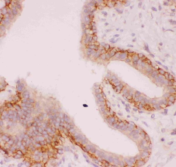
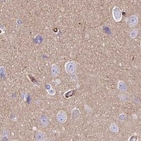
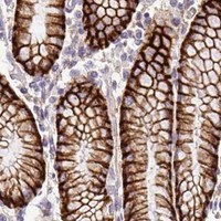

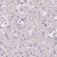


Comprehensive Human Protein Atlas IHC scoring per tissue (reliability: Enhanced — High consistency between antibody staining and RNA expression data.). Rows are taken directly from the HPA tissue chart — click any row's HPA link to view the source.
| Tissue | Cell type | Level | Evidence | Source |
|---|---|---|---|---|
| Colon | Glandular cells | High | Protein (IHC) | HPA → |
| Duodenum | Glandular cells | High | Protein (IHC) | HPA → |
| Epididymis | Glandular cells | High | Protein (IHC) | HPA → |
| Gallbladder | Glandular cells | High | Protein (IHC) | HPA → |
| Kidney | Collecting ducts | High | Protein (IHC) | HPA → |
| Tissue | Cell type | Level | Evidence | Source |
|---|---|---|---|---|
| Bone marrow | Hematopoietic cells | Not detected | Protein (IHC) | HPA → |
| Caudate | Glial cells | Not detected | Protein (IHC) | HPA → |
| Cerebellum | Cells in granular layer | Not detected | Protein (IHC) | HPA → |
| Hippocampus | Glial cells | Not detected | Protein (IHC) | HPA → |
| Liver | Cholangiocytes | Not detected | Protein (IHC) | HPA → |
Use the catalog antibody’s rat kidney IHC protocol as the starting point, then judge staining against SLC9A1’s expected membrane pattern (datasheet PB9151; UniProt P19634).
PB9151 has real SLC9A1 IHC images from paraffin sections of rat and mouse kidney and human mammary cancer tissue (PB9151 image captions). No IF image is supplied (PB9151 catalog).
PB9151 is listed for IHC and WB in human, mouse and rat (PB9151 catalog). Its IHC captions document paraffin sections of rat and mouse kidney and human mammary cancer tissue (PB9151 image captions).
Which to pick: For tissue IHC, choose PB9151 for paraffin sections; its image captions document that processing, but do not report the fixative (PB9151 image captions). No SKU can be recommended for IF/ICC because PB9151 lists IHC and WB only and supplies no IF image (PB9151 catalog). For cross-species IHC, PB9151 lists human, mouse and rat reactivity and shows IHC images from all three species; it has a rabbit host, with clonality unreported (PB9151 catalog; PB9151 image captions).