This website uses cookies to ensure you get the best experience on our website.


- Table of Contents
Plan SORBS1 IHC-P around general cytoplasmic staining (HPA tissue IHC). Use heart cardiomyocytes or smooth muscle cells as positive controls (HPA tissue IHC), and start the catalog antibody at 0.5–1 μg/mL (datasheet A04426-3).

Expected localisation, validated protocols, controls and antibodies — the at-a-glance facts below, then the full design guide.
| Expected localisation | General cytoplasmic tissue staining (HPA tissue IHC) | |
| Staining pattern | Cytoplasmic staining in cardiomyocytes and smooth muscle cells (HPA tissue IHC) | |
| Antigen retrieval | EDTA pH 8.0 HIER, heat-mediated (datasheet A04426-3) | |
| Positive control | Duodenum+4 more · see all | |
| Negative control | Adipose tissue+4 more · see all |
| Fixation | Keep fixation consistent across samples (standard IHC practice; not target-specific) | |
| Caveat | Antibody staining has low consistency with RNA expression (HPA tissue IHC) | |
| Regulation | Intensity regulation is unreported (UniProt) | |
| Isoform / epitope | 12 isoforms; epitope coverage is unspecified (UniProt) |
Compare the catalog antibody’s IHC-P protocol (datasheet: A04426-3) with published SORBS1 IHC methods from two breast cancer studies (PMC7922363; PMC9206459).
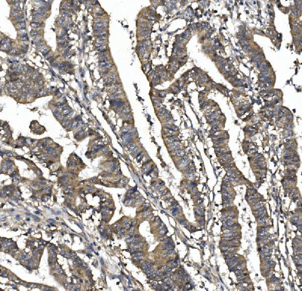
| Sample | Paraffin-embedded human rectal cancer tissue; fixative not specified (datasheet A04426-3) |
| Fixation | Image fixative and duration unreported (datasheet A04426-3); verify before use. |
| Sectioning | 4–5 µm sections on charged slides (standard) |
| Deparaffinisation | Xylene, graded ethanol series to water (standard) |
| Antigen retrieval | Heat retrieval: EDTA pH 8.0 (datasheet A04426-3); 20 min, 95–100 °C (standard) |
| Peroxidase block | 3% H2O2, 10 min, room temperature (standard) |
| Blocking | 10% goat serum (datasheet A04426-3) |
| Primary antibody | Rabbit anti-SORBS1, 0.5-1μg/ml (datasheet A04426-3) |
| Primary incubation | Overnight at 4 °C (datasheet A04426-3) |
| Detection | Streptavidin-biotin complex (SABC), DAB chromogen (datasheet A04426-3) |
| Counterstain | Hematoxylin, blue, dehydrate and mount (standard) |
| Expected result | SORBS1-positive staining in glandular cells of duodenum (HPA tissue IHC: High). HPA tissue profile: General cytoplasmic expression. No signal in the no-primary control. |
SORBS1 is mainly cytoplasmic in tissue IHC, with high staining reported in cardiomyocytes, smooth muscle cells, and duodenal and small intestinal glandular cells (HPA tissue IHC). Junctional and focal adhesion localization is also plausible (UniProt Q9BX66 subcellular location). SORBS1 has no transmembrane segment (UniProt Q9BX66 topology). HPA rates tissue IHC reliability Enhanced but reports low consistency with RNA expression (HPA tissue IHC).
| Clear cytoplasmic stain in cardiomyocytes or smooth muscle cells (HPA: High in both). | This matches the reported tissue IHC pattern. Score the named cells and their intensity, while retaining HPA's low RNA–staining consistency caveat (HPA tissue IHC). |
| Strong stain confined to vessel lumens or extracellular spaces, with little cellular signal. | That distribution does not match HPA's general cytoplasmic pattern (HPA tissue IHC). Treat it as possible detection artefact and compare with a no-primary control (general IHC practice). |
| Strong stain in adipocytes or lymph node non-germinal center cells (HPA: Not detected in these cells). | Unexpected staining raises cross-reactivity or endogenous detection activity as possibilities; check control sections and the detection system before assigning SORBS1 positivity (general IHC practice). |
| A diffuse haze covers cells and surrounding tissue. | Diffuse background prevents reliable cell-specific scoring. Review blocking, antibody concentration, washing, and detection controls (general IHC practice); it does not establish SORBS1 distribution. |
| No signal in heart cardiomyocytes or smooth muscle cells (HPA: High in both). | Check a known-positive section, antibody and detection controls, and retrieval conditions before interpreting the specimen as negative (general IHC practice). HPA's High level is an observed pattern, not a guarantee for every section (HPA tissue IHC). |
| Tissue and cell selection (HPA tissue IHC) | Heart cardiomyocytes and smooth muscle cells provide reported High examples; adipocytes and lymph node non-germinal center cells are reported Not detected (HPA tissue IHC). |
| Antibody evidence (HPA antibody validation) | HPA027559 and HPA043084 have Enhanced IHC ratings; CAB032850 is Approved (HPA antibody validation). These ratings do not remove the tissue profile's RNA-consistency caveat (HPA tissue IHC). |
| IF/ICC: where should signal localize? (HPA subcellular ICC-IF) | Mainly at focal adhesion sites, with plasma membrane signal also supported; centrosome localization is uncertain (HPA subcellular ICC-IF). These cell-image findings need not appear as resolvable puncta in chromogenic tissue IHC. |
| Compartment and topology (UniProt Q9BX66) | SORBS1 is associated with cytoskeleton, junctions, focal adhesions, and sometimes nucleus; it has no transmembrane segment (UniProt Q9BX66 subcellular location and topology). Do not infer a cell-surface-only IHC pattern. |
| Isoforms and epitope scope (UniProt Q9BX66) | UniProt lists 12 isoforms (UniProt Q9BX66). The supplied antibody records give no epitope positions, so isoform-specific staining cannot be predicted from them. |
| Fixation sensitivity | Target-specific fixation sensitivity is unreported in the supplied UniProt and HPA records. Optimize retrieval against controls as general IHC practice, without assuming a SORBS1-specific fixation effect. |
| Situation | Likely cause | Next action |
|---|---|---|
| The known-positive section is blank. | A detection, antibody, or retrieval failure is possible (general IHC practice); cardiomyocytes and smooth muscle cells are reported High (HPA tissue IHC). | Run a known-positive section alongside a detection control; confirm antibody use and compare retrieval conditions (general IHC practice). |
| Stain is uniformly dark across the section. | Excess antibody or detection background may obscure the reported cytoplasmic pattern (general IHC practice; HPA tissue IHC). | Compare a no-primary control, review blocking and washes, and titrate the primary antibody (general IHC practice). |
| Unexpected cells stain strongly. | Cross-reactivity or endogenous detection activity is possible, especially where HPA reports Not detected cells (HPA tissue IHC; general IHC practice). | Compare cell types on the same section and examine no-primary and detection controls before scoring them positive (general IHC practice). |
| Signal appears only in lumens or extracellular material. | The location conflicts with HPA's general cytoplasmic tissue pattern (HPA tissue IHC) and may reflect artefact. | Check tissue morphology and no-primary controls; score cellular staining separately from deposits (general IHC practice). |
| A different antibody gives a different tissue pattern. | HPA validation status varies by antibody, and HPA reports low consistency between staining and RNA expression (HPA antibody validation; HPA tissue IHC). | Record antibody ID and compare staining in the same named cell types with matched controls (general IHC practice). |
| The section shows nuclear staining without the expected cytoplasmic pattern. | UniProt includes nuclear localization, but HPA describes general cytoplasmic tissue IHC (UniProt Q9BX66 subcellular location; HPA tissue IHC). | Check morphology and controls before calling nuclear stain artefactual or SORBS1-positive; document the compartment separately (general IHC practice). |


Comprehensive Human Protein Atlas IHC scoring per tissue (reliability: Enhanced — Low consistency between antibody staining and RNA expression data.). Rows are taken directly from the HPA tissue chart — click any row's HPA link to view the source.
| Tissue | Cell type | Level | Evidence | Source |
|---|---|---|---|---|
| Duodenum | Glandular cells | High | Protein (IHC) | HPA → |
| Heart muscle | Cardiomyocytes | High | Protein (IHC) | HPA → |
| Small intestine | Glandular cells | High | Protein (IHC) | HPA → |
| Smooth muscle | Smooth muscle cells | High | Protein (IHC) | HPA → |
| Adrenal gland | Glandular cells | Medium | Protein (IHC) | HPA → |
| Tissue | Cell type | Level | Evidence | Source |
|---|---|---|---|---|
| Adipose tissue | Adipocytes | Not detected | Protein (IHC) | HPA → |
| Lymph node | Non-germinal center cells | Not detected | Protein (IHC) | HPA → |
| Ovary | Ovarian stroma cells | Not detected | Protein (IHC) | HPA → |
| Parathyroid gland | Glandular cells | Not detected | Protein (IHC) | HPA → |
| Prostate | Glandular cells | Not detected | Protein (IHC) | HPA → |
Troubleshoot SORBS1 staining in paraffin sections by checking retrieval, cell type and subcellular pattern against the available tissue evidence.
The IHC-validated anti-SORBS1 antibody has real staining data from a human paraffin section (catalog image caption); IF/ICC data are absent (catalog applications and images).
A04426-3 lists human IHC reactivity for paraffin sections (catalog applications and reactivity). Its IHC image shows SORBS1 staining in human rectal cancer tissue (catalog image caption).
Which to pick: Choose A04426-3 for human paraffin-section IHC; its caption documents EDTA retrieval at pH 8.0 and primary antibody at 1 μg/ml (catalog image caption). The fixative is unreported (catalog image caption). No IF/ICC or cross-species choice is supported by this payload: A04426-3 lists human reactivity and no IF/ICC application or image (catalog applications, reactivity, and images).