SOX4 / Transcription factor SOX-4 · IHC design guide

Design Immunohistochemistry for SOX4

Plan SOX4 IHC-P around mainly nuclear staining and high signal in cerebral cortex and hippocampal neurons (HPA tissue IHC). Start the catalog antibody at 5 μg/mL (datasheet), and use controls to assess presumed off-target staining (HPA tissue IHC).

Evidence assembled Oct 2026 · For research use; verify linked source records and product datasheet before use
Immunohistochemistry protocol sheet for SOX4 (IHC for SOX4): expected localisation Mainly nuclear staining in most tissues (HPA tissue IHC), antibody A01892, validated IHC image, and IHC protocol steps
Printable SOX4 IHC protocol sheet — expected localisation Mainly nuclear staining in most tissues (HPA tissue IHC), antibody A01892, controls and protocol steps. Open the full SOX4 IHC guide →

SOX4 Immunohistochemistry Experimental Design Guide

Expected localisation, validated protocols, controls and antibodies — the at-a-glance facts below, then the full design guide.

Must know before staining
Expected localisation Mainly nuclear staining in most tissues (HPA tissue IHC)
Staining pattern Mainly nuclear staining across most tissues (HPA tissue IHC)
Antigen retrieval Tris-EDTA pH 9.0 HIER, 95–98 °C, 20 min (rule: nuclear antigen)
Positive control ⓘ Cerebral cortex+4 more · see all
Negative control ⓘ Adipose tissue+4 more · see all
Important caveats
Reasons your staining may differ from the expected pattern.
Fixation Keep fixation consistent across sections. (standard IHC practice; not target-specific)
Caveat Presumed off-target binding may mimic positivity (HPA tissue IHC)
Regulation Tissue-specific: testis, brain, heart (UniProt)
Isoform / epitope No annotated isoforms; one 1–474 chain (UniProt)
Section 1

Recommended SOX4 IHC & IF Protocols

The catalog antibody’s IHC-P protocol is paired with three published SOX4 paraffin-section methods (PMC8145451; PMC3527618; PMC3694951).

Recommended immunohistochemistry (IHC-P) protocol parameters
SampleTissue sections; selected-image fixative not specified (standard IHC workflow)
FixationImage fixative and duration unreported (datasheet A01892); verify before use.
Sectioning4–5 µm sections on charged slides (standard)
DeparaffinisationXylene, graded ethanol series to water (standard)
Antigen retrievalHeat-induced epitope retrieval in Tris-EDTA buffer, pH 9.0, 20 min at 95–98 °C (standard rule: nuclear antigen)
Peroxidase block3% H2O2, 10 min, room temperature (standard)
Blocking10% normal serum of the secondary host, 30 min, room temperature (standard)
Primary antibodyRabbit anti-SOX4, 5 μg/mL (datasheet A01892)
Primary incubationOvernight at 4 °C (standard)
DetectionHRP-polymer secondary, DAB chromogen 5–10 min (standard)
CounterstainHematoxylin, blue, dehydrate and mount (standard)
Expected resultSOX4-positive staining in neuronal cells of cerebral cortex (HPA tissue IHC: High). HPA tissue profile: Mainly nuclear expression in most tissues. No signal in the no-primary control.
💡Decision noteStart with Tris-EDTA pH 9.0 retrieval at 95–98 °C for 20 min (page retrieval rule; UniProt Q06945: nucleus). Compare the published citrate pH 6.0 methods if needed (PMC3527618; PMC3694951).
Section 2

What Is the Expected SOX4 Staining Pattern?

SOX4 should appear mainly in nuclei, with strong staining in cerebral cortex and hippocampal neurons and moderate staining in several other cell types (HPA tissue IHC). Nuclear localisation fits its role as a transcription factor and its lack of a transmembrane segment (UniProt Q06945). Treat the pattern as a guide: HPA rates the tissue staining Approved but reports low consistency with RNA data and disregarded presumed off-target staining (HPA tissue IHC).

What am I looking at on my slide?
Strong, discrete nuclear staining in cerebral cortex or hippocampal neurons.This matches the High neuronal staining reported in both tissues (HPA tissue IHC). Compare stained nuclei with the hematoxylin counterstain and nearby cells; a positive field need not mean every cell stains equally (general IHC practice).
Predominantly cytoplasmic or membranous staining, with little nuclear signal.This conflicts with the mainly nuclear tissue profile (HPA tissue IHC) and nuclear localisation (UniProt Q06945). Review morphology, retrieval and detection controls before assigning this signal to SOX4 (general IHC practice).
Strong staining in a cell population reported as undetected, such as adipocytes.HPA reports SOX4 as Not detected in adipocytes (HPA tissue IHC). Check for nonspecific primary-antibody binding or endogenous detection activity; morphology and controls are needed because one discordant field cannot establish the cause (general IHC practice).
Diffuse colour across nuclei, cytoplasm and extracellular spaces.A field-wide deposit obscures the expected mainly nuclear pattern (HPA tissue IHC). Uneven reagent coverage, inadequate washing or detection background can produce this appearance; compare a section processed without primary antibody (general IHC practice).
No staining in cerebral cortex or hippocampal neurons.Both neuronal populations are reported High by HPA (HPA tissue IHC), so an entirely blank result warrants a run-control check. HPA's Approved rating and RNA discordance mean an individual specimen is a useful reference, not an absolute guarantee of positivity (HPA tissue IHC).
💡Expected SOX4 appearanceCall a result convincing when cerebral cortex or hippocampal neuronal nuclei show clear, relatively strong chromogenic staining against a clean background (HPA tissue IHC); dominant cytoplasmic, membranous or field-wide colour is discordant with the expected nuclear pattern (HPA tissue IHC; UniProt Q06945).
How each factor affects the staining
Tissue and cell choiceCerebral cortex and hippocampal neurons are High; adrenal glandular and bone-marrow hematopoietic cells are Medium (HPA tissue IHC). Score the specified cells, since a tissue name alone does not define its positive population.
Negative reference populationsAdipocytes, ovarian stroma cells and pancreatic exocrine glandular cells are Not detected (HPA tissue IHC). Use these as cell-specific comparisons, while allowing for specimen and assay variation (general IHC practice).
Antibody evidenceHPA029901 is Approved for IHC, but HPA notes low staining–RNA consistency and presumed off-target binding that was disregarded (HPA tissue IHC; HPA antibodies). Interpret unexpected staining with controls and morphology.
RNA versus proteinOvary is RNA tissue enhanced, yet ovarian stroma cells are Not detected by IHC (HPA tissue IHC). RNA enrichment does not establish positive staining in that particular cell population.
IF/ICC Q&A: where should signal appear?Mainly in the nucleoplasm, supported by HPA; additional mitochondrial localisation is uncertain (HPA subcellular ICC-IF). This IF/ICC observation does not change the expected mainly nuclear tissue IHC pattern (HPA tissue IHC).
Why is my staining missing, weak or wrong?
SituationLikely causeNext action
The positive-reference section is blank.The run may have failed, or the sampled cells may differ from HPA's reported High neurons (HPA tissue IHC).Confirm neuronal morphology, section integrity and detection controls; then review the assay's retrieval and primary-antibody steps (general IHC practice).
Colour is widespread in a no-primary control.Signal without primary antibody points to endogenous detection activity or background from detection reagents (general IHC practice).Address the relevant endogenous activity and blocking steps, and repeat with the same detection chemistry (general IHC practice).
Cytoplasm dominates while nuclei remain pale.The distribution disagrees with nuclear SOX4 (UniProt Q06945) and HPA's mainly nuclear tissue pattern (HPA tissue IHC).Check counterstained nuclear boundaries and control sections; optimize the existing IHC workflow before scoring the cytoplasmic colour as SOX4 (general IHC practice).
An apparently negative population stains strongly.Off-target binding is plausible because HPA reports presumed off-target staining, although it was disregarded in its profile (HPA tissue IHC).Compare the exact cell type with HPA's cell-level entry and a no-primary control; seek independent antibody or orthogonal support for a persistent discordance (general IHC practice).
An ovary section seems positive despite a negative stroma reference.Ovary RNA is tissue enhanced, while the supplied Not detected IHC entry specifically concerns ovarian stroma cells (HPA tissue IHC).Identify the stained cell population before calling the result discordant; score stroma separately and document its nuclear versus nonnuclear signal (general IHC practice).
Staining is weak and hard to score.Low contrast can obscure a nuclear pattern; weak signal alone cannot distinguish limited expression from an assay issue (general IHC practice).Compare with High neuronal and Medium cell references (HPA tissue IHC); inspect counterstain, background and detection controls before adjusting routine IHC conditions (general IHC practice).

Sample controls for SOX4 IHC & IF

🧪Run cerebral cortex first; neuronal cells should show strong staining (HPA: High in cerebral cortex neuronal cells). Use adipose tissue as the negative tissue (HPA: Not detected in adipocytes), and assess adjacent non-neuronal cells on the cortex slide as internal background comparators; expected SOX4 signal is nuclear (UniProt Q06945: Nucleus).
Positive control tissue: Cerebral cortex (Neuronal cells, HPA High)
Negative control tissue: Adipose tissue (HPA Not detected)
ICC-IF cell lines (HPA subcellular resource): HPA ICC-IF images show SOX4 in A-431, U-251MG, U2OS, with annotated localisation: Nucleoplasm (supported) (HPA subcellular).
Technical controls: Include a no-primary, secondary-only control and an irrelevant isotype control matched to the primary antibody’s host species and clonality; use SOX4 knockout material, if available, as a biological specificity control (standard IHC practice). Quench endogenous peroxidase and inspect the cerebral cortex no-primary control for background before scoring chromogenic signal (standard IHC practice).
⚠️Feasibility: No target-specific fixation window or fixation effect is reported in the supplied evidence, and the selected A01892 rat-testis IHC caption does not state a fixative (selected-SKU tissue-IHC caption: fixative not stated). Retrieval dependency is unreported; optimize antigen retrieval for paraffin sections empirically (standard IHC practice). The evidence does not establish that frozen sections or IF are easier; cerebral cortex lipofuscin can contribute autofluorescence if IF is used (standard IF practice).

HPA tissue IHC evidence for SOX4

Comprehensive Human Protein Atlas IHC scoring per tissue (reliability: Approved — Low consistency between antibody staining and RNA expression data. Presumed off target binding observed and disregarded.). Rows are taken directly from the HPA tissue chart — click any row's HPA link to view the source.

Positive expression · recommended positive controls

TissueCell typeLevelEvidenceSource
Cerebral cortex Neuronal cells High Protein (IHC) HPA →
Hippocampus Neuronal cells High Protein (IHC) HPA →
Adrenal gland Glandular cells Medium Protein (IHC) HPA →
Bone marrow Hematopoietic cells Medium Protein (IHC) HPA →
Breast Glandular cells Medium Protein (IHC) HPA →

Undetected expression · recommended negative controls

TissueCell typeLevelEvidenceSource
Adipose tissue Adipocytes Not detected Protein (IHC) HPA →
Appendix Glandular cells Not detected Protein (IHC) HPA →
Ovary Ovarian stroma cells Not detected Protein (IHC) HPA →
Pancreas Exocrine glandular cells Not detected Protein (IHC) HPA →
Soft tissue Fibroblasts Not detected Protein (IHC) HPA →
Section 3

Advanced SOX4 IHC Tips

Troubleshoot SOX4 staining in paraffin sections by checking nuclear localisation, tissue controls and the limits of the available antibody evidence.

How should I optimise antigen retrieval when SOX4 nuclear staining is weak?
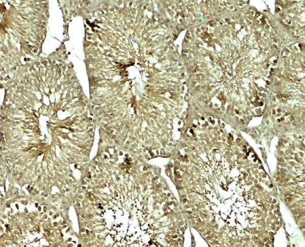
Start with Tris-EDTA at pH 9.0, heated to 95–98 °C for 20 min (page retrieval setting: nuclear antigen). Run matched sections with and without retrieval, then compare nuclear signal and tissue damage before changing heating time (standard IHC practice). The catalog antibody has a rat testis IHC image at 5 µg/mL, but its caption does not report fixation or retrieval, so the image cannot establish retrieval performance under your conditions (catalog antibody A01892 caption). Include a reference section with expected nuclear staining, such as cerebral cortex neuronal cells, and judge improvement by nuclear contrast rather than total brown signal (HPA: high in cerebral cortex neuronal cells; UniProt Q06945: nucleus; standard IHC practice).
Could fixation be causing weak or patchy SOX4 staining in paraffin sections?
SOX4-specific sensitivity to fixative type or fixation duration is unknown from the supplied evidence (catalog antibody A01892 caption: fixative unstated). Record the fixative, fixation interval, section thickness and processing history for each specimen, and compare similarly processed sections before attributing patchiness to the antibody (standard IHC practice). If tissue is available, compare controlled fixation conditions while holding Tris-EDTA pH 9.0 retrieval at 95–98 °C for 20 min constant (page retrieval setting; standard IHC practice). Assess nuclear preservation and morphology alongside staining, since damaged or unevenly processed tissue can produce misleading regional differences (UniProt Q06945: nucleus; standard IHC practice).
Should cytoplasmic or mitochondrial colour count as positive SOX4 staining?
Prioritise a clearly defined nuclear signal in intact cells: SOX4 is annotated in the nucleus, and the tissue profile reports mainly nuclear expression (UniProt Q06945: nucleus; HPA tissue IHC: mainly nuclear). HPA subcellular imaging supports nucleoplasmic localisation, while its additional mitochondrial assignment is uncertain and comes from cellular imaging rather than this chromogenic tissue assay (HPA subcellular: nucleoplasm supported; mitochondria uncertain). Score diffuse cytoplasmic colour separately and examine whether it follows tissue damage, endogenous pigment or detection background (standard IHC practice). SOX4 has no annotated transmembrane segment, so a convincing membrane rim warrants renewed specificity checks rather than automatic inclusion in the nuclear score (UniProt Q06945: topology; standard IHC practice).
How can I investigate suspected epitope masking or an unexpected SOX4 staining pattern?
The supplied SOX4 record lists one chain spanning residues 1–474, no annotated isoforms and one example modified residue, acetyllysine at position 95 (UniProt Q06945: processing, isoforms and modified residues). The catalog image does not identify the antibody epitope or establish whether that modification affects binding (catalog antibody A01892 caption). Request the immunogen or epitope information for the exact antibody, then interpret any overlap with residue 95 as a question to test experimentally (UniProt Q06945: modified residues; standard IHC practice). Compare retrieval conditions on serial sections and use an independent epitope antibody, if available, to assess whether the nuclear pattern is reproducible (standard IHC practice; UniProt Q06945: nucleus).
How should I cross-check SOX4 staining with multiplex immunofluorescence?
Use IF as an orthogonal check of cellular localisation, while evaluating the paraffin IHC result by its chromogenic nuclear signal (UniProt Q06945: nucleus; standard IHC practice). Pair SOX4 with a validated neuronal marker when examining cerebral cortex, where neuronal cells are reported as high, and choose spectrally separated fluorophores after checking tissue autofluorescence in an unstained section (HPA tissue IHC: high in cerebral cortex neuronal cells; standard IF practice). SOX4 has no transmembrane segment and is mainly nucleoplasmic, so optimise permeabilisation for access to an intracellular nuclear epitope after fixation (UniProt Q06945: topology; HPA subcellular: supported nucleoplasm; standard IF practice). Include single-label and no-primary controls before interpreting colocalisation (standard IF practice).
What should I check when SOX4 staining is diffuse or uniformly brown?
Compare the stained section with a no-primary control and inspect whether colour accumulates in nuclei, tissue edges, damaged regions or areas of endogenous pigment (standard IHC practice; UniProt Q06945: nucleus). In a peroxidase and DAB workflow, confirm that the peroxidase block, washes and detection-only control suppress nonspecific colour; these are general chromogenic IHC checks, not evidence of a SOX4-specific requirement (standard IHC practice). Titrate the catalog antibody against its reported 5 µg/mL rat testis image concentration while keeping retrieval and detection constant (catalog antibody A01892 caption; standard IHC practice). Treat broad extranuclear colour cautiously because the HPA tissue assessment notes presumed off-target binding and low staining-to-RNA consistency (HPA tissue IHC: reliability description).
How should I quantify SOX4 staining across paraffin sections? ⚠ ANSWER MARKED FOR VERIFICATION
Define the cell population and score intact nuclei with either the percentage positive or an H-score based on nuclear intensity and percentage; exclude cytoplasmic colour from that nuclear score (UniProt Q06945: nucleus; standard IHC practice). If cells are unevenly distributed, report positive nuclei per mm² of analysed viable tissue as an additional measure (standard IHC practice). Normalise comparisons to the number of evaluable nuclei or analysed viable area within the same defined compartment, and keep retrieval, detection and imaging settings consistent (standard IHC practice). Record scoring thresholds using controls before comparing samples, since the HPA tissue assessment reports low consistency with RNA expression and presumed off-target staining (HPA tissue IHC: reliability description).
What makes a SOX4-positive cell convincing rather than an artefact?
A convincing IHC call shows nuclear staining in morphologically intact cells, supported by a clean no-primary control and reproducibility across comparable sections (UniProt Q06945: nucleus; standard IHC practice). Cerebral cortex and hippocampal neuronal cells are reported as high, whereas adipocytes are reported as not detected, providing cell-specific comparison points rather than universal pass or fail controls (HPA tissue IHC: positive and negative cell profiles). Recheck signals confined to section edges, necrotic areas, unexpected cell populations or the cytoplasm, and exclude colour attributable to endogenous peroxidase or pigment (standard IHC practice). Interpret discordance conservatively because the HPA assessment reports presumed off-target binding and low consistency between staining and RNA expression (HPA tissue IHC: reliability description).
Boster reagents

Best SOX4 / Transcription factor SOX-4 IHC Antibodies

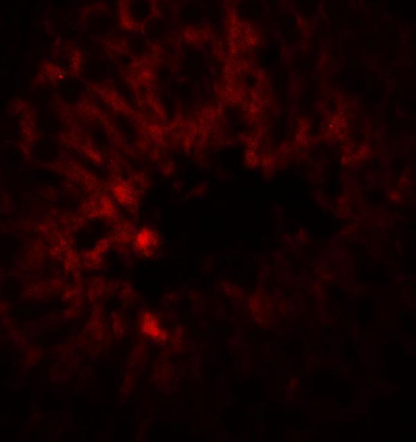
The anti-SOX4 catalog antibody supports IHC-P and IF, with listed human, mouse and rat reactivity (catalog: applications/reactivity). Its IHC and IF images show rat testis (A01892 image captions).

Real IHC data Immunohistochemistry of SOX4 in rat testis tissue with SOX4 antibody at 5 μg/mL.
Anti-Transcription factor SOX-4 SOX4 Antibody
Cat # A01892

A01892 will render with an IHC image of rat testis at 5 μg/mL (A01892 IHC caption). Its IF image also shows rat testis at 20 μg/mL; human and mouse are listed as reactive species without corresponding images in this payload (A01892 IF caption; catalog: reactivity/image captions).

Which to pick: Choose A01892 for paraffin-section IHC because IHC-P is listed and its own IHC caption shows rat testis at 5 μg/mL; the fixative is unreported (catalog: applications; A01892 IHC caption). Choose A01892 for tissue IF based on its rat testis IF image at 20 μg/mL; ICC is not separately documented (A01892 IF caption; catalog: applications). A01892 is the cross-species candidate because human, mouse and rat reactivity is listed; its rabbit host informs secondary-antibody selection (catalog: reactivity/host).

Each figure is that product's own IHC / IF validation image from its datasheet.

References

  1. UniProt Consortium. UniProt entry Q06945 (SOX4_HUMAN, Transcription factor SOX-4).
  2. Human Protein Atlas. SOX4 tissue IHC expression (reliability: Approved).
  3. Human Protein Atlas. SOX4 subcellular location (ICC-IF): Mainly localized to the nucleoplasm. In addition localized to the mitochondria..
  4. Human Protein Atlas. SOX4 antibody validation summary (1 antibodies).
  5. Diagnostic Utility of SOX4 Expression in Adult T-Cell Leukemia/Lymphoma. Diagnostics (Basel, Switzerland) 2021 — PMC8145451.
  6. Clinical and prognostic association of transcription factor SOX4 in gastric cancer. PloS one 2012 — PMC3527618.
  7. Clinical and Prognostic Implications of Transcription Factor SOX4 in Patients with Colon Cancer. PloS one 2013 — PMC3694951.
  8. LncRNA FENDRR attenuates colon cancer progression by repression of SOX4 protein. OncoTargets and therapy 2019 — PMC6549791.
  9. PubMed PMID:8268656 — UniProt-cited evidence.
  10. PubMed PMID:14574404 — UniProt-cited evidence.
  11. PubMed PMID:15489334 — UniProt-cited evidence.