This website uses cookies to ensure you get the best experience on our website.


- Table of Contents
Use this guide to plan chromogenic SOX6 IHC in paraffin sections. It covers nuclear staining in several tissues, including testis (HPA tissue IHC), and a human-reactive IHC-P antibody (datasheet A02877-2).

Expected localisation, validated protocols, controls and antibodies — the at-a-glance facts below, then the full design guide.
| Expected localisation | Nuclear staining in several tissues (HPA tissue IHC) | |
| Staining pattern | Nuclear staining in round or early spermatids (HPA tissue IHC) | |
| Antigen retrieval | EDTA pH 8.0 HIER, heat-mediated (datasheet A02877-2) | |
| Positive control | Skin+4 more · see all | |
| Negative control | Adrenal gland+4 more · see all |
| Fixation | Keep fixation consistent across sections (standard IHC practice; not target-specific) | |
| Caveat | Tissue staining has medium agreement with RNA (HPA tissue IHC) | |
| Regulation | High in round or early spermatids (HPA tissue IHC) | |
| Isoform / epitope | Four isoforms; epitope coverage is undetermined (UniProt) |
Compare the catalog antibody’s IHC-P protocol (datasheet A02877-2) with published SOX6 staining workflows for lung tissue (PMC6943225) and embryonic brain sections (PMC3963837).
| Sample | Paraffin-embedded rat testis tissue; fixative not specified (datasheet A02877-2) |
| Fixation | Image fixative and duration unreported (datasheet A02877-2); verify before use. |
| Sectioning | 4–5 µm sections on charged slides (standard) |
| Deparaffinisation | Xylene, graded ethanol series to water (standard) |
| Antigen retrieval | Heat retrieval: EDTA pH 8.0 (datasheet A02877-2); 20 min, 95–100 °C (standard) |
| Peroxidase block | 3% H2O2, 10 min, room temperature (standard) |
| Blocking | 10% goat serum (datasheet A02877-2) |
| Primary antibody | Rabbit anti-SOX6, 2-5μg/ml (datasheet A02877-2) |
| Primary incubation | Overnight at 4 °C (datasheet A02877-2) |
| Detection | Streptavidin-biotin complex (SABC), DAB chromogen (datasheet A02877-2) |
| Counterstain | Hematoxylin, blue, dehydrate and mount (standard) |
| Expected result | SOX6-positive staining in melanocytes of skin (HPA tissue IHC: Medium). HPA tissue profile: Nuclear expression in several tissues, including testis. No signal in the no-primary control. |
SOX6 is a nuclear and cytoplasmic transcription factor with no transmembrane segment (UniProt P35712). In tissue IHC, expect chiefly nuclear staining in selected cells, especially round or early spermatids in testis, with medium staining reported in melanocytes and several other cell populations (HPA tissue IHC). HPA rates the tissue pattern Enhanced, while describing only medium consistency between staining and RNA expression (HPA tissue IHC).
| Distinct nuclear staining in round or early spermatids, strongest among the listed positive cell populations (HPA: High in testis). | This fits the reported tissue pattern. Judge the signal in the named cells rather than treating every cell in testis as an expected positive; the HPA entry identifies a cell population, not uniform staining across the section (HPA tissue IHC). |
| Predominantly membrane staining, or strong staining confined to cytoplasm without a convincing nuclear pattern. | Review this as a possible localisation artefact. SOX6 has no transmembrane segment and is annotated in the nucleus and cytoplasm (UniProt P35712); HPA describes nuclear tissue expression and supported nucleoplasmic localisation by ICC-IF (HPA tissue IHC; HPA subcellular). Cytoplasmic signal alone is therefore insufficient to call the expected tissue pattern. |
| Strong signal in a cell population reported as undetected, such as adrenal gland glandular cells (HPA: Not detected). | Check the cell identity and detection controls before assigning SOX6 positivity. Cross-reactivity or endogenous detection activity can mimic chromogenic staining (general IHC practice). An HPA negative designation is specific to the listed cell population and does not establish that every cell in that tissue must be blank (HPA tissue IHC). |
| Diffuse colour over cells and surrounding tissue, with nuclei difficult to distinguish. | Treat this as background until a cell-restricted nuclear pattern emerges. Review blocking, washing, chromogen development and the no-primary control (general IHC practice). Diffuse colour does not reproduce the nuclear pattern reported across several tissues (HPA tissue IHC). |
| No nuclear signal in round or early spermatids in an otherwise assessable testis section (HPA: High). | First check that the named cells are present. Then assess the IHC detection run, antibody dilution and antigen retrieval against the applicable antibody instructions (general IHC practice). A failed positive control limits interpretation of negative study sections; the supplied HPA pattern does not identify a SOX6-specific retrieval condition. |
| Compartment to score | Score nuclear staining in the specified cell population as the principal tissue readout (HPA tissue IHC). UniProt also annotates cytoplasm, so a cytoplasmic component can be recorded, but it should be interpreted alongside the documented nuclear tissue pattern (UniProt P35712; HPA tissue IHC). |
| Cell population and intensity | Reference levels differ by cell population: round or early spermatids are High; melanocytes, adipocytes, bone marrow hematopoietic cells, cortical neuronal cells and lymph node germinal center cells are Medium (HPA tissue IHC). Compare like cells when assessing intensity. |
| Reference negatives | HPA reports Not detected in specified populations including adrenal gland glandular cells and heart muscle cardiomyocytes (HPA tissue IHC). Use the named cells when checking unexpected staining; a negative entry does not describe every cell in the organ. |
| Strength of validation | HPA labels the tissue pattern Enhanced but notes medium consistency between antibody staining and RNA expression (HPA tissue IHC). Its antibody records distinguish HPA003908 as IHC Enhanced and HPA001923 as IHC Supported (HPA antibodies); do not transfer either rating to a different antibody. |
| IF/ICC question: where should the signal appear? | The supported ICC-IF location is the nucleoplasm (HPA subcellular). That observation helps assess localisation in IF/ICC, but it does not supply an IF/ICC staining protocol or establish performance for the IHC antibody. |
| Situation | Likely cause | Next action |
|---|---|---|
| Testis positive control has no nuclear signal. | The relevant round or early spermatids may be absent from the viewed area, or the detection run may have failed (HPA: High in round or early spermatids; general IHC practice). | Confirm cell identity on the section, then review the antibody instructions, dilution, retrieval and detection controls before interpreting study negatives (general IHC practice). No SOX6-specific retrieval setting is supplied. |
| An HPA reference negative shows strong colour. | The stained cells may differ from the named negative population; cross-reactivity or endogenous detection activity is also possible (HPA tissue IHC; general IHC practice). | Identify the stained cells, examine a no-primary control and check endogenous-activity blocking where applicable (general IHC practice). Compare only with the corresponding HPA cell population. |
| Colour is diffuse and obscures nuclear boundaries. | Background from blocking, washing or chromogen development may obscure a specific pattern (general IHC practice). | Review those workflow steps and the no-primary control; assess SOX6 only when nuclear signal can be distinguished in the expected cells (general IHC practice; HPA tissue IHC). |
| Signal is predominantly at cell membranes. | That distribution conflicts with the reported nuclear tissue pattern and SOX6's lack of a transmembrane segment (HPA tissue IHC; UniProt P35712). | Check morphology and detection controls, and verify the pattern with an independently validated IHC antibody if available (general IHC practice). Do not score membrane colour alone as the expected SOX6 result. |
| A medium-level reference population looks weaker than testis. | HPA reports Medium staining for melanocytes and several other listed populations, compared with High in round or early spermatids (HPA tissue IHC). | Assess each named cell population against its own reference level and retain a working testis control when available (HPA tissue IHC; general IHC practice). Do not require equal intensity across tissues. |
| Two antibodies yield different tissue patterns. | Their HPA IHC validation ratings differ: HPA003908 is Enhanced and HPA001923 is Supported (HPA antibodies). The ratings alone do not identify why individual slides disagree. | Compare staining in the same named positive and negative cell populations, review controls and antibody-specific instructions, and report unresolved differences (HPA tissue IHC; general IHC practice). |
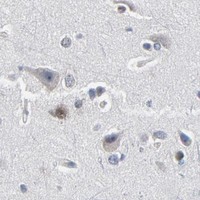
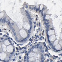
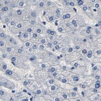
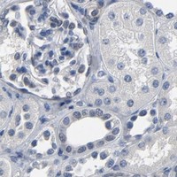


Comprehensive Human Protein Atlas IHC scoring per tissue (reliability: Enhanced — Medium consistency between antibody staining and RNA expression data.). Rows are taken directly from the HPA tissue chart — click any row's HPA link to view the source.
| Tissue | Cell type | Level | Evidence | Source |
|---|---|---|---|---|
| Skin | Melanocytes | Medium | Protein (IHC) | HPA → |
| Testis | Round or early spermatids | High | Protein (IHC) | HPA → |
| Adipose tissue | Adipocytes | Medium | Protein (IHC) | HPA → |
| Bone marrow | Hematopoietic cells | Medium | Protein (IHC) | HPA → |
| Cerebral cortex | Neuronal cells | Medium | Protein (IHC) | HPA → |
| Tissue | Cell type | Level | Evidence | Source |
|---|---|---|---|---|
| Adrenal gland | Glandular cells | Not detected | Protein (IHC) | HPA → |
| Breast | Adipocytes | Not detected | Protein (IHC) | HPA → |
| Bronchus | Respiratory epithelial cells | Not detected | Protein (IHC) | HPA → |
| Cervix | Glandular cells | Not detected | Protein (IHC) | HPA → |
| Endometrium | Cells in endometrial stroma | Not detected | Protein (IHC) | HPA → |
Troubleshoot SOX6 staining in paraffin sections by checking retrieval, nuclear localisation, cell type and controls before interpreting chromogenic signal.
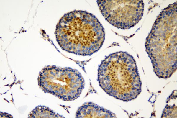
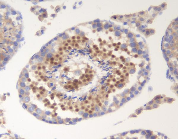
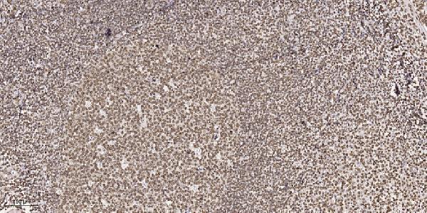
Anti-SOX6 IHC images document paraffin sections of rat and mouse testis and human tonsil (catalog IHC captions). IF is listed for A02877, with no IF image supplied (A02877 catalog).
A02877-2 has IHC images from paraffin sections of rat and mouse testis; its listed reactivity is human, mouse and rat (A02877-2 IHC captions; catalog reactivity). A02877 has a paraffin-section IHC image from human tonsil, lists IF/ICC, and lists human, monkey, mouse and rat reactivity (A02877 IHC caption; catalog applications and reactivity).
Which to pick: For tissue IHC, choose A02877-2 when its rat or mouse testis examples match your sample, or A02877 for the human tonsil example (respective IHC captions). For IF/ICC or the broadest listed species coverage, choose the polyclonal A02877; IF/ICC is listed, but no IF image is supplied (A02877 catalog applications, dilution data, reactivity and image records). Both IHC captions specify paraffin sections; neither reports the fixative (respective IHC captions).