SS18 / Protein SSXT · IHC design guide

Design Immunohistochemistry for SS18

Plan chromogenic SS18 IHC around the general nuclear tissue pattern (HPA tissue IHC). Compare high-staining colon glandular cells with undetected adipocytes (HPA tissue IHC), and titrate the catalog antibody within 1:100–1:300 (datasheet: A03947).

Evidence assembled Oct 2026 · For research use; verify linked source records and product datasheet before use
Immunohistochemistry protocol sheet for SS18 (IHC for SS18): expected localisation General nuclear staining in tissue (HPA tissue IHC), antibody A03947, validated IHC image, and IHC protocol steps
Printable SS18 IHC protocol sheet — expected localisation General nuclear staining in tissue (HPA tissue IHC), antibody A03947, controls and protocol steps. Open the full SS18 IHC guide →

SS18 Immunohistochemistry Experimental Design Guide

Expected localisation, validated protocols, controls and antibodies — the at-a-glance facts below, then the full design guide.

Must know before staining
Expected localisation General nuclear staining in tissue (HPA tissue IHC)
Staining pattern Nuclear staining, including in glandular cells (HPA tissue IHC)
Antigen retrieval Tris-EDTA pH 9.0 HIER, heat-mediated (datasheet A03947)
Positive control ⓘ Colon+4 more · see all
Negative control ⓘ Adipose tissue+4 more · see all
Important caveats
Reasons your staining may differ from the expected pattern.
Fixation Keep fixation consistent across paraffin sections. (standard IHC practice; not target-specific)
Caveat Staining varies by cell type; some are undetected (HPA tissue IHC)
Regulation Low tissue specificity (HPA tissue RNA)
Isoform / epitope 2 isoforms; epitope coverage is unspecified (UniProt)
Section 1

Recommended SS18 IHC & IF Protocols

The catalog antibody’s IHC-P protocol is paired with one published paraffin-section protocol using an SS18-SSX fusion-specific antibody (PMC8204458).

Recommended immunohistochemistry (IHC-P) protocol parameters
SampleParaffin-embedded human tonsil tissue; fixative not specified (datasheet A03947)
FixationImage fixative and duration unreported (datasheet A03947); verify before use.
Sectioning4–5 µm sections on charged slides (standard)
DeparaffinisationXylene, graded ethanol series to water (standard)
Antigen retrievalHeat retrieval: Tris-EDTA pH 9.0 (datasheet A03947); 20 min, 95–100 °C (standard)
Peroxidase block3% H2O2, 10 min, room temperature (standard)
Blocking10% normal serum of the secondary host, 30 min, room temperature (standard)
Primary antibodyRabbit anti-SS18, 1:100-1:300 (datasheet A03947)
Primary incubationOvernight at 4 °C (standard)
DetectionHRP-polymer secondary, DAB chromogen 5–10 min (standard)
CounterstainHematoxylin, blue, dehydrate and mount (standard)
Expected resultSS18-positive staining in glandular cells of colon (HPA tissue IHC: High). HPA tissue profile: General nuclear expression. No signal in the no-primary control.
💡Decision noteStart with heat-mediated Tris-EDTA retrieval at pH 9.0 for the catalog antibody (datasheet A03947); the published fusion-specific protocol does not state retrieval conditions (PMC8204458).
Section 2

What Is the Expected SS18 Staining Pattern?

SS18 should stain mainly in nuclei (UniProt Q15532: nucleus; HPA: general nuclear expression). Expect staining across multiple tissues, including glandular cells in colon, gallbladder and rectum, and spermatogonia in testis (HPA: High). HPA rates its tissue IHC pattern Supported, with medium consistency between staining and RNA expression (HPA: reliability). SS18 has no transmembrane segment (UniProt Q15532: topology).

What am I looking at on my slide?
Nuclear chromogen in glandular cells of colon, gallbladder or rectum.This fits the expected compartment and cell types (UniProt Q15532: nucleus; HPA: High in these glandular cells). Score the named cells and their nuclei, rather than treating every stained structure in the section as equivalent. HPA reports a general nuclear pattern, but its tissue IHC reliability is Supported, with medium consistency against RNA data (HPA: tissue IHC).
Predominantly cytoplasmic chromogen with little or no nuclear staining.This conflicts with the expected IHC pattern (HPA: general nuclear expression; UniProt Q15532: nucleus). Review staining distribution, detection controls and antibody specificity before calling it SS18. Cytosol is an approved additional location in HPA ICC-IF, so a minor cytosolic component alone does not establish an artefact (HPA: subcellular ICC-IF).
Strong staining in adipocytes, cardiomyocytes or skeletal myocytes.These named cell types were not detected in the supplied HPA tissue IHC observations (HPA: adipose tissue, heart muscle and skeletal muscle). Check whether the signal follows cell boundaries, pigment or tissue-wide reagent activity. Cross-reactivity or endogenous detection activity are possibilities to investigate (general IHC practice); this pattern alone cannot identify the cause.
Diffuse chromogen across nuclei, cytoplasm and surrounding tissue.A widespread haze makes nuclear localization difficult to judge against the reported pattern (HPA: general nuclear expression). Compare a reagent control and a known-positive section processed in the same run. Review blocking, washing and detection settings as general IHC troubleshooting steps; HPA does not assign this appearance to an SS18-specific mechanism.
No nuclear signal in a known-positive colon or testis section.This is inconsistent with High staining reported for colon glandular cells and testis spermatogonia (HPA: tissue IHC). First confirm that the expected cells are present and interpretable, then check the run controls and staining workflow (general IHC practice). A failed section does not establish that SS18 is absent from the sample.
💡Expected SS18 appearanceCall an IHC result positive when chromogen is predominantly nuclear in the relevant cells, with High HPA staining reported in colon, gallbladder and rectal glandular cells or testis spermatogonia; strong signal confined to an HPA Not detected cell type warrants investigation (HPA: tissue IHC; UniProt Q15532: nucleus).
How each factor affects the staining
Tissue and cell selectionUse the named cell type when interpreting a section: HPA reports High staining in colon glandular cells and testis spermatogonia, but Not detected in adipocytes and skeletal myocytes (HPA: tissue IHC). The comparison is cell specific; it is not a whole-organ positive or negative call.
IHC antibody evidenceBoth listed antibodies, HPA055741 and HPA059539, have Supported IHC status (HPA: antibodies). The supplied record does not label either IHC result Enhanced. Treat agreement with the expected nuclear pattern as supporting evidence and investigate discordant staining; the status alone does not prove every stained cell is specific.
Isoforms and processingUniProt lists 2 SS18 isoforms and a protein chain spanning residues 2–418, with no signal peptide or propeptide (UniProt Q15532: isoforms and processing). The payload gives no antibody epitope position or isoform-specific IHC result, so staining cannot distinguish the isoforms or support a shedding explanation.
IF/ICC Q: what localization should I expect?A: HPA reports mainly nucleoplasmic signal, with additional cytosolic localization in ICC-IF (HPA: approved subcellular locations). That observation helps interpret a mixed compartment signal; it is not an IHC-P staining protocol. HPA lists BJ, SiHa and U2OS ICC-IF images (HPA: subcellular ICC-IF).
Why is my staining missing, weak or wrong?
SituationLikely causeNext action
Positive-control nuclei remain unstained.The expected cells may be missing or the IHC run may have failed (HPA: High in colon glandular cells and testis spermatogonia; general IHC practice).Verify cell identity and section quality, then review the IHC-validated antibody, retrieval, dilution and detection steps against its supplied protocol (general IHC practice). No SS18-specific retrieval condition is established by these payloads.
Chromogen is mainly cytoplasmic.The result departs from the reported nuclear IHC pattern (HPA: tissue IHC; UniProt Q15532: nucleus).Check nuclear counterstain, reagent controls and whether nuclei carry any specific signal (general IHC practice). Interpret limited cytosolic staining cautiously because HPA also approves cytosol as an additional ICC-IF location (HPA: subcellular ICC-IF).
An HPA Not detected cell type stains strongly.Cross-reactivity or endogenous detection activity may contribute (general IHC practice); HPA reports no detected staining in the specified comparator cells (HPA: tissue IHC).Confirm the cell identity and compare a detection-only control with a known-positive section (general IHC practice). Do not infer the cause from tissue identity alone.
Diffuse background obscures nuclei.Background can prevent assessment of the reported general nuclear pattern (HPA: tissue IHC); its source is undetermined.Review blocking, washes and detection exposure with appropriate reagent controls (general IHC practice). Reassess nuclear localization only after the background is sufficiently controlled.
Expected staining varies among cell types in one section.HPA reports cell-specific levels, including High, Medium, Low and Not detected observations across tissues (HPA: tissue IHC).Record the named cell population and nuclear pattern separately from overall section intensity. Compare like cell types when judging concordance with HPA (HPA: tissue IHC).
A negative result is attributed to fixation.No target-specific SS18 fixation sensitivity is reported in the supplied UniProt or HPA evidence.Check whether known-positive cells stained in the same run, then review routine processing and assay controls (general IHC practice). Keep fixation as an unconfirmed possibility rather than an established SS18-specific explanation.

Sample controls for SS18 IHC & IF

🧪Run colon first; its glandular cells should stain (HPA: High in colon glandular cells). Use adipose tissue as the negative comparator, scoring adipocytes (HPA: Not detected in adipocytes); on the colon slide, use non-glandular cells without staining as an internal background reference, without assuming they are SS18-negative.
Positive control tissue: Colon (Glandular cells, HPA High)
Negative control tissue: Adipose tissue (HPA Not detected)
ICC-IF cell lines (HPA subcellular resource): HPA ICC-IF images show SS18 in BJ [Human fibroblast], SiHa, U2OS, with annotated localisation: Nucleoplasm (approved) (HPA subcellular).
Technical controls: Include a no-primary, secondary-only control; an isotype control matched to the primary antibody’s host species and monoclonal or polyclonal format; and SS18 knockout material or an antibody-compatible peptide block. For chromogenic colon IHC, block endogenous peroxidase and, if using avidin–biotin detection, endogenous biotin.
⚠️Feasibility: A target-specific fixation window or fixation effect is unreported in the supplied evidence; the selected A03947 paraffin-section caption does not state a fixative (datasheet: fixative not stated). Its Tris-EDTA pH 9.0 retrieval step provides a starting condition, but does not establish retrieval dependence (datasheet: Tris-EDTA pH 9.0). The evidence does not establish whether frozen sections or IF are easier; in colon, check that glandular staining follows the expected nuclear pattern rather than luminal deposits (UniProt Q15532: nucleus; HPA: High in colon glandular cells).

HPA tissue IHC evidence for SS18

Comprehensive Human Protein Atlas IHC scoring per tissue (reliability: Supported — Medium consistency between antibody staining and RNA expression data.). Rows are taken directly from the HPA tissue chart — click any row's HPA link to view the source.

Positive expression · recommended positive controls

TissueCell typeLevelEvidenceSource
Colon Glandular cells High Protein (IHC) HPA →
Gallbladder Glandular cells High Protein (IHC) HPA →
Nasopharynx Ciliated cells (cell body) High Protein (IHC) HPA →
Rectum Glandular cells High Protein (IHC) HPA →
Testis Spermatogonia cells High Protein (IHC) HPA →

Undetected expression · recommended negative controls

TissueCell typeLevelEvidenceSource
Adipose tissue Adipocytes Not detected Protein (IHC) HPA →
Heart muscle Cardiomyocytes Not detected Protein (IHC) HPA →
Hippocampus Glial cells Not detected Protein (IHC) HPA →
Kidney Cells in glomeruli Not detected Protein (IHC) HPA →
Ovary Follicle cells Not detected Protein (IHC) HPA →
Section 3

Advanced SS18 IHC Tips

Troubleshoot SS18 staining in paraffin sections by checking retrieval, nuclear localisation, controls and scoring before interpreting biological differences.

What retrieval should I try first when SS18 nuclear staining is weak?
Start with heat-mediated Tris-EDTA at pH 9.0 for paraffin sections (datasheet A03947). The catalog image used this retrieval with primary antibody at 1:200, incubated at 4°C overnight, so reproduce those conditions before changing several variables together (datasheet A03947). Compare a retrieved section with an otherwise matched section receiving less retrieval, and judge nuclear signal alongside tissue integrity (standard IHC practice; UniProt Q15532: nucleus). If nuclei remain faint, adjust heating duration in small steps; excessive retrieval can damage morphology and increase diffuse staining (standard IHC practice). Include a section expected to stain in the same run so a failed run is distinguishable from a low-expressing sample (HPA: high in colon glandular cells; standard IHC practice).
Could fixation explain weak or uneven SS18 staining in my paraffin sections?
SS18-specific fixation sensitivity is unknown from the supplied evidence; the selected paraffin-section caption does not state a fixative (datasheet A03947). Record each specimen’s fixative and fixation duration, then compare sections processed through the same retrieval and detection run (standard IHC practice). Uneven fixation can change morphology and antigen accessibility, so assess nuclear staining in well-preserved areas before altering antibody concentration (standard IHC practice; UniProt Q15532: nucleus). Use the documented Tris-EDTA retrieval at pH 9.0 as the starting condition and keep the primary at the caption’s 1:200 while investigating processing differences (datasheet A03947). Do not assign an SS18-specific fixation effect without a controlled comparison (standard IHC practice).
How should I assess cytoplasmic SS18 staining when the expected signal is nuclear?
Prioritise staining in intact nuclei: SS18 is annotated as nuclear, and tissue IHC shows general nuclear expression (UniProt Q15532: subcellular location; HPA: tissue IHC profile). Additional cytosolic localisation is reported in cell imaging, so some cytoplasmic signal is biologically plausible but needs independent support in the tissue being examined (HPA: approved nucleoplasm and cytosol locations). Compare nuclear and cytoplasmic compartments in the same cells against a no-primary control and surrounding tissue (standard IHC practice). Strong diffuse cytoplasm with little nuclear staining warrants a check of retrieval, antibody concentration and detection background before interpretation (standard IHC practice; UniProt Q15532: nucleus). Document compartment-specific results separately rather than merging them into one positive score (standard IHC practice).
Can this stain distinguish SS18 isoforms or detect an SS18 fusion specifically?
Do not assign isoform identity from this stain without an antibody epitope map or isoform-specific validation; the supplied record lists 2 SS18 isoforms (UniProt Q15532: isoforms). Check whether the antibody’s documented epitope lies in sequence shared by both isoforms before making an isoform claim (standard IHC practice; UniProt Q15532: isoforms). SS18-SSX1 and SS18-SSX2 fusion genes are reported in synovial sarcomas, whereas native SS18 is fairly ubiquitously expressed (UniProt Q15532: tissue specificity). Accordingly, nuclear positivity alone cannot establish a fusion; use an independently validated fusion-specific assay for that question (UniProt Q15532: tissue specificity; standard IHC practice). Record the epitope and validation evidence used for any more specific interpretation (standard IHC practice).
How can IF help resolve an ambiguous SS18 IHC localisation result?
Use IF as a separate validation experiment and multiplex SS18 with an independently validated marker for the cell type under study, such as glandular cells when assessing colon (HPA: high in colon glandular cells; standard IF practice). Select fluorophores after checking unstained tissue for autofluorescence, and place the weaker channel where background is lower (standard IF practice). SS18 is primarily nucleoplasmic, with additional cytosolic localisation reported, so fixation-compatible permeabilisation should allow antibody access to intracellular nuclear antigen (HPA: approved nucleoplasm and cytosol locations; standard IF practice). Its exact antibody epitope is not supplied, so do not infer a membrane-facing epitope or transfer the paraffin IHC conditions directly to IF (datasheet A03947; standard IF practice). Include single-channel and no-primary controls when judging colocalisation (standard IF practice).
What should I check when SS18 chromogenic staining appears diffuse or widespread?
First distinguish expected widespread expression from haze: SS18 is fairly ubiquitously expressed, and tissue IHC reports general nuclear expression (UniProt Q15532: tissue specificity; HPA: tissue IHC profile). Inspect a no-primary control for secondary-reagent or detection background, and compare nuclear staining with diffuse cytoplasmic or extracellular colour (standard IHC practice). Confirm adequate endogenous peroxidase blocking before DAB detection and rinse thoroughly between steps (standard chromogenic IHC practice). If the control is clean but test sections are dark, titrate around the documented 1:200 primary dilution while holding retrieval at Tris-EDTA pH 9.0 constant (datasheet A03947; standard IHC practice). Judge changes against preserved nuclear detail in the same tissue (standard IHC practice).
How should I quantify SS18 staining across paraffin-section samples? ⚠ ANSWER MARKED FOR VERIFICATION
Define the scored compartment as nuclei before collecting data, consistent with the reported general nuclear tissue pattern (HPA: tissue IHC profile). For each annotated cell population, report the percentage of positive nuclei and, if intensity is reproducible, an H-score calculated from proportions at intensity grades 0–3, giving a range of 0–300 (standard IHC scoring practice). Normalise counts to the number of evaluable nuclei in that population, or report positive-cell density per mm² of viable annotated tissue when cell number is unavailable (standard IHC scoring practice). Apply identical thresholds and imaging conditions across samples, and exclude folds, edges and necrotic areas from scoring (standard IHC practice). Report cytoplasmic staining separately if assessed (HPA: additional cytosol location; standard IHC practice).
How can I separate true SS18 positivity from staining artefact?
Look for reproducible nuclear signal in intact cells: SS18 is nuclear, and HPA describes general nuclear tissue expression (UniProt Q15532: subcellular location; HPA: tissue IHC profile). Colon glandular cells offer a reported high-expression comparison, while adipocytes are reported as not detected; evaluate each cell type rather than treating the whole section uniformly (HPA: high in colon glandular cells; HPA: not detected in adipocytes). Staining confined to section edges, folds or necrotic material should prompt review of section quality and controls (standard IHC practice). Colour in a no-primary control or persistent granular signal after inadequate peroxidase blocking supports a detection artefact (standard chromogenic IHC practice). Nuclear SS18 staining alone does not establish an SS18 fusion (UniProt Q15532: tissue specificity; standard IHC practice).
Boster reagents

Best SS18 / Protein SSXT IHC Antibodies

Three anti-SS18 antibodies have human paraffin-section IHC images (catalog IHC captions); two list IF, one lists ICC, and all list human, mouse, and rat reactivity (catalog applications and reactivity).

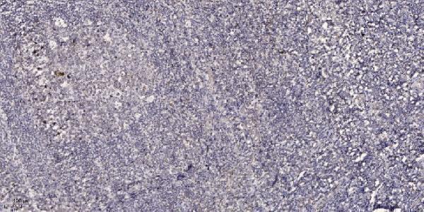
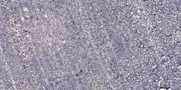
Real IHC data Immunohistochemical analysis of paraffin-embedded human tonsil. 1, Antibody was diluted at 1:200 (4° overnight). 2, Tris-EDTA, pH9.0 was used for antigen retrieval. 3, Secondary antibody was diluted at 1:200 (room temperature, 45min).
Anti-SYT SS18 Antibody
Cat # A03947
Real IHC data Immunohistochemical analysis of paraffin-embedded human tonsil. 1, Antibody was diluted at 1:200(4° overnight). 2, Tris-EDTA,pH9.0 was used for antigen retrieval. 3,Secondary antibody was diluted at 1:200(room temperature, 45min).
Anti-SSXT SS18 Antibody
Cat # A30497
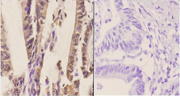
Real IHC data Immunohistochemistry (IHC) analyzes of SYT (A7) pAb in paraffin-embedded human colon carcinoma tissue at 1:50.showing cytoplasmic and nucleus staining. Negative control (the right)Using PBS instead of primary antibody, secondary antibody is Goat Anti-Rabbit IgG-biotin followed by avidin-peroxidase.
Anti-SYT (A7) SS18 Antibody
Cat # A03947-1

A03947 and A30497 each show IHC staining of paraffin-embedded human tonsil (respective IHC image captions). A03947-1 shows IHC staining of paraffin-embedded human colon carcinoma, with cytoplasmic and nuclear staining (A03947-1 IHC image caption).

Which to pick: For tissue IHC, A03947 and A30497 have human tonsil examples, while A03947-1 has a human colon carcinoma example; all three captions specify paraffin sections but do not report the fixative (respective IHC image captions). For IF, consider A03947 or A30497; for ICC, choose A30497, the only SKU listing ICC (catalog applications). All three list human, mouse, and rat reactivity, but their supplied IHC images show human tissue only (catalog reactivity; respective IHC image captions).

Each figure is that product's own IHC / IF validation image from its datasheet.

References

  1. UniProt Consortium. UniProt entry Q15532 (SSXT_HUMAN, Protein SSXT).
  2. Human Protein Atlas. SS18 tissue IHC expression (reliability: Supported).
  3. Human Protein Atlas. SS18 subcellular location (ICC-IF): Mainly localized to the nucleoplasm. In addition localized to the cytosol..
  4. Human Protein Atlas. SS18 antibody validation summary (2 antibodies).
  5. Primary ovarian synovial sarcoma in an adolescent mimicking a germ cell tumor: A case report and diagnostic challenge. Gynecologic oncology reports 2026 — PMC13396617.
  6. Utility of Immunohistochemistry With Antibodies to SS18-SSX Chimeric Proteins and C-Terminus of SSX Protein for Synovial Sarcoma Differential Diagnosis. The American journal of surgical pathology 2024 — PMC12054528.
  7. Usefulness of SS18-SSX antibody as a diagnostic marker for pulmonary metastatic synovial sarcoma. Diagnostic pathology 2021 — PMC8204458.
  8. The Recent Advances in Molecular Diagnosis of Soft Tissue Tumors. International journal of molecular sciences 2023 — PMC10051446.
  9. PubMed PMID:7951320 — UniProt-cited evidence.
  10. PubMed PMID:11368913 — UniProt-cited evidence.
  11. PubMed PMID:15489334 — UniProt-cited evidence.