This website uses cookies to ensure you get the best experience on our website.


- Table of Contents
Plan paraffin-section ST6GAL1 IHC with hepatocytes and tonsillar germinal-center cells as high-staining references (HPA tissue IHC). This guide covers the cytoplasmic Golgi pattern (HPA tissue IHC), a catalog antibody dilution of 1:100–1:300 (datasheet A30620), and interpretation of a potentially secreted form (UniProt).

Expected localisation, validated protocols, controls and antibodies — the at-a-glance facts below, then the full design guide.
| Expected localisation | Cytoplasmic Golgi pattern (HPA tissue IHC) | |
| Staining pattern | Hepatocyte and germinal-center cell Golgi staining (HPA tissue IHC) | |
| Antigen retrieval | EDTA pH 8.0 HIER, heat-mediated (datasheet A30620) | |
| Positive control | Liver+4 more · see all | |
| Negative control | Adipose tissue+4 more · see all |
| Fixation | Matched tissue-IHC evidence does not establish the fixation claim. Validate the specimen-specific method before use. (selected-SKU IHC image A30620) | |
| Caveat | A secreted variant may alter tissue staining (HPA tissue IHC) | |
| Regulation | Liver and lymphoid tissue group enrichment (HPA tissue IHC) | |
| Isoform / epitope | 2 isoforms; cytoplasmic versus luminal epitope coverage is unknown (UniProt) |
The catalog antibody’s paraffin-section protocol uses EDTA pH 8.0 retrieval (datasheet A30620). Published chromogenic protocols describe TNBC cryosections and lymphoma cytospins (PMC9663294; PMC4027875).
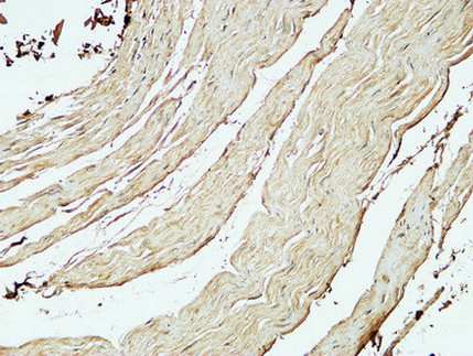
| Sample | Paraffin-embedded Human stomach tissue; fixative not specified (datasheet A30620) |
| Fixation | Image fixative and duration unreported (datasheet A30620); verify before use. |
| Sectioning | 4–5 µm sections on charged slides (standard) |
| Deparaffinisation | Xylene, graded ethanol series to water (standard) |
| Antigen retrieval | Heat retrieval: EDTA pH 8.0 (datasheet A30620); 20 min, 95–100 °C (standard) |
| Peroxidase block | 3% H2O2, 10 min, room temperature (standard) |
| Blocking | 10% normal serum of the secondary host, 30 min, room temperature (standard) |
| Primary antibody | Rabbit anti-ST6GAL1, 1:100 - 1:300 (datasheet A30620) |
| Primary incubation | Overnight at 4 °C (standard) |
| Detection | HRP-polymer secondary, DAB chromogen 5–10 min (standard) |
| Counterstain | Hematoxylin, blue, dehydrate and mount (standard) |
| Expected result | ST6GAL1-positive staining in hepatocytes of liver (HPA tissue IHC: High). HPA tissue profile: Cytoplasmic expression with a Golgi pattern in several tissues, including liver and lymphoid tissues. No signal in the no-primary control. |
ST6GAL1 is a Golgi stack membrane protein with a lumenal catalytic region and a reported secreted form (UniProt P15907 topology and location). In IHC, expect cytoplasmic staining with a Golgi pattern, especially in hepatocytes and tonsillar germinal center cells (HPA tissue IHC: High in both). HPA rates the tissue evidence Enhanced but notes only medium consistency with RNA expression and a possible mismatch between RNA and protein location (HPA tissue IHC).
| Hepatocytes or tonsillar germinal center cells show concentrated cytoplasmic staining near the nucleus. | This fits the Golgi pattern reported across tissues and the High staining recorded in these two cell populations (HPA tissue IHC). Judge the distribution within the expected cells as well as signal strength; a dark deposit alone does not establish the expected pattern (general IHC practice). |
| The dominant deposit is nuclear, or it sharply outlines cell surfaces without a Golgi-like cytoplasmic component. | That distribution does not match the reported Golgi stack membrane location or HPA tissue pattern (UniProt P15907 location; HPA tissue IHC). Treat it as suspect and compare it with a known-positive section and appropriate detection controls before assigning it to ST6GAL1 (general IHC practice). |
| Strong staining appears in cells recorded as Not detected, such as adipocytes or cardiomyocytes. | That conflicts with the supplied tissue observations (HPA tissue IHC: Not detected in adipocytes and cardiomyocytes). Cross-reactivity or endogenous chromogenic activity is possible; a no-primary control can help distinguish detection-system signal from primary-antibody-associated staining (general IHC practice). |
| Color is spread uniformly across cells or tissue, obscuring a discrete Golgi-like pattern. | Diffuse background limits interpretation even where ST6GAL1 is expected (HPA tissue IHC: cytoplasmic Golgi pattern). Review the no-primary control, blocking, washes and chromogen development before scoring positive cells (general IHC practice); diffuse color by itself is not evidence of a secreted form. |
| No convincing stain appears in hepatocytes or tonsillar germinal center cells. | These are useful positive comparators because HPA records High staining in both (HPA tissue IHC). A blank result there raises a run-level or antibody-use question before a negative result elsewhere is interpreted; inspect the control section, detection reagents and section quality (general IHC practice). |
| Golgi topology | Residues 10–26 form the membrane span; residues 27–406 face the lumen (UniProt P15907 topology). This supports an intracellular Golgi pattern but does not identify the catalog antibody's epitope or specify an antigen-retrieval condition. |
| Secreted form | UniProt reports secretion into body fluid by similarity alongside the membrane-bound Golgi form (UniProt P15907 location). HPA warns that secreted variants can complicate agreement between tissue RNA and protein locations (HPA tissue IHC); interpret staining against cell morphology and controls. |
| Tissue-dependent signal | HPA records High staining in liver hepatocytes and tonsillar germinal center cells, Medium in several other listed populations, and Not detected in selected cells (HPA tissue IHC). Those categories are observed patterns, not universal intensity thresholds for every specimen. |
| Antibody evidence | The supplied catalog antibody, CAB080031, has an Enhanced IHC validation status (HPA antibodies). HPA's tissue profile also carries an Enhanced reliability label with medium RNA–protein consistency (HPA tissue IHC); neither label proves that every deposit in a new section is specific. |
| IF/ICC Q&A: what location should be expected? | A Golgi-associated pattern is biologically plausible from the reported membrane location (UniProt P15907 location), but HPA lists no main ICC-IF location or cell-line images here (HPA subcellular). This record cannot establish an observed IF/ICC pattern. |
| Situation | Likely cause | Next action |
|---|---|---|
| Known-positive liver or tonsil section is blank. | The result conflicts with HPA's High staining in hepatocytes or germinal center cells (HPA tissue IHC); a run or detection failure is possible (general IHC practice). | Check that the control contains the expected cells, then verify the primary-antibody step, retrieval used, detection reagents and chromogen development against the applicable IHC-P workflow (general IHC practice). No target-specific retrieval setting is supplied. |
| The section shows widespread faint color with little cellular definition. | Background can mask the cytoplasmic Golgi pattern reported by HPA (HPA tissue IHC); excess detection signal or incomplete washing are general possibilities (general IHC practice). | Compare a no-primary control, assess wash and blocking steps, and review chromogen development before lowering the staining score or changing conditions (general IHC practice). |
| Unexpected cells stain strongly, including cells HPA records as Not detected. | Cross-reactivity or endogenous chromogenic activity may explain the discrepancy (HPA tissue IHC: selected cells Not detected; general IHC practice). HPA's category does not guarantee every individual specimen will be blank. | Confirm cell identity on the counterstained section and inspect no-primary and known-positive controls; if unexpected signal persists only with primary antibody, reassess specificity (general IHC practice). |
| Staining is predominantly nuclear or forms a uniform surface rim. | That distribution conflicts with the reported Golgi membrane location and cytoplasmic Golgi tissue pattern (UniProt P15907 location; HPA tissue IHC). | Review section morphology and control staining, then reassess primary-antibody specificity and detection background before interpreting the compartment as ST6GAL1 (general IHC practice). |
| A Medium-staining tissue looks weaker than the liver or tonsil control. | Different intensity is consistent with the supplied HPA categories: several listed populations are Medium, while hepatocytes and tonsillar germinal center cells are High (HPA tissue IHC). | Score the expected cell population and Golgi-like distribution within that tissue; compare sections processed in the same run rather than imposing one intensity cutoff across tissues (general IHC practice). |
| ICC-IF images do not show a clear Golgi pattern. | HPA supplies no ICC-IF cell-line images or main subcellular location for this record, although UniProt places the membrane-bound form in Golgi stacks (HPA subcellular; UniProt P15907 location). | Treat the IF result as unconfirmed by this payload and use the separate IF/ICC guide for assay-specific controls and interpretation; do not transfer an IHC-P staining result into an IF validation claim. |


Comprehensive Human Protein Atlas IHC scoring per tissue (reliability: Enhanced — Medium consistency between antibody staining and RNA expression data. At least one protein variant secreted, tissue location of RNA and protein might differ and correlation is complex.). Rows are taken directly from the HPA tissue chart — click any row's HPA link to view the source.
| Tissue | Cell type | Level | Evidence | Source |
|---|---|---|---|---|
| Liver | Hepatocytes | High | Protein (IHC) | HPA → |
| Tonsil | Germinal center cells | High | Protein (IHC) | HPA → |
| Appendix | Glandular cells | Medium | Protein (IHC) | HPA → |
| Bone marrow | Hematopoietic cells | Medium | Protein (IHC) | HPA → |
| Bronchus | Respiratory epithelial cells | Medium | Protein (IHC) | HPA → |
| Tissue | Cell type | Level | Evidence | Source |
|---|---|---|---|---|
| Adipose tissue | Adipocytes | Not detected | Protein (IHC) | HPA → |
| Adrenal gland | Glandular cells | Not detected | Protein (IHC) | HPA → |
| Breast | Glandular cells | Not detected | Protein (IHC) | HPA → |
| Cerebellum | Cells in granular layer | Not detected | Protein (IHC) | HPA → |
| Epididymis | Glandular cells | Not detected | Protein (IHC) | HPA → |
Use Golgi-pattern staining and cell-type controls to troubleshoot ST6GAL1 in paraffin-section chromogenic IHC (UniProt P15907; HPA tissue IHC).
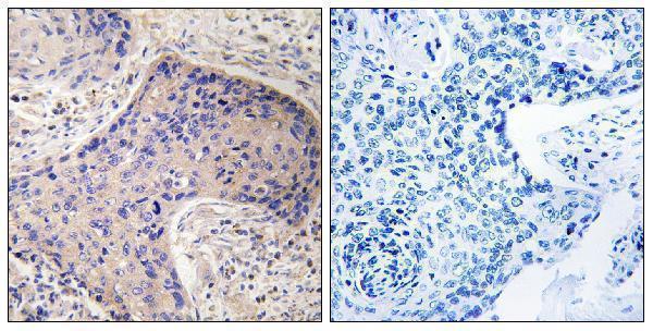
The anti-ST6GAL1 catalog antibody has real IHC images from human stomach and prostate carcinoma paraffin sections (catalog IHC captions). IF/ICC is listed, but no IF image is supplied (catalog applications; IF image payload).
A30620 has IHC images from human stomach and prostate carcinoma paraffin sections (catalog IHC captions). Its listed applications include IHC, IF and ICC, and its listed reactivity covers human, mouse and rat (catalog applications; catalog reactivity).
Which to pick: Choose A30620 for paraffin-section IHC: its human stomach image reports 1:200 antibody dilution and EDTA retrieval at pH 8.0; the fixative is unreported (catalog stomach IHC caption). A30620 is also the listed IF/ICC option, although no IF image is supplied (catalog applications; IF image payload). For mouse or rat work, A30620 lists reactivity with both species, but the supplied tissue images are human; it is polyclonal (catalog reactivity; catalog IHC captions; catalog dilution record).