This website uses cookies to ensure you get the best experience on our website.


- Table of Contents
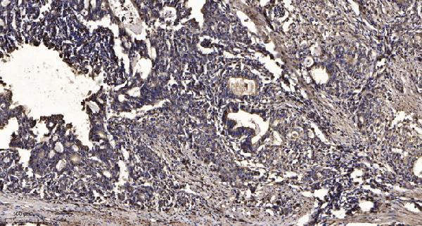
Plan chromogenic STK38L staining in paraffin sections using the reported cytoplasmic and membranous tissue pattern (HPA tissue IHC). Compare results with the catalog antibody’s human tonsil IHC image (datasheet A06774), and interpret staining with its specificity limitation in mind (HPA tissue IHC).

Expected localisation, validated protocols, controls and antibodies — the at-a-glance facts below, then the full design guide.
| Expected localisation | Cytoplasmic and membranous tissue staining (HPA tissue IHC) | |
| Staining pattern | Glandular and endothelial cells; cytoplasmic/membranous (HPA tissue IHC) | |
| Antigen retrieval | Tris-EDTA pH 9.0 HIER, heat-mediated (datasheet A06774) | |
| Positive control | Adrenal gland+4 more · see all | |
| Negative control | Adipose tissue+4 more · see all |
| Fixation | Keep fixation consistent across sections (standard IHC practice; not target-specific). Selected-image fixative and duration unreported (datasheet A06774); verify before use. | |
| Caveat | Antibody staining may reflect more than one gene (HPA tissue IHC) | |
| Regulation | Expression regulation is unspecified (UniProt) | |
| Isoform / epitope | 2 isoforms; epitope differences are unspecified (UniProt) |
The catalog antibody protocol uses Tris-EDTA pH 9.0 retrieval (datasheet A06774). The published human PDAC tissue microarray protocol uses sodium citrate pH 6.0 (PMC5667982).
| Sample | Paraffin-embedded human tonsil tissue; fixative not specified (datasheet A06774) |
| Fixation | Image fixative and duration unreported (datasheet A06774); verify before use. |
| Sectioning | 4–5 µm sections on charged slides (standard) |
| Deparaffinisation | Xylene, graded ethanol series to water (standard) |
| Antigen retrieval | Heat retrieval: Tris-EDTA pH 9.0 (datasheet A06774); 20 min, 95–100 °C (standard) |
| Peroxidase block | 3% H2O2, 10 min, room temperature (standard) |
| Blocking | 10% normal serum of the secondary host, 30 min, room temperature (standard) |
| Primary antibody | Rabbit anti-STK38L, 1:50 recommended; image 1:200 (datasheet A06774) |
| Primary incubation | Overnight at 4 °C (standard) |
| Detection | HRP-polymer secondary, DAB chromogen 5–10 min (standard) |
| Counterstain | Hematoxylin, blue, dehydrate and mount (standard) |
| Expected result | STK38L-positive staining in glandular cells of adrenal gland (HPA tissue IHC: Medium). HPA tissue profile: Cytoplasmic and membranous expression in several different tissue types. No signal in the no-primary control. |
STK38L is expected in the cytoplasm and may appear along cell membranes or the actin cytoskeleton (UniProt Q9Y2H1; HPA tissue IHC). Look for staining in selected glandular, respiratory epithelial, endothelial, and neuronal cells (HPA tissue IHC). It has no transmembrane segment (UniProt Q9Y2H1 topology). Treat the pattern as provisional: HPA rates tissue IHC Approved but reports low agreement with RNA and cautions that the antibodies may target proteins from multiple genes (HPA tissue IHC).
| Cytoplasmic staining, sometimes with a cell-edge accent, in bronchial respiratory epithelium, colon endothelium, or caudate neurons. | This fits the reported compartments and Medium staining in those cell types (HPA tissue IHC). A cell-edge accent is compatible with membrane association; it does not imply that STK38L spans the membrane (UniProt Q9Y2H1 topology). |
| Strong nuclear-only staining, with little cytoplasmic signal. | A nuclear-only pattern conflicts with the reported cytoplasmic, cytoskeletal, and membrane locations (UniProt Q9Y2H1; HPA tissue IHC). Treat it as a suspected artefact and review morphology, counterstain, and detection controls before assigning it to STK38L (general IHC practice). |
| Prominent staining in adipocytes, liver cholangiocytes, or pancreatic exocrine cells. | HPA lists these cells as Not detected (HPA tissue IHC). Check for antibody cross-reactivity or endogenous detection activity before calling the signal target-specific; a single positive-looking section cannot resolve HPA's multi-gene targeting caution (HPA tissue IHC; general IHC practice). |
| Diffuse colour across stroma, empty spaces, and several unrelated cell types. | A widespread deposit without clear cellular boundaries is background rather than a convincing cytoplasmic pattern (general IHC practice; HPA tissue IHC). Compare it with a no-primary control and inspect blocking, washing, and chromogen development (general IHC practice). |
| No staining in bronchial respiratory epithelium despite intact tissue morphology. | HPA reports Medium staining there, so an absent signal calls for a technical check (HPA tissue IHC). Compare a known-positive section in the same run and review retrieval, antibody dilution, and detection according to the antibody's validated IHC workflow (general IHC practice). |
| Cell type and compartment | HPA reports cytoplasmic and membranous staining across several tissues, including Medium signal in bronchial respiratory epithelium and colon endothelium (HPA tissue IHC). UniProt also places STK38L with the actin cytoskeleton and at membranes (UniProt Q9Y2H1). |
| Antibody evidence and specificity | The tissue IHC antibody HPA038623 is Approved, while HPA reports low staining-to-RNA consistency and cautions about multi-gene targeting (HPA antibodies; HPA tissue IHC). Interpret an apparently correct pattern with controls and avoid treating the rating as proof of specificity. |
| Isoforms and epitope coverage | UniProt lists 2 STK38L isoforms, but the supplied sources do not map antibody epitopes or establish isoform-specific staining (UniProt Q9Y2H1; HPA antibodies). An uneven cell pattern alone cannot identify which isoform is present. |
| Chromogenic detection activity | Endogenous enzyme activity can create colour unrelated to primary-antibody binding in enzyme-based IHC (general IHC practice). A no-primary control helps distinguish this from cellular staining; the supplied sources do not identify a STK38L-specific detection interference. |
| Situation | Likely cause | Next action |
|---|---|---|
| Positive-control epithelium has little or no colour. | Weak detection or a missed workflow step is possible; bronchial respiratory epithelium is reported at Medium staining (HPA tissue IHC; general IHC practice). | Run a known-positive section alongside the sample. Check the IHC-validated antibody's retrieval, dilution, and detection instructions, then inspect whether the control recovers (general IHC practice). |
| Nuclei dominate the staining. | Nuclear-only colour does not match the reported STK38L locations (UniProt Q9Y2H1; HPA tissue IHC). Counterstain or nonspecific detection may be obscuring the cellular pattern (general IHC practice). | Inspect a no-primary control and compare nuclear colour with the counterstain. Score STK38L only where a reproducible cytoplasmic or membrane-associated signal is clear (general IHC practice; HPA tissue IHC). |
| HPA-undetected cells appear strongly positive. | Adipocytes and pancreatic exocrine cells are listed as Not detected; cross-reactivity or endogenous detection activity is possible (HPA tissue IHC; general IHC practice). | Compare a no-primary control and an HPA-reported positive cell population in the same run. Keep the result provisional if the discrepancy persists, given HPA's multi-gene targeting caution (HPA tissue IHC). |
| Brown colour spreads beyond cell boundaries. | Diffuse chromogen deposit can reflect background from detection, insufficient washing, or overdevelopment (general IHC practice). It does not establish the cellular pattern reported by HPA (HPA tissue IHC). | Review the no-primary control, washing, blocking, and chromogen development. Reassess only after cell boundaries and cytoplasmic localisation can be judged (general IHC practice). |
| Does an IF/ICC image settle an uncertain IHC result? | HPA reports supported cytosolic ICC-IF localisation, but cautions that the antibodies may target proteins from multiple genes (HPA subcellular). The ICC-IF antibody HPA051324 is marked Supported; the tissue IHC antibody HPA038623 is Approved (HPA antibodies). | Use the cytosolic IF observation as a compartment check, then assess IHC cell type, controls, and antibody specificity on the stained section. IF/ICC protocol selection belongs in its separate guide (HPA subcellular; HPA tissue IHC; general IHC practice). |


Comprehensive Human Protein Atlas IHC scoring per tissue (reliability: Approved — Low consistency between antibody staining and RNA expression data. Caution, targets protein from more than one gene.). Rows are taken directly from the HPA tissue chart — click any row's HPA link to view the source.
| Tissue | Cell type | Level | Evidence | Source |
|---|---|---|---|---|
| Adrenal gland | Glandular cells | Medium | Protein (IHC) | HPA → |
| Appendix | Glandular cells | Medium | Protein (IHC) | HPA → |
| Breast | Glandular cells | Medium | Protein (IHC) | HPA → |
| Bronchus | Respiratory epithelial cells | Medium | Protein (IHC) | HPA → |
| Caudate | Neuronal cells | Medium | Protein (IHC) | HPA → |
| Tissue | Cell type | Level | Evidence | Source |
|---|---|---|---|---|
| Adipose tissue | Adipocytes | Not detected | Protein (IHC) | HPA → |
| Liver | Cholangiocytes | Not detected | Protein (IHC) | HPA → |
| Oral mucosa | Squamous epithelial cells | Not detected | Protein (IHC) | HPA → |
| Pancreas | Exocrine glandular cells | Not detected | Protein (IHC) | HPA → |
| Parathyroid gland | Glandular cells | Not detected | Protein (IHC) | HPA → |
Troubleshoot chromogenic STK38L staining in paraffin sections using the catalog antibody’s tissue image, protein annotation, and cell level staining evidence.
A06774 has a paraffin-section IHC image from human tonsil (image caption) and lists IF plus Human, Mouse and Rat reactivity (catalog: applications and reactivity).
A06774 will render with an IHC image of paraffin-embedded human tonsil (image caption). Its catalog lists IHC and IF applications and Human, Mouse and Rat reactivity; no IF image is supplied (catalog: applications, reactivity and image captions).
Which to pick: For tissue IHC, choose A06774: its own image caption documents paraffin-embedded human tonsil with Tris-EDTA retrieval at pH 9.0 and primary antibody at 1:200 overnight at 4 °C; the fixative is unreported (image caption). For IF, A06774 is listed at 1:50, but ICC-specific validation and an IF image are unreported (catalog: IF dilution, applications and image captions). For cross-species work, A06774 is a rabbit polyclonal listed as reactive with Human, Mouse and Rat, while its IHC image documents human tissue only (catalog: host, dilution_raw and reactivity; image caption).