STK39 / STE20/SPS1-related proline-alanine-rich protein kinase · IHC design guide

Design Immunohistochemistry for STK39

Plan STK39 paraffin IHC around predominantly cytoplasmic staining with a lesser nuclear signal (HPA tissue IHC). Compare high-staining glandular or neuronal cells with unstained bone marrow hematopoietic cells (HPA tissue IHC), and start catalog antibody A02516 at 2.5 μg/mL (datasheet: A02516).

Evidence assembled Oct 2026 · For research use; verify linked source records and product datasheet before use
Immunohistochemistry protocol sheet for STK39 (IHC for STK39): expected localisation Predominantly cytoplasmic; lesser nuclear staining (HPA tissue IHC), antibody A02516, validated IHC image, and IHC protocol steps
Printable STK39 IHC protocol sheet — expected localisation Predominantly cytoplasmic; lesser nuclear staining (HPA tissue IHC), antibody A02516, controls and protocol steps. Open the full STK39 IHC guide →

STK39 Immunohistochemistry Experimental Design Guide

Expected localisation, validated protocols, controls and antibodies — the at-a-glance facts below, then the full design guide.

Must know before staining
Expected localisation Predominantly cytoplasmic; lesser nuclear staining (HPA tissue IHC)
Staining pattern Glandular and neuronal cells: cytoplasmic, some nuclear (HPA tissue IHC)
Antigen retrieval Tris-EDTA pH 9.0 HIER, 95–98 °C, 20 min (rule: nuclear antigen)
Positive control ⓘ Appendix+4 more · see all
Negative control ⓘ Bone marrow+3 more · see all
Important caveats
Reasons your staining may differ from the expected pattern.
Fixation Keep fixation consistent across sections. (standard IHC practice; not target-specific)
Caveat Caspase cleavage can add nuclear signal (UniProt)
Regulation Expression regulation not specified (UniProt)
Isoform / epitope 2 isoforms; epitope coverage is unspecified (UniProt)
Section 1

Recommended STK39 IHC & IF Protocols

The catalog antibody’s IHC-P protocol (datasheet) is accompanied by published STK39 IHC methods for lung and breast cancer sections (PMC5308657; PMC7057922).

Recommended immunohistochemistry (IHC-P) protocol parameters
SampleTissue sections; selected-image fixative not specified (standard IHC workflow)
FixationImage fixative and duration unreported (datasheet A02516); verify before use.
Sectioning4–5 µm sections on charged slides (standard)
DeparaffinisationXylene, graded ethanol series to water (standard)
Antigen retrievalHeat-induced epitope retrieval in Tris-EDTA buffer, pH 9.0, 20 min at 95–98 °C (standard rule: nuclear antigen)
Peroxidase block3% H2O2, 10 min, room temperature (standard)
Blocking10% normal serum of the secondary host, 30 min, room temperature (standard)
Primary antibodyRabbit anti-STK39, 2.5 μg/mL (datasheet A02516)
Primary incubationOvernight at 4 °C (standard)
DetectionHRP-polymer secondary, DAB chromogen 5–10 min (standard)
CounterstainHematoxylin, blue, dehydrate and mount (standard)
Expected resultSTK39-positive staining in glandular cells of appendix (HPA tissue IHC: High). HPA tissue profile: Ubiquitous cytoplasmic and to a lesser extent nuclear expression. No signal in the no-primary control.
💡Decision noteStart with Tris-EDTA pH 9.0 HIER at 95–98 °C for 20 min (page retrieval rule); compare the published citrate conditions (PMC5308657; PMC7057922).
Section 2

What Is the Expected STK39 Staining Pattern?

In paraffin-section IHC, expect widespread cytoplasmic STK39 staining with lesser nuclear staining (HPA tissue IHC: Enhanced reliability; medium staining–RNA consistency). High signal is reported in selected glandular cells and neurons (HPA tissue IHC). STK39 has no transmembrane segment, and UniProt lists cytoplasmic localization plus nuclear localization when caspase-cleaved (UniProt Q9UEW8 topology and subcellular location).

What am I looking at on my slide?
Clear cytoplasmic staining in appendix, colon, or duodenal glandular cells, or in cerebral-cortex neurons.This matches cell populations scored High by HPA. Lesser nuclear staining can fit the tissue pattern; judge intensity in the named cells rather than across the whole section (HPA tissue IHC: High in these cells; cytoplasmic with lesser nuclear expression).
Staining is confined to cell borders or extracellular material, with little intracellular signal.This is discordant with the reported cytoplasmic and lesser nuclear IHC pattern and with a protein lacking a transmembrane segment. Check the detection control and compare a known-positive section before calling it STK39 (HPA tissue IHC; UniProt Q9UEW8 topology).
Strong signal appears mainly in bone-marrow hematopoietic cells or oral-mucosa squamous cells.HPA reports STK39 as Not detected in those cells. Cross-reactivity or endogenous chromogen activity is possible; inspect a detection-only control. An unexpected signal alone does not identify its cause (HPA tissue IHC: Not detected; standard chromogenic IHC practice).
Uniform haze covers cells, stroma, and otherwise unstained areas.A diffuse field cannot resolve the cell and compartment pattern reported by HPA. Treat it as background until blocking, washing, and detection controls show a cell-specific signal (HPA tissue IHC: cytoplasmic with lesser nuclear expression; standard IHC practice).
No staining is visible in appendix glandular cells or cerebral-cortex neurons.Both are HPA High examples, so first check the positive control and the IHC staining workflow. Absence on one section does not overturn the reference pattern; HPA reports only medium consistency between antibody staining and RNA expression (HPA tissue IHC).
💡Expected STK39 appearanceCall a positive result when selected HPA High glandular or neuronal cells show clear cytoplasmic staining, possibly with lesser nuclear signal; broad border-only, extracellular, or uniform background signal is discordant (HPA tissue IHC; UniProt Q9UEW8 topology).
How each factor affects the staining
Tissue and cell selectionAppendix, colon, and duodenal glandular cells and cerebral-cortex neurons are High IHC examples; thyroid glandular cells are Low, while skeletal-muscle myocytes are Not detected. Select and score the named cells (HPA tissue IHC).
Assay-specific localizationTissue IHC describes predominantly cytoplasmic staining with lesser nuclear expression. ICC-IF reports mainly nucleoplasmic signal, with vesicles and cytosol also reported. Use the IHC tissue pattern for this paraffin-section readout; IF/ICC has its own guide (HPA tissue IHC; HPA subcellular ICC-IF).
Topology and processingSTK39 has no transmembrane segment or signal peptide; UniProt lists nuclear localization when caspase-cleaved. Nuclear signal can be plausible, but staining alone cannot establish cleavage or its cause (UniProt Q9UEW8 topology, processing, and subcellular location).
Antibody validationThree listed antibodies have Enhanced IHC status; one has Supported ICC status. Validation supports the reported patterns but does not prove that every signal in a new specimen is specific (HPA antibodies: HPA061803, HPA062802, CAB046022).
Isoforms and epitope coverageUniProt lists 2 isoforms and a kinase domain at residues 63–337. The supplied record does not locate the catalog antibody's epitope, so isoform detection and any retrieval requirement remain unresolved (UniProt Q9UEW8 isoforms and domains).
Why is my staining missing, weak or wrong?
SituationLikely causeNext action
Known-positive glandular cells are blank.The control, primary incubation, retrieval, or detection step may have failed; HPA does not establish a STK39-specific retrieval condition (HPA tissue IHC: High in appendix glandular cells; standard IHC practice).Confirm the positive section and detection controls, then review the documented IHC-P workflow and reagent performance. Do not infer STK39 fixation sensitivity from this result (standard IHC practice).
The slide has diffuse brown haze.Background from inadequate blocking, washing, or detection control is possible; haze is not the reported cell-resolved pattern (standard IHC practice; HPA tissue IHC).Inspect a detection-only control, improve blocking and washing as appropriate, and score only resolved intracellular staining (standard chromogenic IHC practice).
Strong staining outlines membranes or extracellular spaces.That distribution conflicts with the HPA IHC pattern and STK39's lack of a transmembrane segment (HPA tissue IHC; UniProt Q9UEW8 topology).Compare the positive tissue and detection control; withhold a positive call until intracellular staining is resolved (HPA tissue IHC; standard IHC practice).
A reported negative cell population stains strongly.Cross-reactivity or endogenous detection activity may contribute; HPA reports bone-marrow hematopoietic cells as Not detected (HPA tissue IHC; standard chromogenic IHC practice).Check the cell identity and detection-only control, then compare an HPA High population stained in the same run (HPA tissue IHC; standard IHC practice).
Nuclear staining exceeds cytoplasmic staining in tissue IHC.HPA tissue IHC reports lesser nuclear expression, although UniProt permits nuclear localization when caspase-cleaved; the slide cannot establish cleavage (HPA tissue IHC; UniProt Q9UEW8 subcellular location).Check nuclear detail and background against controls, then record the compartment distribution without assigning a processing state (standard IHC practice; UniProt Q9UEW8).
ICC-IF images look more nuclear than the IHC section.HPA's ICC-IF summary is mainly nucleoplasmic, whereas its tissue IHC summary is predominantly cytoplasmic (HPA subcellular ICC-IF; HPA tissue IHC).Interpret each assay against its own HPA reference pattern. Use the separate IF/ICC guide for that assay; this IHC section supplies no IF/ICC protocol (HPA subcellular ICC-IF; HPA tissue IHC).

Sample controls for STK39 IHC & IF

🧪Run cerebellum first; cells in the granular layer should stain (HPA: High in cerebellar granular-layer cells). Run oral mucosa as the negative tissue, where squamous epithelial cells are not detected (HPA: Not detected in oral-mucosa squamous epithelial cells); on the cerebellar slide, use any unstained neighboring cells as background comparators without assigning them a presumed negative identity (HPA: High in cerebellar granular-layer cells).
Positive control tissue: Appendix (Glandular cells, HPA High)
Negative control tissue: Bone marrow (HPA Not detected)
ICC-IF cell lines (HPA subcellular resource): HPA ICC-IF images show STK39 in BJ [Human fibroblast], Hep-G2, U2OS, with annotated localisation: Nucleoplasm (supported) (HPA subcellular).
Technical controls: Include a no-primary (secondary-only) control, an isotype control matched to the primary antibody’s host species and clonality, and STK39-knockout material if available (standard IHC controls). For chromogenic cerebellar IHC, block endogenous peroxidase and inspect pigment before scoring (standard IHC practice).
⚠️Feasibility: A target-specific fixation window or fixation effect is unreported, and the selected A02516 brain IHC caption does not state the fixative (A02516 caption: fixative not stated). Antigen-retrieval dependence is unreported; optimize retrieval on matched paraffin sections (standard IHC practice). The supplied evidence does not establish whether frozen sections or IF are easier; brain pigment can complicate cerebellar chromogenic scoring (standard histology practice).

HPA tissue IHC evidence for STK39

Comprehensive Human Protein Atlas IHC scoring per tissue (reliability: Enhanced — Medium consistency between antibody staining and RNA expression data.). Rows are taken directly from the HPA tissue chart — click any row's HPA link to view the source.

Positive expression · recommended positive controls

TissueCell typeLevelEvidenceSource
Appendix Glandular cells High Protein (IHC) HPA →
Cerebellum Cells in granular layer High Protein (IHC) HPA →
Cerebral cortex Neuronal cells High Protein (IHC) HPA →
Colon Glandular cells High Protein (IHC) HPA →
Duodenum Glandular cells High Protein (IHC) HPA →

Undetected expression · recommended negative controls

TissueCell typeLevelEvidenceSource
Bone marrow Hematopoietic cells Not detected Protein (IHC) HPA →
Oral mucosa Squamous epithelial cells Not detected Protein (IHC) HPA →
Ovary Ovarian stroma cells Not detected Protein (IHC) HPA →
Skeletal muscle Myocytes Not detected Protein (IHC) HPA →
Section 3

Advanced STK39 IHC Tips

Troubleshoot STK39 staining in paraffin sections by checking retrieval, cell identity, compartment, and controls before comparing signal intensity.

How should I retrieve STK39 in paraffin sections with weak staining?
Use Tris-EDTA at pH 9.0 for heat-induced retrieval at 95–98 °C for 20 minutes (page retrieval rule: nuclear antigen). Allow sections to cool in buffer, then compare them with a concurrently processed positive control; the catalog image documents STK39 staining in human brain at 2.5 µg/mL (catalog caption: A02516). If staining remains weak, trial a milder retrieval condition on serial sections while holding antibody concentration and detection constant (standard IHC practice). Score cytoplasmic and nuclear signal separately because tissue IHC shows mainly cytoplasmic staining, whereas nuclear staining is less prominent (HPA tissue IHC profile).
Could fixation explain inconsistent STK39 staining across paraffin blocks?
Target-specific fixation sensitivity is unknown: the selected human brain IHC caption does not state a fixative (catalog caption: A02516). Record each block’s fixative, fixation duration, and processing history, then compare sections of similar thickness using the same retrieval and detection runs (standard IHC practice). Excessive fixation or uneven processing can reduce accessible epitopes in paraffin sections, but neither effect has been established specifically for STK39 (standard IHC practice; catalog caption: A02516). Include a concurrently processed tissue control and a no-primary control before assigning a weak or patchy result to fixation (standard IHC practice).
Should STK39 staining be cytoplasmic or nuclear in tissue sections?
Expect predominantly cytoplasmic staining with a smaller nuclear component in tissue IHC (HPA tissue IHC profile). STK39 is annotated in the cytoplasm and nucleus, with nuclear localisation noted when it is caspase-cleaved (UniProt Q9UEW8 subcellular annotation). By comparison, cell-based IF reports mainly nucleoplasmic signal with additional vesicular and cytosolic signal, so compartment proportions need not match across applications (HPA subcellular profile; HPA tissue IHC profile). Assess compartments within identified cells rather than treating diffuse staining over the entire section as localisation, and check that counterstained nuclei remain morphologically intact (standard IHC practice).
How might isoforms or epitope masking affect STK39 IHC?
STK39 has 2 annotated isoforms and a protein kinase domain spanning residues 63–337 (UniProt Q9UEW8 isoforms and domains). Its reported modifications include phosphorylation at residues 231, 235, 309, and 354, plus acetylation at 349 (UniProt Q9UEW8 modified residues). Without a mapped antibody epitope, staining cannot be assigned to either isoform or a particular modification state (UniProt Q9UEW8 isoforms; catalog caption: A02516). Compare retrieval conditions on serial sections and, if an epitope map becomes available, check whether its sequence is shared by both isoforms before interpreting regional differences (standard IHC practice).
How can IF help check STK39 cell identity and compartment?
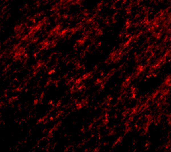
For brain IF, multiplex STK39 with a neuronal marker because neuronal cells in cerebral cortex show high tissue IHC staining (HPA tissue IHC: cerebral cortex). Choose a far-red STK39 channel when shorter-wavelength tissue autofluorescence obscures signal, and include single-label controls to assess bleed-through (standard IF practice). STK39 has no transmembrane segment and is annotated in the cytoplasm and nucleus, so permeabilise to access intracellular epitopes rather than assuming an extracellular epitope (UniProt Q9UEW8 topology and subcellular annotation). Optimise permeabilisation on matched sections and compare nuclear and cytosolic signal with the IHC pattern; IF localisation alone does not establish tissue IHC specificity (standard IF practice; HPA tissue IHC profile).
What should I check when STK39 chromogenic staining is diffuse?
Start with a no-primary control and inspect whether colour persists around vessels, damaged regions, or section edges (standard chromogenic IHC practice). Block endogenous peroxidase before HRP and DAB detection, and adjust protein blocking or antibody concentration if background persists (standard chromogenic IHC practice). The catalog caption reports human brain staining at 2.5 µg/mL, but gives no fixative or detection details, so treat that concentration as image context rather than a universal working concentration (catalog caption: A02516). Compare background with the expected mainly cytoplasmic, lesser nuclear cell-associated pattern before calling widespread haze STK39 positive (HPA tissue IHC profile; standard IHC practice).
How should I quantify STK39 across paraffin tissue sections? ⚠ ANSWER MARKED FOR VERIFICATION
Define the cell population and compartment before scoring because tissue IHC reports mainly cytoplasmic and lesser nuclear expression (HPA tissue IHC profile). For each compartment, record the percentage of positive cells and intensity categories, then calculate an H-score consistently across sections (standard IHC scoring practice). Normalise counts to the number of eligible cells, or report positive-cell density per mm² of viable tissue when cell segmentation is unreliable (standard IHC scoring practice). Keep section thickness, retrieval, imaging, and threshold settings consistent, and compare like cell types because STK39 staining differs across annotated tissues and cells (standard IHC practice; HPA tissue IHC profile).
How can I distinguish true STK39 signal from staining artefact?
A credible positive signal should follow identifiable cells and a plausible cytoplasmic or lesser nuclear distribution in tissue IHC (HPA tissue IHC profile). High staining in cerebral cortex neuronal cells or cerebellar granular-layer cells provides context, while skeletal-muscle myocytes are reported as not detected (HPA tissue IHC: cerebral cortex, cerebellum, skeletal muscle). Treat staining restricted to section edges, necrotic areas, or no-primary controls as suspect, and check endogenous peroxidase when DAB persists without primary antibody (standard chromogenic IHC practice). Nuclear staining warrants careful cell identification: UniProt notes nuclear localisation when STK39 is caspase-cleaved, but staining alone cannot establish cleavage (UniProt Q9UEW8 subcellular annotation; standard IHC interpretation).
Boster reagents

Best STK39 / STE20/SPS1-related proline-alanine-rich protein kinase IHC Antibodies

The catalog includes STK39 antibodies with human brain IHC images and an A431 cell IF image (catalog image captions); listed reactivity covers human, mouse, and rat (catalog reactivity).

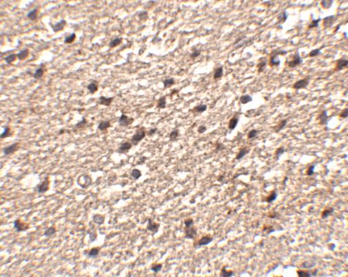
Real IHC data Immunohistochemistry of Stk39 in human brain tissue with Stk39 antibody at 2.5 μg/mL.
Anti-Stk39 Antibody
Cat # A02516
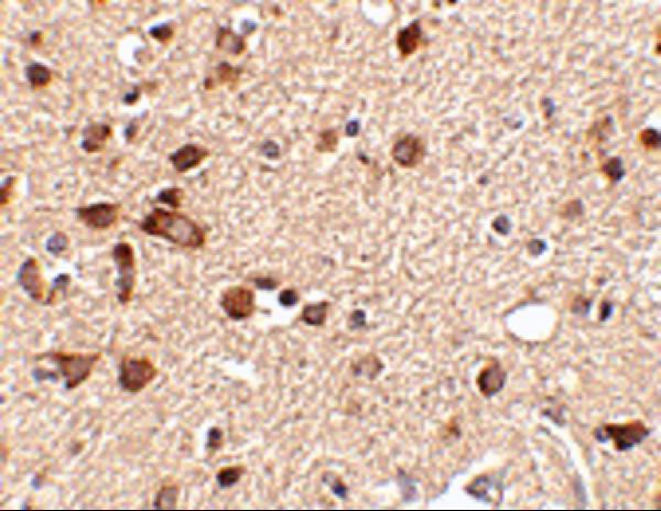
Real IHC data Immunohistochemistry of Stk39 in human brain tissue with Stk39 antibody at 2.5 μg/mL.
Anti-Stk39 Antibody
Cat # A02516-1
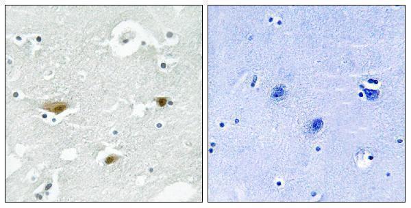
Real IHC data Immunohistochemistry analysis of paraffin-embedded human brain, using STK39 (Phospho-Ser325) Antibody. The picture on the right is blocked with the phospho peptide.
Anti-Phospho-SPAK (S325) STK39 Antibody
Cat # A02516S325
Real IF data IF analysis of SPAK/STK39 using anti-SPAK/STK39 antibody (A02516-3). SPAK/STK39 was detected in immunocytochemical section of A431 cells. Enzyme antigen retrieval was performed using IHC enzyme antigen retrieval reagent (AR0022) for 15 mins. The cells were blocked with 10% goat serum. And then incubated with 5μg/mL rabbit anti-SPAK/STK39 Antibody (A02516-3) overnight at 4°C. DyLight®488 Conjugated Goat Anti-Rabbit IgG (BA1127) was used as secondary antibody at 1:100 dilution and incubated for 30 minutes at 37°C. The section was counterstained with DAPI. Visualize using a fluorescence microscope and filter sets appropriate for the label used.
Anti-SPAK/STK39 Antibody ®
Cat # A02516-3

A02516 and A02516-1 have human brain IHC images at 2.5 μg/mL (catalog IHC image captions); A02516 also lists IF (catalog applications). A02516S325 has a phospho-Ser325 IHC image from paraffin-embedded human brain (catalog IHC image caption); A02516-3 has an IF/ICC image from A431 cells (catalog IF image caption).

Which to pick: For tissue IHC, choose A02516 or A02516-1 for human brain staining at 2.5 μg/mL (catalog IHC image captions), or A02516S325 for phospho-Ser325 staining in paraffin-embedded human brain (catalog IHC image caption); the captions do not report the fixative. For IF/ICC, choose A02516-3: its A431 cell image documents IF/ICC at 5 μg/mL (catalog IF image caption). For broader species coverage, A02516-1 lists human, mouse, and rat reactivity (catalog reactivity), but its IHC image documents human brain only (catalog IHC image caption); A02516S325 also lists all three species and is described as polyclonal (catalog reactivity and dilution data).

Each figure is that product's own IHC / IF validation image from its datasheet.

References

  1. UniProt Consortium. UniProt entry Q9UEW8 (STK39_HUMAN, STE20/SPS1-related proline-alanine-rich protein kinase).
  2. Human Protein Atlas. STK39 tissue IHC expression (reliability: Enhanced).
  3. Human Protein Atlas. STK39 subcellular location (ICC-IF): Mainly localized to the nucleoplasm. In addition localized to vesicles and cytosol..
  4. Human Protein Atlas. STK39 antibody validation summary (3 antibodies).
  5. STK39-mediated amplification of γ-H2A.X promotes homologous recombination and contributes to PARP inhibitor resistance. Nucleic acids research 2024 — PMC11662673.
  6. Role of high expression levels of STK39 in the growth, migration and invasion of non-small cell type lung cancer cells. Oncotarget 2016 — PMC5308657.
  7. STK39 is a novel kinase contributing to the progression of hepatocellular carcinoma by the PLK1/ERK signaling pathway. Theranostics 2021 — PMC7797677.
  8. MicroRNA‑299‑5p inhibits cell metastasis in breast cancer by directly targeting serine/threonine kinase 39. Oncology reports 2020 — PMC7057922.
  9. PubMed PMID:10980603 — UniProt-cited evidence.
  10. PubMed PMID:24722188 — UniProt-cited evidence.
  11. PubMed PMID:15815621 — UniProt-cited evidence.