TAF9 / Transcription initiation factor TFIID subunit 9 · IHC design guide

Design Immunohistochemistry for TAF9

Plan chromogenic TAF9 IHC-P around the nuclear staining reported in most tissues (HPA tissue IHC). Use colon glandular cells as a positive tissue reference (HPA tissue IHC) and 5 μg/mL as the catalog antibody's IHC starting concentration (datasheet).

Evidence assembled Oct 2026 · For research use; verify linked source records and product datasheet before use
Immunohistochemistry protocol sheet for TAF9 (IHC for TAF9): expected localisation Nuclear staining in most tissues (HPA tissue IHC), antibody A05319, validated IHC image, and IHC protocol steps
Printable TAF9 IHC protocol sheet — expected localisation Nuclear staining in most tissues (HPA tissue IHC), antibody A05319, controls and protocol steps. Open the full TAF9 IHC guide →

TAF9 Immunohistochemistry Experimental Design Guide

Expected localisation, validated protocols, controls and antibodies — the at-a-glance facts below, then the full design guide.

Must know before staining
Expected localisation Nuclear staining in most tissues (HPA tissue IHC)
Staining pattern Nuclear signal in most tissues; high in gut glandular cells (HPA tissue IHC)
Antigen retrieval Tris-EDTA pH 9.0 HIER, 95–98 °C, 20 min (rule: nuclear antigen)
Positive control ⓘ Colon+4 more · see all
Negative control ⓘ Adipose tissue+4 more · see all
Important caveats
Reasons your staining may differ from the expected pattern.
Fixation Matched tissue-IHC evidence does not establish the fixation claim. Validate the specimen-specific method before use. (selected-SKU IHC image A05319)
Caveat Staining has medium agreement with RNA expression (HPA tissue IHC)
Regulation No specific expression regulator annotated (UniProt)
Isoform / epitope 0 annotated isoforms; one 1–264 chain (UniProt)
Section 1

Recommended TAF9 IHC & IF Protocols

The catalog antibody’s IHC-P protocol is followed by one published TAF9 xenograft IHC protocol (PMC12372275).

Recommended immunohistochemistry (IHC-P) protocol parameters
SampleTissue sections; selected-image fixative not specified (standard IHC workflow)
FixationImage fixative and duration unreported (datasheet A05319); verify before use.
Sectioning4–5 µm sections on charged slides (standard)
DeparaffinisationXylene, graded ethanol series to water (standard)
Antigen retrievalHeat-induced epitope retrieval in Tris-EDTA buffer, pH 9.0, 20 min at 95–98 °C (standard rule: nuclear antigen)
Peroxidase block3% H2O2, 10 min, room temperature (standard)
Blocking10% normal serum of the secondary host, 30 min, room temperature (standard)
Primary antibodyRabbit anti-TAF9, 5 μg/mL (datasheet A05319)
Primary incubationOvernight at 4 °C (standard)
DetectionHRP-polymer secondary, DAB chromogen 5–10 min (standard)
CounterstainHematoxylin, blue, dehydrate and mount (standard)
Expected resultTAF9-positive staining in glandular cells of colon (HPA tissue IHC: High). HPA tissue profile: Nuclear expression in most tissues. No signal in the no-primary control.
💡Decision noteStart with Tris-EDTA pH 9.0 HIER at 95–98 °C for 20 min for nuclear TAF9 (UniProt Q16594; page retrieval rule); compare citrate pH 6 (PMC12372275).
Section 2

What Is the Expected TAF9 Staining Pattern?

TAF9 is a nuclear transcription factor component with no transmembrane segment (UniProt Q16594: subcellular location, function, topology). In paraffin sections, expect nuclear staining across many tissues, with high staining reported in colon, duodenum, rectum and small-intestine glandular cells and placental decidual cells (HPA: tissue IHC). HPA rates the tissue profile Approved, with medium consistency between antibody staining and RNA expression (HPA: tissue IHC reliability).

What am I looking at on my slide?
Distinct nuclear staining in intestinal glandular cells or placental decidual cells, with little surrounding background.This matches the reported compartment and high-staining cell populations (UniProt Q16594: nucleus; HPA: high in colon, duodenum, rectum and small-intestine glandular cells and placental decidual cells). Compare cells within the same section before treating intensity differences as biological; HPA reports low tissue RNA specificity and only medium staining–RNA consistency (HPA: tissue IHC).
Predominantly cytoplasmic, membranous or extracellular signal instead of a nuclear pattern.These compartments conflict with the annotated nuclear location and lack of a transmembrane segment (UniProt Q16594: subcellular location, topology). Treat the result as suspect and check controls and detection background before assigning it to TAF9 (general IHC practice).
Strong staining in a cell population reported as not detected, such as adipocytes or cardiomyocytes.This disagrees with those HPA tissue observations (HPA: adipocytes and cardiomyocytes not detected). Cross-reactivity or endogenous detection activity are possibilities, not proven explanations; compare a known-positive section and a no-primary control processed alongside it (general IHC practice).
Diffuse colour over nuclei, cytoplasm and empty tissue spaces, with poor cellular boundaries.The distribution cannot be scored confidently as nuclear TAF9 (UniProt Q16594: nucleus). Check the no-primary control, blocking, washes and detection steps for nonspecific background (general IHC practice). Do not interpret an overall darker slide as higher TAF9 expression without resolvable nuclear signal.
No nuclear signal in a section expected to show high staining, such as colon glandular cells.A blank result conflicts with the reported high staining in that cell population (HPA: colon glandular cells High). First check tissue preservation, the IHC-validated antibody's documented IHC-P conditions and detection controls (general IHC practice). A single negative section does not establish absence of TAF9.
💡Expected TAF9 appearanceCall a result positive when identifiable nuclei stain in the expected cells, especially high-staining intestinal glandular or placental decidual cells (HPA: tissue IHC; UniProt Q16594: nucleus); isolated cytoplasmic or diffuse background colour is a suspect positive (general IHC practice).
How each factor affects the staining
Tissue and cell selectionUse a reported high-staining population to judge whether the assay can reveal nuclear signal: intestinal glandular cells or placental decidual cells (HPA: tissue IHC). A reported not-detected population is a comparison, not proof that every cell in that tissue must be blank (HPA: tissue IHC).
Compartment and tissue caveatThe expected compartment is nuclear (UniProt Q16594: subcellular location; HPA: nuclear expression in most tissues). HPA also lists medium staining in cerebral-cortex neuropil (HPA: tissue IHC); that observation alone does not establish a general cytoplasmic pattern.
IHC evidence strengthThe tissue profile is Approved, with medium consistency between antibody staining and RNA expression (HPA: tissue IHC reliability). HPA072658 is Approved for IHC (HPA: antibody validation). Use the observed pattern as a reference while evaluating controls and individual sections.
IF/ICC cross-check?For an IF/ICC localisation question, HPA reports enhanced nucleoplasmic localisation and ICC validation for HPA028930 and HPA053429 (HPA: subcellular ICC-IF; antibody validation). This supports a nuclear expectation; IF/ICC preparation and detection belong in the separate guide.
Why is my staining missing, weak or wrong?
SituationLikely causeNext action
Known-positive glandular cells have no visible nuclear signal.The assay may have failed, or the sampled section may not reproduce the reference pattern; high staining is reported in colon glandular cells (HPA: tissue IHC).Run a known-positive section alongside the sample and review the catalog antibody's IHC-P instructions, reagent application and detection controls (general IHC practice).
Nuclei stain, but the whole section is hazy.Background can obscure cellular localisation (general IHC practice); the intended pattern is nuclear (UniProt Q16594: nucleus).Compare with a no-primary control, then review blocking, washes and chromogen development before scoring nuclear intensity (general IHC practice).
Colour is strongest outside nuclei.The pattern conflicts with nuclear localisation and no transmembrane segment (UniProt Q16594: subcellular location, topology).Check whether the signal persists in a no-primary control and whether a known-positive section yields distinct nuclear staining (general IHC practice).
Adipocytes or cardiomyocytes show strong staining.Both cell populations are reported as not detected (HPA: tissue IHC); nonspecific binding or endogenous detection activity may explain a mismatch (general IHC practice).Inspect localisation and the no-primary control, then compare with a reported high-staining cell population before calling the result TAF9 (general IHC practice; HPA: tissue IHC).
A low-staining tissue appears negative while the positive control stains.Low staining may approach the assay's visual detection limit (general IHC practice); HPA reports low staining in thyroid glandular cells, among others (HPA: tissue IHC).Report the sampled cells and assay conditions, and avoid extending that negative observation to all tissues (general IHC practice; HPA: nuclear expression in most tissues).
IHC and IF/ICC appear to disagree on localisation.HPA reports nuclear tissue expression and enhanced nucleoplasmic ICC-IF localisation (HPA: tissue IHC; subcellular ICC-IF). A discordant image needs method-specific control review (general IHC/IF practice).Evaluate each method with its own controls and score whether signal resolves within nuclei; use the separate IF/ICC guide for that method's workflow (general IHC/IF practice).

Sample controls for TAF9 IHC & IF

🧪Run colon first: glandular cells should show TAF9 staining (HPA: High in colon glandular cells). Use adipose tissue as the negative tissue, focusing on adipocytes (HPA: Not detected in adipocytes); the colon rows do not identify a confirmed internal negative cell type, so assess any cells lacking nuclear signal as background references only (HPA: colon glandular-cell row; UniProt Q16594: nucleus).
Positive control tissue: Colon (Glandular cells, HPA High)
Negative control tissue: Adipose tissue (HPA Not detected)
ICC-IF cell lines (HPA subcellular resource): HPA ICC-IF images show TAF9 in A-549, SK-MEL-30, U2OS, MCF-7, U-251MG, A-431, PC-3, with annotated localisation: Nucleoplasm (enhanced) (HPA subcellular).
Technical controls: Include a no-primary (secondary-only) control, an isotype control matched to the primary antibody’s host and clonality, and a biological specificity control using TAF9 depletion or a cognate peptide block if available (standard IHC practice). For chromogenic colon IHC, block endogenous peroxidase and, if using biotin-based detection, assess endogenous biotin background (standard IHC practice).
⚠️Feasibility: A target-specific fixation window or fixation effect is unreported, and the A05319 mouse-liver IHC caption does not state its fixative (supplied application evidence; selected-SKU caption: fixative not stated). Antigen-retrieval dependence is unreported; optimize retrieval against the control slides (supplied application evidence; standard IHC practice). The supplied evidence does not establish whether frozen sections or IF are easier; for colon chromogenic sections, check inflammatory-cell peroxidase background when interpreting staining (supplied application evidence; standard IHC practice).

HPA tissue IHC evidence for TAF9

Comprehensive Human Protein Atlas IHC scoring per tissue (reliability: Approved — Medium consistency between antibody staining and RNA expression data.). Rows are taken directly from the HPA tissue chart — click any row's HPA link to view the source.

Positive expression · recommended positive controls

TissueCell typeLevelEvidenceSource
Colon Glandular cells High Protein (IHC) HPA →
Duodenum Glandular cells High Protein (IHC) HPA →
Placenta Decidual cells High Protein (IHC) HPA →
Rectum Glandular cells High Protein (IHC) HPA →
Small intestine Glandular cells High Protein (IHC) HPA →

Undetected expression · recommended negative controls

TissueCell typeLevelEvidenceSource
Adipose tissue Adipocytes Not detected Protein (IHC) HPA →
Adrenal gland Glandular cells Not detected Protein (IHC) HPA →
Bone marrow Hematopoietic cells Not detected Protein (IHC) HPA →
Caudate Glial cells Not detected Protein (IHC) HPA →
Cerebellum Cells in granular layer Not detected Protein (IHC) HPA →
Section 3

Advanced TAF9 IHC Tips

Troubleshoot paraffin-section TAF9 IHC by checking nuclear staining, section processing, controls and cell-specific scoring (UniProt Q16594; HPA tissue IHC).

How should I adjust retrieval when nuclear TAF9 staining is weak?
Use Tris-EDTA pH 9.0 HIER at 95–98 °C for 20 min for paraffin-section TAF9 IHC (page retrieval rule: nuclear antigen). If nuclear staining is weak, compare adjacent sections processed with the same fixation, antibody concentration and detection settings before changing retrieval (standard IHC practice). Check for nuclear staining in colon glandular cells as a reference, while recognizing that the HPA tissue-IHC assessment reports medium consistency with RNA expression (HPA: High in colon glandular cells; HPA: Approved). If retrieval changes are needed, test a shorter heating time on matched sections and assess tissue damage alongside staining (standard IHC practice).
Could fixation explain weak TAF9 staining in paraffin sections?
Target-specific fixation sensitivity is unknown from the supplied evidence; the A05319 mouse liver IHC caption does not state a fixative (A05319 tissue-IHC caption). Record the fixative, fixation duration and processing schedule for each specimen before comparing staining across blocks (standard IHC practice). Test matched sections with identical retrieval and detection conditions, then compare nuclear signal with tissue morphology and the no-primary control (standard IHC practice; UniProt Q16594: nucleus). Avoid assigning weak staining to fixation alone when section thickness, antibody concentration or chromogen development also differ between runs (standard IHC practice). Treat an observed fixation effect as specific to the tested specimens and workflow until it is reproduced (standard IHC practice).
What staining pattern should count as TAF9 in tissue IHC?
Score predominantly nuclear staining because TAF9 is annotated in the nucleus and HPA reports enhanced nucleoplasmic localisation (UniProt Q16594: nucleus; HPA subcellular: nucleoplasm). Compare signal within intact nuclei against nearby cells and the hematoxylin counterstain, using the same viewing and scoring conditions across sections (standard IHC practice). HPA reports nuclear expression in most tissues, with high staining in colon and duodenal glandular cells (HPA tissue IHC). Strong diffuse cytoplasmic or membranous colour without convincing nuclear enrichment warrants a specificity and background check, since TAF9 has no annotated transmembrane segment (UniProt Q16594 topology; standard IHC practice).
How can epitope accessibility affect TAF9 IHC interpretation?
The supplied UniProt record lists 0 isoforms and a 1–264 chain, so an isoform-specific staining explanation is unsupported here (UniProt Q16594). It also lists acetyllysine at residue 5 and phosphoserines at 149, 152, 155 and 158 (UniProt Q16594: modified residues). Their effect on antibody binding cannot be assigned without the catalog antibody’s epitope and validation data (standard IHC interpretation). If staining changes after retrieval, compare matched sections and antibody controls before attributing the change to a modification or inaccessible epitope (standard IHC practice). Report the antibody identifier and retrieval conditions so the observation can be reproduced (standard IHC practice).
How should I follow up an IHC pattern with multiplex IF?
Use IF as a separate validation experiment and check whether TAF9 signal overlaps nuclei, consistent with its nucleoplasmic localisation (HPA subcellular: nucleoplasm). In colon sections, multiplex with a glandular epithelial marker to identify the expected cell population, since HPA reports high TAF9 staining in colon glandular cells (HPA tissue IHC; standard IF practice). Choose spectrally separated fluorophores and consider a far-red channel if tissue autofluorescence obscures shorter-wavelength signal (standard IF practice). Apply a controlled permeabilisation step appropriate for access to a nuclear epitope; TAF9 has no annotated transmembrane segment (UniProt Q16594 topology; standard IF practice). Include single-stain and no-primary controls to assess bleed-through and background (standard IF practice).
How can I separate TAF9 nuclear signal from chromogenic background?
Inspect no-primary and secondary-only controls for nonspecific colour, and compare them with nuclear staining in the test section (standard IHC practice; UniProt Q16594: nucleus). Include a peroxidase block before DAB development when using a peroxidase detection system, and keep development time consistent across samples (standard chromogenic IHC practice). If colour follows section edges, folds or damaged areas, assess tissue morphology before scoring those regions (standard IHC practice). Titrate the catalog antibody and detection reagents on matched sections, retaining the condition that gives clear nuclear signal with acceptable background (standard IHC practice). The A05319 mouse liver caption reports 5 µg/mL, but does not specify fixation (A05319 tissue-IHC caption).
What is a defensible way to quantify TAF9 IHC? ⚠ ANSWER MARKED FOR VERIFICATION
Define the cell population and score nuclear staining, consistent with the reported TAF9 localisation (UniProt Q16594: nucleus; HPA subcellular: nucleoplasm). Record the percentage of positive nuclei and an intensity category, then calculate a nuclear H-score if those categories are prespecified (standard IHC quantification practice). Normalise positive-cell counts to the total eligible nuclei in the same annotated region, or report positive nuclei per mm² when tissue area is the denominator (standard IHC quantification practice). Exclude folds, edges and necrotic areas using the same rule for every section, and keep retrieval, imaging and threshold settings consistent (standard IHC practice). Report cell type because HPA staining levels differ across tissue cell populations (HPA tissue IHC).
When is apparent TAF9 positivity likely to be an artefact?
A credible result shows nuclear enrichment in intact cells, consistent with UniProt nuclear localisation and HPA nucleoplasmic staining (UniProt Q16594; HPA subcellular). Compare the cell population with the reference pattern: HPA reports high staining in colon glandular cells but no detection in adipocytes (HPA tissue IHC). Diffuse cytoplasmic colour, edge-restricted staining and colour concentrated in necrotic tissue require review of morphology and controls before interpretation (standard IHC practice; UniProt Q16594: nucleus). Check no-primary and peroxidase-block controls when DAB colour could reflect nonspecific detection or endogenous enzyme activity (standard chromogenic IHC practice). HPA’s Approved rating has medium consistency with RNA expression, so treat staining as supporting evidence rather than proof of identity (HPA tissue IHC).
Boster reagents

Best TAF9 / Transcription initiation factor TFIID subunit 9 IHC Antibodies

A05319 has IHC and IF images from mouse liver tissue (catalog image captions). Human, mouse, and rat are listed as reactive species (catalog reactivity).

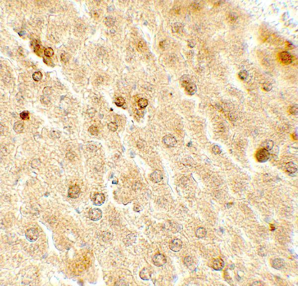
Real IHC data Immunohistochemistry of TAF9 in mouse liver tissue with TAF9 antibody at 5 μg/mL.
Anti-TAF9 Antibody
Cat # A05319

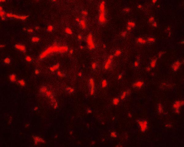
A05319 shows IHC staining in mouse liver tissue at 5 μg/mL (A05319 IHC image caption). The same SKU shows IF staining in mouse liver tissue at 20 μg/mL (A05319 IF image caption).

Which to pick: For paraffin-section tissue IHC, pick A05319: IHC-P is listed, and its own IHC image shows mouse liver tissue at 5 μg/mL (catalog applications; A05319 IHC image caption). For IF, A05319 has a mouse liver image; ICC-specific validation is unreported (A05319 IF image caption; catalog applications). For cross-species planning, A05319 is a rabbit antibody listed as reactive with human, mouse, and rat; no clone or IHC fixative is reported (catalog host, clone, and reactivity; A05319 IHC image caption).

Each figure is that product's own IHC / IF validation image from its datasheet.

References

  1. UniProt Consortium. UniProt entry Q16594 (TAF9_HUMAN, Transcription initiation factor TFIID subunit 9).
  2. Human Protein Atlas. TAF9 tissue IHC expression (reliability: Approved).
  3. Human Protein Atlas. TAF9 subcellular location (ICC-IF): Localized to the nucleoplasm..
  4. Human Protein Atlas. TAF9 antibody validation summary (4 antibodies).
  5. Activation of TAF9 via Danshensu-Induced Upregulation of HDAC1 Expression Alleviates Non-alcoholic Fatty Liver Disease. Frontiers in pharmacology 2021 — PMC8678612.
  6. LncRNA938/ TAF9/TTK axis promotes EMT and serves as a therapeutic target in hepatoblastoma. Journal of translational medicine 2025 — PMC12372275.
  7. hsa_circ_0077837 Alleviated the Malignancy of Non-Small Cell Lung Cancer by Regulating the miR-1178-3p/APITD1 Axis. Journal of oncology 2022 — PMC8926487.
  8. PubMed PMID:7761466 — UniProt-cited evidence.
  9. PubMed PMID:7597030 — UniProt-cited evidence.
  10. PubMed PMID:7667268 — UniProt-cited evidence.