This website uses cookies to ensure you get the best experience on our website.


- Table of Contents
Plan TAP1 paraffin-section IHC using its observed granular cytoplasmic pattern (HPA tissue IHC) and ER membrane location (UniProt). This guide covers fixation, staining controls and cell-type-aware scoring (standard IHC practice).

Expected localisation, validated protocols, controls and antibodies — the at-a-glance facts below, then the full design guide.
| Expected localisation | Granular cytoplasm (HPA tissue IHC); ER membrane (UniProt) | |
| Staining pattern | Granular cytoplasmic staining across multiple cell types (HPA tissue IHC) | |
| Antigen retrieval | EDTA pH 8.0 HIER, heat-mediated (datasheet PB9823) | |
| Positive control | Adrenal gland+4 more · see all | |
| Negative control | None in HPA (detected in all 45 tissues); use no-primary + isotype controls |
| Fixation | Keep paraffin-section fixation consistent (standard IHC practice; not target-specific) | |
| Caveat | High hematopoietic-cell staining may complicate scoring (HPA tissue IHC) | |
| Regulation | Expression regulation not specified (UniProt) | |
| Isoform / epitope | 2 isoforms; epitope coverage is unspecified (UniProt) |
The catalog antibody protocol uses EDTA pH 8.0 retrieval (datasheet: PB9823). The two published IHC protocols below provide tissue-specific staining conditions (PMC5706630; PMC7171417).
| Sample | Paraffin-embedded human intestinal cancer tissue; fixative not specified (datasheet PB9823) |
| Fixation | Image fixative and duration unreported (datasheet PB9823); verify before use. |
| Sectioning | 4–5 µm sections on charged slides (standard) |
| Deparaffinisation | Xylene, graded ethanol series to water (standard) |
| Antigen retrieval | Heat retrieval: EDTA pH 8.0 (datasheet PB9823); 20 min, 95–100 °C (standard) |
| Peroxidase block | 3% H2O2, 10 min, room temperature (standard) |
| Blocking | 10% goat serum (datasheet PB9823) |
| Primary antibody | Rabbit anti-TAP1, 0.5-1μg/ml (datasheet PB9823) |
| Primary incubation | Overnight at 4 °C (datasheet PB9823) |
| Detection | Streptavidin-biotin complex (SABC), DAB chromogen (datasheet PB9823) |
| Counterstain | Hematoxylin, blue, dehydrate and mount (standard) |
| Expected result | TAP1-positive staining in glandular cells of adrenal gland (HPA tissue IHC: High). HPA tissue profile: General cytoplasmic expression with a granular pattern. No signal in the no-primary control. |
TAP1 is an endoplasmic reticulum (ER) membrane protein with 10 transmembrane segments (UniProt Q03518 topology). In paraffin-section IHC, expect granular cytoplasmic staining (HPA tissue IHC: general cytoplasmic granular pattern). Interpret signal in the context of the cells present: UniProt reports high expression in professional antigen-presenting cells and lymphocyte subsets, while HPA reports high staining in several epithelial, glandular, endothelial, and hematopoietic cell populations (UniProt Q03518 tissue specificity; HPA tissue IHC: High). HPA rates the tissue IHC profile Approved, with medium consistency between staining and RNA data (HPA tissue IHC: reliability).
| Granular cytoplasmic staining in glandular cells or marrow hematopoietic cells, with recognizable tissue structure. | This fits the reported tissue pattern and an ER-associated target (HPA tissue IHC: general cytoplasmic granular pattern; HPA tissue IHC: High in glandular and bone marrow hematopoietic cells; UniProt Q03518 subcellular location). Compare cells within the same section before assigning a score: HPA reports low tissue specificity at the RNA level, so staining need not be confined to one lineage (HPA tissue IHC: RNA specificity). |
| Predominantly nuclear, extracellular, or sharply cell-surface staining without a granular cytoplasmic component. | Treat this as a compartment mismatch requiring verification: the reported IHC pattern is cytoplasmic and granular, and UniProt places TAP1 in the ER membrane (HPA tissue IHC: profile; UniProt Q03518 subcellular location). Check the antibody's IHC validation and the slide controls before calling the pattern TAP1-specific (HPA antibodies: IHC Approved for CAB009516; general IHC practice). |
| Strong colour appears mainly in a cell population unexpected for the chosen tissue, while expected cells remain unstained. | Consider cross-reactivity or endogenous detection activity as possible causes, then compare a reagent control and tissue morphology (general IHC practice). Do not classify a cell as wrong solely because it is absent from HPA's short positive list: HPA reports low tissue RNA specificity and lists no negative cell populations here (HPA tissue IHC: RNA specificity; HPA tissue IHC: negative list). |
| A diffuse haze covers cells and surrounding tissue, obscuring granular detail. | Score the field cautiously: haze does not reproduce the reported granular cytoplasmic profile (HPA tissue IHC: profile). Review blocking, washing, antibody concentration, and chromogen development with appropriate controls; these are general IHC background checks, not documented TAP1-specific effects (general IHC practice). |
| No detectable staining in a section expected to contain HPA high-staining cells. | First confirm that the expected cells are actually present and that the run's positive control developed (general IHC practice). HPA reports high staining in adrenal gland glandular cells and bone marrow hematopoietic cells, among other examples; a blank result there merits review of antibody suitability and the IHC workflow, but does not by itself prove TAP1 absence (HPA tissue IHC: High; HPA antibodies: IHC Approved for CAB009516). |
| ER location and membrane topology | TAP1 occupies the ER membrane and has 10 transmembrane segments (UniProt Q03518 subcellular location and topology). Use that location to interpret a granular cytoplasmic pattern; topology alone does not identify which part a particular antibody recognizes (HPA tissue IHC: profile; UniProt Q03518 topology). |
| Cell populations represented on the section | HPA reports High staining in glandular cells of adrenal gland, appendix, breast, and duodenum; bronchial respiratory epithelial cells; colonic endothelial cells; cerebellar Purkinje cells; and bone marrow hematopoietic cells (HPA tissue IHC: High). UniProt additionally reports high expression in monocytes, dendritic cells, and T, B, and NK lymphocyte subsets (UniProt Q03518 tissue specificity). |
| Strength of the supplied validation | The tissue IHC profile is Approved with medium staining-to-RNA consistency, and CAB009516 has an IHC Approved designation (HPA tissue IHC: reliability; HPA antibodies: CAB009516). These designations support using the reported pattern as a comparison, while still requiring slide-level controls for a new experiment (general IHC practice). |
| Isoforms and processing | UniProt lists 2 isoforms, a chain spanning residues 1–748, and no annotated signal peptide, propeptide, or glycosylation sites (UniProt Q03518 isoforms and processing). Those annotations do not establish differential IHC staining, shedding, or an antibody's isoform coverage without its mapped epitope (UniProt Q03518 annotations). |
| IF/ICC Q&A: what localisation should an image show? | An ER pattern is supported; centriolar satellite localisation is uncertain (HPA subcellular ICC-IF: main location). HPA lists HaCaT and SK-MEL-30 ICC-IF images and rates HPA072354 ICC Supported (HPA subcellular ICC-IF: image lines; HPA antibodies: HPA072354). Interpret IF/ICC using its separate guide rather than transferring an IHC slide score to an image (general microscopy practice). |
| Situation | Likely cause | Next action |
|---|---|---|
| The positive tissue and run control are both blank. | A shared staining-run problem is plausible when independent expected signals fail together (general IHC practice). | Check reagent sequence, detection and chromogen performance, and the run control before interpreting the sample; then repeat with the catalog antibody's IHC-P instructions (general IHC practice). |
| The run control stains, but expected cells in the sample are blank. | The sampled area may lack the expected cells, or staining may be below detection in this preparation; a blank area alone does not establish absent TAP1 (general IHC practice). | Locate the relevant cells on the counterstained section, compare an HPA high-staining reference population, and review the antibody's IHC-P conditions (HPA tissue IHC: High; general IHC practice). |
| Granular signal is weak or uneven across a section. | Target-specific fixation effects are not established by the supplied assay evidence. Verify with a matched IHC source before attributing a result to fixation. | Inspect section integrity and run controls, then verify the catalog IHC-P retrieval and detection steps against its instructions (general IHC practice). |
| All tissue compartments show brown haze. | Nonspecific reagent background or excessive detection development can obscure the reported granular pattern (general IHC practice; HPA tissue IHC: profile). | Compare reagent controls, review blocking and washes, and adjust development using the validated IHC-P workflow (general IHC practice). |
| A surprising cell population stains intensely. | Cross-reactivity or endogenous detection activity is possible, but HPA's low tissue RNA specificity makes an unlisted cell type insufficient evidence of error (general IHC practice; HPA tissue IHC: RNA specificity). | Check cell identity and compartment, inspect reagent controls, and seek concordance with an independently validated antibody if the result is consequential (general IHC practice). |
| Nuclear or extracellular colour dominates the slide. | That distribution conflicts with TAP1's ER membrane location and HPA's granular cytoplasmic IHC profile (UniProt Q03518 subcellular location; HPA tissue IHC: profile). | Review slide morphology and controls, confirm the antibody's IHC validation, and withhold a TAP1-positive call until a compatible cytoplasmic pattern is demonstrated (HPA antibodies: CAB009516 IHC Approved; general IHC practice). |

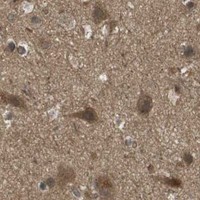
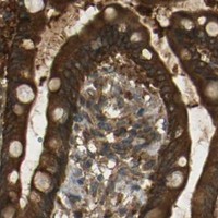
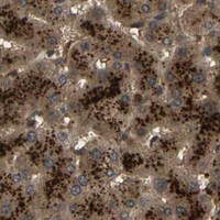
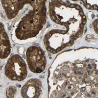


Comprehensive Human Protein Atlas IHC scoring per tissue (reliability: Approved — Medium consistency between antibody staining and RNA expression data.). Rows are taken directly from the HPA tissue chart — click any row's HPA link to view the source.
| Tissue | Cell type | Level | Evidence | Source |
|---|---|---|---|---|
| Adrenal gland | Glandular cells | High | Protein (IHC) | HPA → |
| Appendix | Glandular cells | High | Protein (IHC) | HPA → |
| Bone marrow | Hematopoietic cells | High | Protein (IHC) | HPA → |
| Breast | Glandular cells | High | Protein (IHC) | HPA → |
| Bronchus | Respiratory epithelial cells | High | Protein (IHC) | HPA → |
| Tissue | Cell type | Level | Evidence | Source |
|---|---|---|---|---|
| None in HPA: TAP1 is detected in all 45 scored tissues. Use a no-primary (secondary-only) and an isotype control instead. | ||||
Troubleshoot TAP1 staining in paraffin sections by checking retrieval, cell identity, and the expected endoplasmic reticulum pattern before scoring chromogenic signal.
The catalog includes one human-reactive anti-TAP1 antibody with an IHC figure from a paraffin-embedded human intestinal cancer section; no IF figure is supplied (catalog: PB9823; IHC image caption).
PB9823 is listed for human IHC and has an image showing staining in a paraffin-embedded human intestinal cancer section (catalog: applications and reactivity; IHC image caption). No IF validation image is supplied for PB9823 (catalog: IF images).
Which to pick: Choose PB9823 for human tissue IHC in paraffin sections; its own caption documents that preparation, while the fixative is unreported (catalog: reactivity; IHC image caption). No SKU in the payload is listed for IF/ICC, so there is no supported IF/ICC pick (catalog: applications). PB9823 is listed as human-reactive only, so there is no supported cross-species pick (catalog: reactivity).