TBC1D4 / TBC1 domain family member 4 · IHC design guide

Design Immunohistochemistry for TBC1D4

This guide helps plan chromogenic TBC1D4 IHC in paraffin sections using the catalog antibody at 2–5 μg/mL (datasheet A02004-3). Expect cytoplasmic staining in many tissues, and consider colon glandular cells and skeletal myocytes as contrasting controls (HPA tissue IHC).

Evidence assembled Oct 2026 · For research use; verify linked source records and product datasheet before use
Immunohistochemistry protocol sheet for TBC1D4 (IHC for TBC1D4): expected localisation Cytoplasmic staining in most tissues (HPA tissue IHC), antibody A02004-3, validated IHC image, and IHC protocol steps
Printable TBC1D4 IHC protocol sheet — expected localisation Cytoplasmic staining in most tissues (HPA tissue IHC), antibody A02004-3, controls and protocol steps. Open the full TBC1D4 IHC guide →

TBC1D4 Immunohistochemistry Experimental Design Guide

Expected localisation, validated protocols, controls and antibodies — the at-a-glance facts below, then the full design guide.

Must know before staining
Expected localisation Cytoplasmic staining in most tissues (HPA tissue IHC)
Staining pattern Cytoplasmic signal in glandular and neuronal cells (HPA tissue IHC)
Antigen retrieval EDTA pH 8.0 HIER, heat-mediated (datasheet A02004-3)
Positive control ⓘ Adrenal gland+4 more · see all
Negative control ⓘ Adipose tissue+3 more · see all
Important caveats
Reasons your staining may differ from the expected pattern.
Fixation Matched tissue-IHC evidence does not establish the fixation claim. Validate the specimen-specific method before use. (selected-SKU IHC image A02004-3)
Caveat Skeletal muscle RNA enrichment contrasts with absent myocyte staining (HPA tissue IHC)
Regulation Lower islet levels in type 2 diabetes (UniProt)
Isoform / epitope 5 isoforms; check antibody epitope coverage (UniProt)
Section 1

Recommended TBC1D4 IHC & IF Protocols

The catalog antibody protocol uses EDTA pH 8.0 heat retrieval (datasheet: A02004-3). One published tumor tissue IHC protocol provides a comparison (PMC13612446).

Recommended immunohistochemistry (IHC-P) protocol parameters
SampleParaffin-embedded human gallbladder adenocarcinoma tissue; fixative not specified (datasheet A02004-3)
FixationImage fixative and duration unreported (datasheet A02004-3); verify before use.
Sectioning4–5 µm sections on charged slides (standard)
DeparaffinisationXylene, graded ethanol series to water (standard)
Antigen retrievalHeat retrieval: EDTA pH 8.0 (datasheet A02004-3); 20 min, 95–100 °C (standard)
Peroxidase block3% H2O2, 10 min, room temperature (standard)
Blocking10% goat serum (datasheet A02004-3)
Primary antibodyRabbit anti-TBC1D4, 2-5μg/ml (datasheet A02004-3)
Primary incubationOvernight at 4 °C (datasheet A02004-3)
DetectionStreptavidin-biotin complex (SABC), DAB chromogen (datasheet A02004-3)
CounterstainHematoxylin, blue, dehydrate and mount (standard)
Expected resultTBC1D4-positive staining in glandular cells of adrenal gland (HPA tissue IHC: High). HPA tissue profile: Cytoplasmic expression in most tissues. No signal in the no-primary control.
💡Decision noteStart with EDTA pH 8.0 heat retrieval for the catalog antibody (datasheet: A02004-3); the published protocol specifies retrieval reagent H-3300 without stating its pH (PMC13612446).
Section 2

What Is the Expected TBC1D4 Staining Pattern?

TBC1D4 is cytoplasmic and has no transmembrane segment (UniProt O60343). In paraffin sections, expect cytoplasmic staining in selected glandular cells and neurons, with intensity varying by tissue (HPA: tissue IHC). HPA describes cytoplasmic expression in most tissues, but rates its tissue IHC data Approved with low agreement between staining and RNA expression, pending external verification (HPA: tissue IHC reliability).

What am I looking at on my slide?
Cytoplasmic staining in adrenal or colon glandular cells, or cerebral cortex neurons (HPA: High in these cells).This matches the reported cell types and compartment (HPA: tissue IHC; UniProt O60343: cytoplasm). Compare intensity within the same section before scoring: HPA reports tissue patterns, not a universal intensity threshold (HPA: tissue IHC).
Predominantly nuclear or sharply membrane-restricted signal in otherwise positive cells.Treat this as discordant with the reported cytoplasmic pattern (HPA: tissue IHC; UniProt O60343: cytoplasm, no transmembrane segment). Review morphology and detection controls; the pattern alone cannot identify its cause (general IHC practice).
Strong signal in adipocytes or parathyroid glandular cells.HPA reports TBC1D4 as not detected in those cells (HPA: tissue IHC). Check for nonspecific binding or endogenous detection activity before calling the signal TBC1D4; a negative HPA observation does not prove biological absence (general IHC practice; HPA: Approved, pending external verification).
Uniform color over cells and surrounding section, obscuring cell boundaries.This is poor contrast for interpreting a cytoplasmic target (UniProt O60343: cytoplasm). Review blocking, washes and detection controls as general IHC checks; diffuse background cannot establish which cell types express TBC1D4 (general IHC practice).
No cytoplasmic signal in adrenal glandular cells or cerebral cortex neurons.These are reported High IHC contexts (HPA: tissue IHC). Check section integrity and the IHC-validated antibody's documented conditions before interpreting a negative result; HPA's Approved rating and low RNA agreement warrant restraint (HPA: tissue IHC reliability; general IHC practice).
💡Expected TBC1D4 appearanceCall a section positive when selected glandular cells or neurons show discernible cytoplasmic staining, potentially High in the listed tissues (HPA: tissue IHC); isolated nuclear, membrane-restricted or cell-free color is discordant with the expected pattern (UniProt O60343: cytoplasm, no transmembrane segment; general IHC practice).
How each factor affects the staining
Cell and tissue contextHPA reports High staining in adrenal, appendix, breast, colon, duodenal and epididymal glandular cells; cerebral cortex neurons; and fallopian tube ciliated cell bodies (HPA: tissue IHC). Choose a documented positive cell population when judging assay performance.
Protein versus RNA and isoformsSkeletal myocytes are not detected by HPA IHC despite skeletal muscle enhanced RNA (HPA: tissue IHC). UniProt reports isoform 1 highly expressed in skeletal muscle and heart (UniProt O60343: tissue specificity). These observations do not establish why the assays differ.
Compartment and protein structureTBC1D4 is cytoplasmic, lacks a transmembrane segment and is annotated as one chain of residues 1–1298, with no signal peptide or propeptide (UniProt O60343: subcellular location, topology, processing). These annotations support a cytoplasmic readout, not a claim about epitope exposure after fixation.
Strength of tissue evidenceHPA labels tissue IHC Approved while describing low agreement with RNA expression and pending external verification (HPA: tissue IHC reliability). Treat its High and not-detected entries as reported staining observations, especially when selecting positive and comparison tissues.
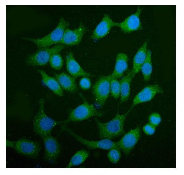
IF/ICC Q: What should a companion cell image show?A cytosolic pattern is consistent with HPA's enhanced ICC-IF localization and UniProt's cytoplasmic annotation (HPA: subcellular ICC-IF; UniProt O60343). UniProt also notes perinuclear cytoplasm for isoform 2 in a myoblastic cell line; that observation does not set an IHC requirement (UniProt O60343).
Why is my staining missing, weak or wrong?
SituationLikely causeNext action
Known-positive glandular cells are unstained.The stain may have failed, or the sampled cells may be unsuitable for interpretation (general IHC practice); HPA reports High staining in adrenal and colon glandular cells (HPA: tissue IHC).Confirm morphology and section quality, then check the IHC-validated antibody's documented IHC-P conditions and detection controls (general IHC practice). Do not infer TBC1D4 absence from this slide alone.
Only nuclei carry the strongest signal.That compartment disagrees with the reported cytoplasmic location (UniProt O60343; HPA: tissue IHC). The appearance alone does not identify a specific artefact.Inspect adjacent cytoplasm and controls; reassess antibody specificity and detection background before scoring the nuclei as positive (general IHC practice).
Color appears in adipocytes or parathyroid glandular cells.These populations are not detected in HPA tissue IHC (HPA: tissue IHC); nonspecific binding or endogenous detection activity is possible (general IHC practice).Compare with an appropriate detection control and a documented positive population. Record the discrepancy without treating HPA's negative observation as absolute proof of absence (general IHC practice; HPA: tissue IHC reliability).
Diffuse color makes weak cellular staining hard to score.Background from blocking, washing or detection can reduce contrast (general IHC practice). It cannot be assigned to TBC1D4 from appearance alone.Review blocking, washes and detection controls, then score only clearly cellular cytoplasmic signal against the background (general IHC practice; UniProt O60343: cytoplasm).
Skeletal myocytes show no stain although RNA suggests expression.HPA reports skeletal muscle enhanced RNA but myocytes not detected by IHC (HPA: tissue IHC); UniProt describes skeletal muscle isoform expression (UniProt O60343). The supplied evidence does not resolve the difference.Use an HPA High IHC tissue as the positive comparison and report the myocyte result as observed. Do not declare the assay failed solely from this mismatch (HPA: tissue IHC; general IHC practice).
A positive tissue shows color outside expected cell populations.The pattern may reflect background or an unverified cell-specific signal; HPA's tissue IHC has low agreement with RNA and awaits external verification (HPA: tissue IHC reliability).Check cell identity by morphology and compare detection controls. Restrict the positive call to convincing cytoplasmic cellular staining, and document any unexplained distribution (general IHC practice; UniProt O60343: cytoplasm).

Sample controls for TBC1D4 IHC & IF

🧪Run adrenal gland first; its glandular cells should stain (HPA: High in adrenal glandular cells). Use parathyroid gland as the negative tissue (HPA: Not detected in parathyroid glandular cells); on the adrenal slide, assess adjacent nonglandular cells as internal negative candidates that should show only background signal.
Positive control tissue: Adrenal gland (Glandular cells, HPA High)
Negative control tissue: Adipose tissue (HPA Not detected)
ICC-IF cell lines (HPA subcellular resource): HPA ICC-IF images show TBC1D4 in CACO-2, U-251MG, U2OS, HEL, JURKAT, with annotated localisation: Cytosol (enhanced) (HPA subcellular).
Technical controls: Include no-primary (secondary-only), matched rabbit IgG isotype, and a TBC1D4 knockout specimen as a biological negative (selected A02004-3 caption: rabbit primary antibody). Block endogenous peroxidase and assess endogenous biotin background in adrenal tissue because the reported detection uses a biotinylated secondary, streptavidin complex, and DAB (selected A02004-3 caption).
⚠️Feasibility: The selected A02004-3 paraffin-section caption does not report a fixative, and no supplied source reports a TBC1D4-specific fixation window or fixation effect (selected A02004-3 caption). That caption reports heat retrieval in EDTA at pH 8.0; it does not establish that retrieval is required under other conditions (selected A02004-3 caption). The evidence does not establish whether frozen sections or IF are easier; with the reported biotin-based DAB method, check adrenal sections for endogenous biotin and peroxidase background (selected A02004-3 caption).

HPA tissue IHC evidence for TBC1D4

Comprehensive Human Protein Atlas IHC scoring per tissue (reliability: Approved — Low consistency between antibody staining and RNA expression data. Pending external verification.). Rows are taken directly from the HPA tissue chart — click any row's HPA link to view the source.

Positive expression · recommended positive controls

TissueCell typeLevelEvidenceSource
Adrenal gland Glandular cells High Protein (IHC) HPA →
Appendix Glandular cells High Protein (IHC) HPA →
Breast Glandular cells High Protein (IHC) HPA →
Cerebral cortex Neuronal cells High Protein (IHC) HPA →
Colon Glandular cells High Protein (IHC) HPA →

Undetected expression · recommended negative controls

TissueCell typeLevelEvidenceSource
Adipose tissue Adipocytes Not detected Protein (IHC) HPA →
Parathyroid gland Glandular cells Not detected Protein (IHC) HPA →
Skeletal muscle Myocytes Not detected Protein (IHC) HPA →
Soft tissue Chondrocytes Not detected Protein (IHC) HPA →
Section 3

Advanced TBC1D4 IHC Tips

Troubleshoot TBC1D4 staining in paraffin sections by checking retrieval, cytoplasmic localisation, cell identity and the controls behind each interpretation.

What should I try first when TBC1D4 staining is weak in paraffin sections?
Start with heat-mediated retrieval in EDTA at pH 8.0 (datasheet A02004-3). The selected paraffin-section example used this retrieval before incubation with 2 μg/ml primary antibody overnight at 4°C, so those conditions provide a documented starting point (datasheet A02004-3). If staining remains weak, vary heating duration on matched sections while holding antibody concentration and detection conditions constant (standard IHC practice). Compare signal in glandular cells of adrenal gland or colon, where staining is reported as high, alongside a no-primary control (HPA: High in adrenal and colon glandular cells; standard IHC practice). Excessive retrieval can damage morphology, so judge cellular detail as well as signal (standard IHC practice).
How can I troubleshoot possible fixation effects on TBC1D4 staining?
Target-specific fixation sensitivity is unknown: the selected example describes a paraffin-embedded section but does not state its fixative (datasheet A02004-3). Record each specimen’s fixative and processing history, then compare sections processed in the same way before attributing a signal difference to TBC1D4 expression (standard IHC practice). Keep retrieval at EDTA pH 8.0 and the primary antibody at the documented 2 μg/ml while assessing those sections (datasheet A02004-3). Check morphology and a no-primary control for each processing group, because tissue damage and nonspecific detection can complicate scoring (standard IHC practice). Neither cytoplasmic localisation nor phosphorylation establishes a fixation effect for this antibody (UniProt O60343; standard IHC practice).
Should TBC1D4 staining appear at the cell membrane or around the nucleus?
Assess the cytoplasm first: tissue IHC reports cytoplasmic expression in most tissues, while cellular imaging places TBC1D4 in the cytosol (HPA: tissue IHC profile; HPA: subcellular summary). TBC1D4 has no transmembrane segment, so membrane-only chromogenic staining deserves scrutiny rather than automatic acceptance (UniProt O60343 topology). Isoform 2 has shown a cytoplasmic perinuclear pattern in a myoblastic cell line, but that observation does not establish a universal tissue pattern (UniProt O60343 subcellular annotation). Compare stained cells with adjacent unstained cells and a no-primary section, preserving the same counterstain and imaging settings (standard IHC practice). Record diffuse cytoplasmic and perinuclear staining separately when reviewing heterogeneous sections (standard IHC practice).
Could isoforms or epitope accessibility explain different staining across tissues?
TBC1D4 has 5 annotated isoforms, and the supplied antibody example does not identify the recognized epitope or establish which isoforms it detects (UniProt O60343 isoforms; datasheet A02004-3). Isoform 1 is highly expressed in skeletal muscle and heart, whereas isoform 2 is strongly expressed in several other tissues, so tissue comparisons need isoform context (UniProt O60343 tissue specificity). The protein also contains PID regions and a Rab-GAP TBC domain and has annotated phosphorylation sites; their effect on this antibody’s IHC staining is untested here (UniProt O60343 domains and modified residues; datasheet A02004-3). Check the antibody’s epitope documentation before assigning staining to one isoform (standard IHC practice). Score comparable cellular compartments across samples (standard IHC practice).
How should I investigate a conflicting TBC1D4 immunofluorescence result?
Treat IF/ICC as a separate application: the supplied staining example documents chromogenic IHC in a paraffin section, without reporting IF validation for this antibody (datasheet A02004-3). For a comparison, multiplex TBC1D4 with a marker identifying the cell type being assessed; glandular cells in adrenal gland or colon provide documented IHC contexts (HPA: High in adrenal and colon glandular cells). Choose fluorophores after examining unstained tissue for autofluorescence, and include single-colour controls to check channel bleed-through (standard IF practice). Because TBC1D4 is cytosolic and has no transmembrane segment, assess permeabilisation appropriate for an intracellular epitope rather than interpreting surface-only signal as expected (HPA: subcellular summary; UniProt O60343 topology; standard IF practice). Compare compartment patterns, while keeping IHC and IF scores distinct (standard IHC/IF practice).
What can cause diffuse brown background in TBC1D4 IHC?
The documented paraffin-section example used 10% goat serum blocking, a biotinylated secondary antibody, a streptavidin–biotin complex and DAB (datasheet A02004-3). Run a no-primary control through the same detection sequence to locate background introduced after the primary antibody step (standard IHC practice). Include a peroxidase block in the chromogenic workflow and check whether residual enzyme activity contributes to brown staining; this is general IHC practice, not TBC1D4-specific evidence (standard IHC practice). With biotin-based detection, assess endogenous biotin where staining persists in the control (standard IHC practice). Compare background against cytoplasmic signal in the intended cell population before adjusting primary concentration or detection time (HPA: tissue IHC profile; standard IHC practice).
How should I score heterogeneous TBC1D4 staining across sections? ⚠ ANSWER MARKED FOR VERIFICATION
Define the cell population and cytoplasmic compartment before scoring, because tissue IHC describes predominantly cytoplasmic TBC1D4 expression (HPA: tissue IHC profile). An H-score combines the percentage of cells at each intensity; alternatively, report percentage positive cells or positive-cell density per mm² when those measures fit the question (standard IHC practice). Normalize cell counts to the number of evaluable cells of the specified type, and area-based counts to the evaluable tissue area (standard IHC practice). Apply the same threshold, counterstain review and exclusion rules to all sections, with observers unaware of group where feasible (standard IHC practice). Report glandular and other cell populations separately rather than pooling their scores (standard IHC practice).
When is TBC1D4 staining convincing rather than an artefact?
A convincing result shows cytoplasmic staining in identifiable cells and a clean no-primary control, consistent with the reported tissue profile (HPA: cytoplasmic expression in most tissues; standard IHC practice). High staining has been reported in adrenal and colon glandular cells, but HPA rates its tissue IHC evidence as Approved with low consistency against RNA data and pending external verification (HPA: tissue IHC). Investigate membrane-only signal, staining confined to cut edges or necrotic areas, and brown deposits compatible with endogenous enzyme activity before calling a section positive (UniProt O60343 topology; standard IHC practice). Identify the stained cell type on the counterstain rather than assigning colour to nearby cells (standard IHC practice). Treat an unexpected pattern as provisional until controls and an independent line of evidence support it (standard IHC practice).
Boster reagents

Best TBC1D4 / TBC1 domain family member 4 IHC Antibodies

The catalog includes human-reactive antibodies with paraffin-section IHC images and an IF/ICC image in Caco-2 cells (catalog applications and image captions).

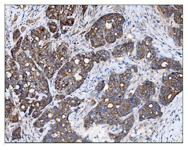
Real IHC data IHC analysis of AS160/TBC1D4 using anti-AS160/TBC1D4 antibody (A02004-3). AS160/TBC1D4 was detected in a paraffin-embedded section of human gallbladder adenocarcinoma tissue. Heat mediated antigen retrieval was performed in EDTA buffer (pH 8.0, epitope retrieval solution). The tissue section was blocked with 10% goat serum. The tissue section was then incubated with 2 μg/ml rabbit anti-AS160/TBC1D4 Antibody (A02004-3) overnight at 4°C. Biotinylated goat anti-rabbit IgG was used as secondary antibody and incubated for 30 minutes at 37°C. The tissue section was developed using Strepavidin-Biotin-Complex (SABC) (Catalog # SA1022) with DAB as the chromogen.
Anti-AS160/TBC1D4 Antibody ®
Cat # A02004-3
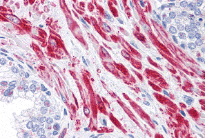
Real IHC data Immunohistochemistry of TBC1D4 in human prostate tissue with TBC1D4 antibody at 10 μg/mL.
Anti-TBC1 domain family member 4 TBC1D4 Antibody
Cat # A02004

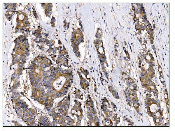
A02004-3 has paraffin-section IHC images from human gallbladder, colonic, lung and ovarian tumors, plus an IF/ICC image in Caco-2 cells (A02004-3 image captions). A02004 is validated for human IHC-P and has an IHC image from prostate tissue (A02004 applications and image caption).

Which to pick: For tissue IHC, A02004-3 has documented paraffin-section staining with 2 μg/mL antibody; its caption does not report the fixative (A02004-3 IHC caption). A02004 is another human IHC-P option with a prostate tissue image, although its caption does not report section processing or fixation (A02004 applications and image caption). For IF/ICC, choose A02004-3 for its Caco-2 image; if human and mouse IF reactivity is needed, M02004-1 is a rabbit monoclonal listed for both species, with no IHC validation in the payload (catalog applications, reactivity and image captions).

Each figure is that product's own IHC / IF validation image from its datasheet.

References

  1. UniProt Consortium. UniProt entry O60343 (TBCD4_HUMAN, TBC1 domain family member 4).
  2. Human Protein Atlas. TBC1D4 tissue IHC expression (reliability: Approved).
  3. Human Protein Atlas. TBC1D4 subcellular location (ICC-IF): Localized to the cytosol..
  4. Human Protein Atlas. TBC1D4 antibody validation summary (2 antibodies).
  5. Breast cancer-associated fibroblast-derived exosomal miR-1290 promotes triple-negative breast cancer cell proliferation. Signal Transduction and Targeted Therapy 2026 — PMC13612446.
  6. Leptin reduces the expression and increases the phosphorylation of the negative regulators of GLUT4 traffic TBC1D1 and TBC1D4 in muscle of ob/ob mice. PloS one 2012 — PMC3253781.
  7. PubMed PMID:18771725 — UniProt-cited evidence.
  8. PubMed PMID:19077034 — UniProt-cited evidence.
  9. PubMed PMID:9628581 — UniProt-cited evidence.