TLN1 / Talin-1 · IHC design guide

Design Immunohistochemistry for TLN1

Plan chromogenic TLN1 IHC in paraffin sections using the cytoplasmic tissue pattern and cell type staining as references (HPA tissue IHC). This guide covers fixation consistency, the catalog antibody's IHC conditions, and controls for interpreting the signal.

Evidence assembled Oct 2026 · For research use; verify linked source records and product datasheet before use
Immunohistochemistry protocol sheet for TLN1 (IHC for TLN1): expected localisation Cytoplasmic staining in tissue sections (HPA tissue IHC), antibody A02859-1, validated IHC image, and IHC protocol steps
Printable TLN1 IHC protocol sheet — expected localisation Cytoplasmic staining in tissue sections (HPA tissue IHC), antibody A02859-1, controls and protocol steps. Open the full TLN1 IHC guide →

TLN1 Immunohistochemistry Experimental Design Guide

Expected localisation, validated protocols, controls and antibodies — the at-a-glance facts below, then the full design guide.

Must know before staining
Expected localisation Cytoplasmic staining in tissue sections (HPA tissue IHC)
Staining pattern General cytoplasmic staining; high in breast glandular cells (HPA tissue IHC)
Antigen retrieval Citrate pH 6 HIER, heat-mediated (datasheet A02859-1)
Positive control ⓘ Appendix+4 more · see all
Negative control ⓘ Heart muscle
Important caveats
Reasons your staining may differ from the expected pattern.
Fixation Keep fixation consistent across paraffin sections. (standard IHC practice; not target-specific)
Caveat Staining varies by cell type; cardiomyocytes are not detected (HPA tissue IHC)
Regulation Expression varies by tissue (HPA tissue IHC)
Isoform / epitope 2 isoforms; epitope coverage needs mapping (UniProt)
Section 1

Recommended TLN1 IHC & IF Protocols

The catalog antibody’s IHC-P protocol (datasheet: A02859-1) is accompanied by published TLN1 protocols for glioma and breast tissues (PMC12903198; PMC8937232).

Recommended immunohistochemistry (IHC-P) protocol parameters
SampleParaffin-embedded human Lung cancer tissue; fixative not specified (datasheet A02859-1)
FixationImage fixative and duration unreported (datasheet A02859-1); verify before use.
Sectioning4–5 µm sections on charged slides (standard)
DeparaffinisationXylene, graded ethanol series to water (standard)
Antigen retrievalHeat retrieval: Citrate pH 6, 20 min (datasheet A02859-1)
Peroxidase block3% H2O2, 10 min, room temperature (standard)
Blocking10% goat serum (datasheet A02859-1)
Primary antibodyRabbit anti-TLN1, 2μg/ml (datasheet A02859-1)
Primary incubationOvernight at 4 °C (datasheet A02859-1)
DetectionStreptavidin-biotin complex (SABC), DAB chromogen (datasheet A02859-1)
CounterstainHematoxylin, blue, dehydrate and mount (standard)
Expected resultTLN1-positive staining in lymphoid tissue of appendix (HPA tissue IHC: High). HPA tissue profile: General cytoplasmic expression. No signal in the no-primary control.
💡Decision noteStart with heat-mediated citrate retrieval at pH 6 (datasheet: A02859-1); the breast tissue protocol specifies microwave heating for 10 min (PMC8937232).
Section 2

What Is the Expected TLN1 Staining Pattern?

TLN1 should appear mainly as cytoplasmic staining in positive cells, with signal near cell–matrix contacts where resolution permits (HPA: general cytoplasmic expression; UniProt Q9Y490: focal adhesions). High staining is reported in several epithelial and hematopoietic cell populations (HPA: tissue IHC). TLN1 has no transmembrane segment (UniProt Q9Y490: topology). HPA rates its tissue IHC profile Supported, with medium consistency between staining and RNA expression (HPA: tissue IHC reliability).

What am I looking at on my slide?
Cytoplasmic stain in breast glandular cells, bronchial respiratory epithelial cells, or bone marrow hematopoietic cells.This matches reported High staining in those cell populations (HPA: tissue IHC). A contact-associated accent can fit TLN1's focal adhesion localization (UniProt Q9Y490: subcellular location), but routine chromogenic sections may not resolve individual adhesions (general IHC practice).
Signal appears predominantly nuclear, with little cytoplasmic staining in otherwise positive cells.A predominantly nuclear pattern conflicts with the reported general cytoplasmic IHC profile and cytosolic localization (HPA: tissue IHC; HPA: subcellular ICC-IF). Consider nonspecific staining or a reading artifact; nuclear signal alone does not establish TLN1 detection.
A cell population stains strongly where the expected population is unstained.Compare the named cells, not the tissue alone: cardiomyocytes were Not detected, whereas bone marrow hematopoietic cells were High (HPA: tissue IHC). An unexpected cell pattern warrants checks for cross-reactivity or endogenous detection activity (general IHC practice); it is not proof of either.
Diffuse color covers the section, including spaces outside identifiable cells.This obscures the cell-specific cytoplasmic pattern reported for TLN1 (HPA: tissue IHC). Background can arise from detection reagents, insufficient blocking or washing, or excess primary antibody (general IHC practice). Interpret cell-specific staining only after that background is controlled.
No staining appears in a known-positive cell population on the same run.Absence in cells reported High, such as appendix lymphoid tissue cells, raises a run-level concern (HPA: tissue IHC). Check tissue preservation, retrieval conditions, primary antibody use, and detection controls (general IHC practice). A negative run cannot establish TLN1 absence.
💡Expected TLN1 appearanceCall a section positive when identifiable cells show cytoplasmic signal, ideally with contact-associated accent where visible, in a population reported High; diffuse section-wide color or predominantly nuclear signal is suspect (HPA: tissue IHC; UniProt Q9Y490: focal adhesions; general IHC practice).
How each factor affects the staining
Cell population and reference tissueHPA reports High staining in appendix lymphoid tissue cells and breast glandular cells, Low in prostate glandular cells, and Not detected in cardiomyocytes (HPA: tissue IHC). Score the named cell population rather than assigning one intensity to an entire organ.
Subcellular interpretationTLN1 is a cytoskeletal protein at focal adhesions and other contact regions, with no transmembrane segment (UniProt Q9Y490: function, location, topology). Membrane-adjacent accent can be compatible with TLN1; a membrane-only interpretation should be checked against the cytoplasmic IHC profile (HPA: tissue IHC).
Antibody evidenceHPA lists IHC as Supported for HPA004748 and CAB002006 (HPA: antibody validation). This supports using their reported patterns as comparisons, but HPA rates overall tissue IHC consistency with RNA as Medium (HPA: tissue IHC reliability).
Isoforms and epitope coverageUniProt lists two TLN1 isoforms (UniProt Q9Y490: isoforms). The supplied evidence gives no antibody epitope or isoform-specific staining result, so neither isoform coverage nor a reason for a discrepant stain can be inferred from that listing.
IF/ICC: where should TLN1 appear?HPA reports supported localization to cytosol and focal adhesion sites, with additional plasma membrane and centriolar satellite localization (HPA: subcellular ICC-IF). These are IF/ICC observations; use the tissue IHC profile to judge paraffin-section staining (HPA: tissue IHC).
Why is my staining missing, weak or wrong?
SituationLikely causeNext action
Positive-control cells show no chromogenic signal.A run or detection problem is possible when cells reported High are blank (HPA: tissue IHC; general IHC practice).Verify the positive control's named cell population, primary antibody application, detection reagents, and retrieval steps before interpreting study sections (general IHC practice). No TLN1-specific retrieval condition is established by the supplied evidence.
Cardiomyocytes stain strongly.That differs from HPA's Not detected result for cardiomyocytes (HPA: tissue IHC); cell misidentification, nonspecific staining, or endogenous detection activity is possible (general IHC practice).Confirm the stained cells on the counterstained section and inspect a no-primary control for detection background (general IHC practice). Treat the HPA result as a reference observation, not a universal exclusion rule.
The whole section has hazy color.Diffuse background may conceal TLN1's reported cytoplasmic pattern (HPA: tissue IHC). Excess primary antibody or inadequate blocking or washing can contribute (general IHC practice).Compare a no-primary control, review blocking and wash steps, and optimize primary antibody concentration using the catalog antibody's IHC-P instructions (general IHC practice). Do not score haze as cellular signal.
Only nuclei appear positive.A nuclear-only result does not fit the reported cytoplasmic IHC profile or supported cytosolic ICC-IF localization (HPA: tissue IHC; HPA: subcellular ICC-IF).Recheck cell boundaries and counterstain, then compare positive and no-primary controls (general IHC practice). Withhold a TLN1-positive call until a credible cellular pattern is present.
A focal adhesion pattern is not visibly punctate in IHC.TLN1 localizes to focal adhesions (UniProt Q9Y490: subcellular location), but section geometry and chromogenic resolution can obscure small structures (general IHC practice).Judge identifiable cells for the expected cytoplasmic pattern and appropriate reference intensity (HPA: tissue IHC). Do not require visible individual adhesions for a positive paraffin-section call.
A low-staining tissue seems negative beside a strong control.Cell populations vary: prostate glandular cells are Low while breast glandular cells are High in HPA tissue IHC (HPA: tissue IHC).Compare like cell populations and the same-run control before changing the score. Record weak but cellular cytoplasmic staining separately from background (HPA: tissue IHC; general IHC practice).

Sample controls for TLN1 IHC & IF

🧪Run appendix first: its lymphoid tissue is the cell population expected to stain (HPA: High in appendix lymphoid tissue). Use heart muscle cardiomyocytes as the negative tissue (HPA: Not detected in cardiomyocytes); cells without specific signal on the appendix slide should show only background DAB staining, but the supplied HPA rows do not identify a named internal-negative cell type (HPA: appendix lymphoid tissue High).
Positive control tissue: Appendix (Lymphoid tissue, HPA High)
Negative control tissue: Heart muscle (HPA Not detected)
ICC-IF cell lines (HPA subcellular resource): HPA ICC-IF images show TLN1 in A-431, U-251MG, U2OS, with annotated localisation: Focal adhesion sites (supported), Cytosol (supported) (HPA subcellular).
Technical controls: Include a no-primary, secondary-only control and a rabbit IgG isotype control matched to the primary antibody’s clonality if known (caption: rabbit anti-Talin 1 antibody). A TLN1 knockout specimen, if available, provides a biological negative; with the caption’s biotin-based DAB detection, block endogenous peroxidase and check for endogenous biotin staining in appendix tissue (caption: SABC with DAB).
⚠️Feasibility: A target-specific fixation window or fixation effect is unreported, and the selected SKU’s paraffin-section caption does not state the fixative (caption: A02859-1, fixative not stated). The reported IHC conditions use heat retrieval in citrate buffer at pH 6 for 20 minutes; retrieval dependence has not been established by a comparison in the supplied evidence (caption: A02859-1). HPA shows TLN1 at focal adhesion sites and in the cytosol by ICC-IF, but the evidence does not establish whether IF or frozen sections are easier than paraffin IHC; endogenous peroxidase or biotin can complicate interpretation with the caption’s SABC/DAB method (HPA: supported focal adhesion sites and cytosol; caption: SABC with DAB).

HPA tissue IHC evidence for TLN1

Comprehensive Human Protein Atlas IHC scoring per tissue (reliability: Supported — Medium consistency between antibody staining and RNA expression data.). Rows are taken directly from the HPA tissue chart — click any row's HPA link to view the source.

Positive expression · recommended positive controls

TissueCell typeLevelEvidenceSource
Appendix Lymphoid tissue High Protein (IHC) HPA →
Bone marrow Hematopoietic cells High Protein (IHC) HPA →
Breast Glandular cells High Protein (IHC) HPA →
Bronchus Respiratory epithelial cells High Protein (IHC) HPA →
Cerebellum Granular cells - cytoplasm/membrane High Protein (IHC) HPA →

Undetected expression · recommended negative controls

TissueCell typeLevelEvidenceSource
Heart muscle Cardiomyocytes Not detected Protein (IHC) HPA →
Section 3

Advanced TLN1 IHC Tips

Troubleshoot TLN1 staining in paraffin sections by checking retrieval, cell compartment, controls, and scoring before interpreting differences between samples.

What retrieval should I try first when TLN1 staining is weak?
Start with heat-mediated citrate retrieval at pH 6 for 20 minutes on paraffin sections (datasheet A02859-1). If staining remains weak, compare a modestly longer heating step on adjacent sections while keeping antibody concentration and detection constant (standard IHC practice). The pictured lung cancer section used 2 µg/mL primary antibody overnight at 4 °C, providing a same-SKU starting condition (datasheet A02859-1). Evaluate cytoplasmic and cell-contact staining against a no-primary control, because TLN1 is associated with the cytoskeleton and focal adhesions (UniProt Q9Y490; HPA subcellular; standard IHC practice).
Could fixation explain variable TLN1 staining between paraffin sections?
The selected paraffin-section caption does not state a fixative, so target-specific TLN1 sensitivity to fixation is unknown (datasheet A02859-1). Record each specimen’s fixative, fixation duration, section age, and retrieval run before attributing a staining difference to biology (standard IHC practice). Compare sections processed together using citrate retrieval at pH 6 for 20 minutes and the same primary concentration, 2 µg/mL, as the illustrated condition (datasheet A02859-1; standard IHC practice). If a processing change tracks with weaker staining, repeat the comparison with matched controls; neither tissue expression patterns nor TLN1 topology establish a fixation effect (standard IHC practice; UniProt Q9Y490).
Where should convincing TLN1 staining appear in tissue sections?
Look primarily for cytoplasmic staining, with enrichment near cell contacts when those structures are resolvable in the section (HPA tissue IHC; UniProt Q9Y490). TLN1 is a cytoskeletal protein associated with focal adhesions, ruffle membranes, and the cell surface; it has no transmembrane segment (UniProt Q9Y490). At 20 minutes of citrate pH 6 retrieval, compare the staining pattern with nearby structures and a no-primary control before calling a thin rim specific (datasheet A02859-1; standard IHC practice). Predominantly nuclear color or uniform staining across unrelated tissue components warrants a background check rather than a TLN1 localisation call (UniProt Q9Y490; standard IHC practice).
Can this stain distinguish TLN1 isoforms or modified epitopes?
Do not assign an isoform from this chromogenic stain alone: the record lists 2 TLN1 isoforms, while the supplied antibody evidence gives no isoform-specific epitope (UniProt Q9Y490; datasheet A02859-1). The FERM region spans residues 86–403 and the I/LWEQ region spans 2293–2533, but neither range identifies where this antibody binds (UniProt Q9Y490; datasheet A02859-1). Document the exact antibody and retrieval condition before comparing specimens, because epitope accessibility can change during paraffin-section processing (standard IHC practice). Likewise, staining cannot establish phosphorylation at residues such as 405 or 620 without a modification-specific reagent and controls (UniProt Q9Y490; standard IHC practice).
How can I check TLN1 localisation with a separate IF experiment?
Use IF as a separate localisation check and multiplex TLN1 with a validated marker for the cell population being examined, plus single-channel controls (standard IF practice). Choose spectrally separated fluorophores after inspecting unstained tissue for autofluorescence, especially before interpreting faint signal near cell boundaries (standard IF practice). Because TLN1 is cytosolic and concentrated at focal adhesions rather than spanning the membrane, permeabilise a test section to make intracellular epitopes accessible (HPA subcellular; UniProt Q9Y490; standard IF practice). The paraffin IHC image used citrate retrieval at pH 6 for 20 minutes; those conditions are IHC evidence and do not establish an IF protocol (datasheet A02859-1).
How should I troubleshoot diffuse brown signal or stained tissue edges?
First compare a no-primary section with the test section and inspect whether DAB color follows tissue edges, folds, or damaged regions (standard IHC practice). Block endogenous peroxidase for chromogenic detection and use an appropriate serum or protein block; the illustrated section used 10% goat serum (standard IHC practice; datasheet A02859-1). The pictured assay used 2 µg/mL primary overnight at 4 °C and biotin-based DAB detection, so excessive primary or detection background should be checked independently (datasheet A02859-1; standard IHC practice). Retain staining as plausible TLN1 only when its cellular pattern exceeds control background and fits cytoplasmic or contact-associated localisation (HPA tissue IHC; UniProt Q9Y490; standard IHC practice).
What is a defensible way to score TLN1 IHC across samples? ⚠ ANSWER MARKED FOR VERIFICATION
Define the cell population and tissue compartment before scoring, then apply identical acquisition and threshold settings to every slide (standard IHC practice). For cellular staining, report percentage of positive cells and an H-score using intensity categories 0–3; for sparse contact-associated signal, report positive area or puncta density per mm² (standard IHC practice). Normalise each measure to eligible nucleated cells or viable tissue area, and exclude folds, necrosis, and section edges using the same rule across samples (standard IHC practice). Report the scoring compartment because HPA describes general cytoplasmic expression, while focal-adhesion enrichment may be less resolvable in tissue sections (HPA tissue IHC; HPA subcellular; standard IHC practice).
When is a TLN1-positive result likely to be an artefact?
A credible result shows cellular cytoplasmic or contact-associated staining above the no-primary control, consistent with TLN1’s cytoskeletal and focal-adhesion localisation (UniProt Q9Y490; HPA subcellular; standard IHC practice). Check the cell population: HPA reports high staining in appendix lymphoid tissue and bone-marrow hematopoietic cells, but no detected staining in heart-muscle cardiomyocytes (HPA tissue IHC). Treat isolated nuclear color, edge-only staining, necrotic deposits, or signal reproduced by a no-primary section as possible artefact (UniProt Q9Y490; standard IHC practice). If brown signal persists without primary antibody, investigate endogenous enzyme activity or detection reagents before assigning TLN1 positivity; DAB was the illustrated chromogen (standard IHC practice; datasheet A02859-1).
Boster reagents

Best TLN1 / Talin-1 IHC Antibodies

TLN1 staining is shown in human and mouse paraffin sections (catalog IHC captions) and HeLa cells by IF/ICC (A02859-2 IF caption).

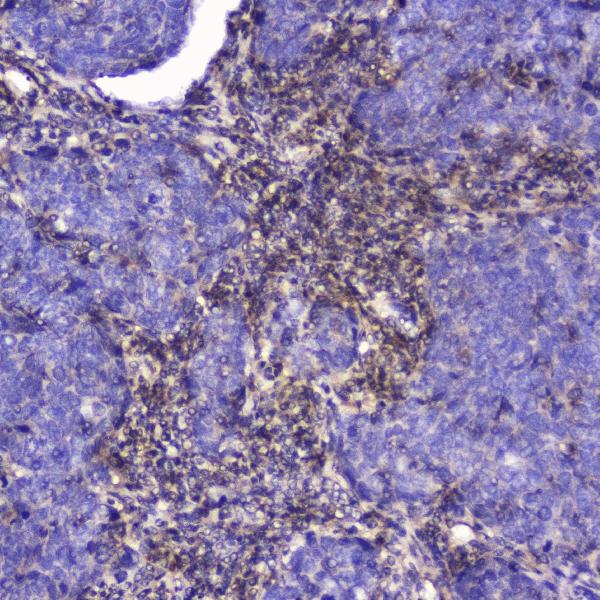
Real IHC data IHC analysis of Talin 1 using anti-Talin 1 antibody (A02859-1). Talin 1 was detected in paraffin-embedded section of human Lung cancer tissue. Heat mediated antigen retrieval was performed in citrate buffer (pH6, epitope retrieval solution) for 20 mins. The tissue section was blocked with 10% goat serum. The tissue section was then incubated with 2μg/ml rabbit anti-Talin 1 Antibody (A02859-1) overnight at 4℃. Biotinylated goat anti-rabbit IgG was used as secondary antibody and incubated for 30 minutes at 37℃. The tissue section was developed using Strepavidin-Biotin-Complex (SABC)(Catalog # SA1022) with DAB as the chromogen.
Anti-Talin 1/TLN1 Antibody ®
Cat # A02859-1

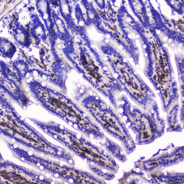
A02859-1 has IHC images from human lung cancer, intestinal cancer and tonsil, plus mouse small intestine (A02859-1 IHC captions). A02859-2 has IHC images from human bladder urothelial carcinoma, lung cancer, tonsil and testicular germ cell tumor, plus IF/ICC data from HeLa cells (A02859-2 IHC and IF captions).

Which to pick: For tissue IHC, choose A02859-1 when using citrate pH 6 retrieval for paraffin sections (A02859-1 IHC captions); choose A02859-2 when using citrate pH 6 retrieval for paraffin sections (A02859-2 IHC captions). For IF/ICC, choose A02859-2, which lists both applications and has a HeLa cell IF image (A02859-2 applications and IF caption). Both list human, mouse and rat reactivity for cross-species planning (catalog reactivity); neither IHC image caption reports the tissue fixative (catalog IHC captions).

Each figure is that product's own IHC / IF validation image from its datasheet.

References

  1. UniProt Consortium. UniProt entry Q9Y490 (TLN1_HUMAN, Talin-1).
  2. Human Protein Atlas. TLN1 tissue IHC expression (reliability: Supported).
  3. Human Protein Atlas. TLN1 subcellular location (ICC-IF): Mainly localized to the cytosol and focal adhesion sites. In addition localized to the plasma membrane and centriolar satellites..
  4. Human Protein Atlas. TLN1 antibody validation summary (2 antibodies).
  5. Oncogenic role of talin-1 in glioma: Association with poor prognosis and regulation of the TGF-beta signaling pathway. Translational oncology 2026 — PMC12903198.
  6. Binding blockade between TLN1 and integrin β1 represses triple-negative breast cancer. eLife 2022 — PMC8937232.
  7. Disulfidptosis-Related Genes as Novel Biomarkers and Therapeutic Targets in Dilated Cardiomyopathy. Journal of inflammation research 2025 — PMC12306564.
  8. Talin1 targeting potentiates anti-angiogenic therapy by attenuating invasion and stem-like features of glioblastoma multiforme. Oncotarget 2015 — PMC4694986.
  9. PubMed PMID:10610730 — UniProt-cited evidence.
  10. PubMed PMID:10470851 — UniProt-cited evidence.
  11. PubMed PMID:12168954 — UniProt-cited evidence.