This website uses cookies to ensure you get the best experience on our website.


- Table of Contents
Plan TMEM106B paraffin IHC using 2.5 μg/mL as the catalog antibody’s starting concentration (datasheet: IHC-P). Compare neuronal staining in cerebral cortex with adipocytes in adipose tissue, and assess the reported cytoplasmic pattern (HPA tissue IHC).

Expected localisation, validated protocols, controls and antibodies — the at-a-glance facts below, then the full design guide.
| Expected localisation | Cytoplasmic staining in several tissues (HPA tissue IHC) | |
| Staining pattern | Neuronal cytoplasmic staining in cortex and caudate (HPA tissue IHC) | |
| Antigen retrieval | Citrate pH 6.0 HIER, 95–98 °C, 20 min (rule: cytoplasmic / membrane antigen) | |
| Positive control | Adrenal gland+4 more · see all | |
| Negative control | Adipose tissue+4 more · see all |
| Fixation | Keep fixation consistent across sections (standard IHC practice; not target-specific); Matched tissue-IHC evidence does not establish the fixation claim. Validate the specimen-specific method before use. (selected-SKU IHC image A03541) | |
| Caveat | Staining and RNA expression have low consistency (HPA tissue IHC) | |
| Regulation | Low tissue RNA specificity; no regulator specified (HPA tissue RNA) | |
| Isoform / epitope | No isoforms; cytoplasmic vs lumenal epitope matters (UniProt) |
The catalog antibody’s IHC-P protocol is accompanied by published TMEM106B staining protocols for frontal lobe sections (PMC10626444; PMC11181561).
| Sample | Tissue sections; selected-image fixative not specified (standard IHC workflow) |
| Fixation | Image fixative and duration unreported (datasheet A03541); verify before use. |
| Sectioning | 4–5 µm sections on charged slides (standard) |
| Deparaffinisation | Xylene, graded ethanol series to water (standard) |
| Antigen retrieval | Heat-induced epitope retrieval in citrate buffer, pH 6.0, 20 min at 95–98 °C (standard rule: cytoplasmic / membrane antigen) |
| Peroxidase block | 3% H2O2, 10 min, room temperature (standard) |
| Blocking | 10% normal serum of the secondary host, 30 min, room temperature (standard) |
| Primary antibody | Rabbit anti-TMEM106B, 2.5 μg/mL (datasheet A03541) |
| Primary incubation | Overnight at 4 °C (standard) |
| Detection | HRP-polymer secondary, DAB chromogen 5–10 min (standard) |
| Counterstain | Hematoxylin, blue, dehydrate and mount (standard) |
| Expected result | TMEM106B-positive staining in glandular cells of adrenal gland (HPA tissue IHC: Medium). HPA tissue profile: Cytoplasmic expression in several tissues. No signal in the no-primary control. |
TMEM106B is a late endosome and lysosome membrane protein; its cytoplasmic tail spans residues 2–96, its membrane segment spans 97–117, and its lumenal region spans 118–274 (UniProt Q9NUM4 topology). In paraffin section IHC, expect cytoplasmic staining in several tissues, including neurons (HPA: tissue IHC). Treat the pattern as a guide rather than definitive proof: HPA rates the IHC antibody Approved but reports low consistency between staining and RNA expression (HPA: reliability).
| Cytoplasmic granular staining in cerebral cortex or hippocampal neurons, with appreciable cell-to-cell variation (HPA: Medium in neuronal cells; HPA: cytoplasmic expression). | This fits the observed IHC cell population and the late endosome/lysosome location (HPA: tissue IHC; UniProt Q9NUM4 subcellular location). At chromogenic resolution, a granular impression is compatible with intracellular organelles; individual lysosomes need not be distinguishable (standard IHC interpretation). |
| Predominantly nuclear staining, with little cytoplasmic staining in an HPA-positive cell population (HPA: tissue IHC). | A nuclear-dominant pattern conflicts with the reported endosome/lysosome membrane location and cytoplasmic IHC profile (UniProt Q9NUM4 subcellular location; HPA: tissue IHC). Check localization and controls before assigning it to TMEM106B (standard IHC practice). |
| Strong, uniform staining in adipocytes or bronchial respiratory epithelial cells (HPA: Not detected in those cells). | This differs from the HPA reference observations and warrants a check for antibody cross-reactivity or endogenous detection activity (HPA: tissue IHC; standard IHC troubleshooting). A 'Not detected' entry is an observation under HPA conditions, not proof of universal absence (HPA: tissue IHC). |
| Diffuse color across cells and surrounding tissue, obscuring cell boundaries (standard IHC interpretation). | Interpret the run cautiously: nonspecific binding, endogenous enzyme activity, or excess chromogen development can obscure a localized signal (standard chromogenic IHC practice). The expected intracellular pattern offers a spatial check, but does not identify which workflow step failed (UniProt Q9NUM4 subcellular location; standard IHC practice). |
| No detectable staining in cerebral cortex neurons or cerebellar Purkinje cells (HPA: Medium in those cells). | First investigate the run and tissue quality; a negative result in one section does not overturn the reference pattern (HPA: tissue IHC; standard IHC practice). HPA's reported low antibody-staining/RNA consistency adds reason to avoid calling a biological absence from this result alone (HPA: reliability). |
| Organelle location and topology | TMEM106B occupies late endosome and lysosome membranes, with a cytoplasmic N-terminal region and lumenal C-terminal region (UniProt Q9NUM4 topology and subcellular location). Interpret cytoplasmic IHC color in that context; the HPA tissue images establish a cytoplasmic pattern, not resolution of individual membrane faces (HPA: tissue IHC). |
| Cell population selected for assessment | Cerebral cortex and hippocampal neuronal cells, cerebellar Purkinje cells, and caudate neuronal cells have Medium HPA IHC staining (HPA: tissue IHC). Adipocytes and bronchial respiratory epithelial cells are listed as Not detected; compare the named cells, not whole organs indiscriminately (HPA: tissue IHC). |
| Strength of the tissue evidence | The tissue IHC antibody is rated Approved, with low consistency between antibody staining and RNA expression (HPA: reliability). This supports using its reported cell patterns as references while checking unexpected results with appropriate controls; it does not establish that every stained structure is specific (HPA: reliability; standard IHC interpretation). |
| Surface staining | A small fraction of TMEM106B resides at the cell surface, although late endosome and lysosome membranes are its principal reported locations (UniProt Q9NUM4 subcellular location). A small peripheral component is therefore possible; isolated strong, continuous surface staining without the expected cytoplasmic pattern deserves review (UniProt Q9NUM4 subcellular location; standard IHC interpretation). |
| IF/ICC Q&A: where should signal localize? | Endosomes and lysosomes are the supported main locations in HPA ICC-IF; images are listed for A-549, U-251MG, and U2OS (HPA: subcellular). This answers localization for the separate IF/ICC guide, without supplying an IF protocol or implying that cultured-cell fluorescence predicts paraffin section intensity (HPA: subcellular; standard assay interpretation). |
| Situation | Likely cause | Next action |
|---|---|---|
| No signal in an expected-positive neuronal population (HPA: Medium in cerebral cortex neurons). | The section or staining run may have failed; a single negative section cannot distinguish assay failure from sample variation (standard IHC troubleshooting; HPA: tissue IHC). | Check tissue preservation, section integrity, retrieval and detection against the antibody's validated IHC-P instructions; include a known-positive section in the same run (standard IHC practice). Avoid inferring TMEM106B-specific fixation sensitivity: none is supplied here. |
| Nuclear color dominates while expected cytoplasmic color is absent (HPA: cytoplasmic expression). | The location conflicts with the reference endosome/lysosome membrane assignment; nonspecific nuclear signal or a detection artifact is possible (UniProt Q9NUM4 subcellular location; standard IHC troubleshooting). | Compare a no-primary control and an expected-positive tissue; reassess blocking, antibody concentration, and image interpretation under the same detection settings (standard IHC practice). |
| Uniform staining appears across HPA Not detected cells, such as adipocytes (HPA: tissue IHC). | Cross-reactivity or endogenous enzyme activity could account for color outside the reference pattern (standard chromogenic IHC troubleshooting; HPA: tissue IHC). | Run no-primary and detection-only controls; if background persists, address endogenous detection activity according to the detection chemistry and reassess the named cell population (standard IHC practice). |
| Diffuse background prevents scoring of cytoplasmic staining (HPA: cytoplasmic expression). | Nonspecific binding, insufficient washing, or excessive chromogen development can reduce contrast (standard chromogenic IHC troubleshooting). | Review blocking, wash steps, antibody concentration, and development time using the validated IHC-P workflow; judge the result by whether expected cells become distinguishable (standard IHC practice; HPA: tissue IHC). |
| A strong continuous rim is the only staining in an expected-positive cell (HPA: tissue IHC). | UniProt reports a small cell-surface fraction, but surface-only dominance does not match its principal endosome/lysosome location (UniProt Q9NUM4 subcellular location). | Inspect the same cell population for cytoplasmic staining and compare with a no-primary control; report peripheral-only color separately until specificity is supported (standard IHC interpretation; UniProt Q9NUM4 subcellular location). |
| HPA Medium cells stain weakly while another tissue stains more strongly (HPA: tissue IHC). | The HPA levels describe observed reference staining, and its low staining/RNA consistency limits any claim that intensity alone measures TMEM106B abundance (HPA: tissue IHC and reliability). | Score compartment and named cell type alongside intensity, compare sections stained in the same run, and document discordance with HPA rather than assigning a biological difference from color alone (standard IHC scoring; HPA: tissue IHC). |


Comprehensive Human Protein Atlas IHC scoring per tissue (reliability: Approved — Low consistency between antibody staining and RNA expression data.). Rows are taken directly from the HPA tissue chart — click any row's HPA link to view the source.
| Tissue | Cell type | Level | Evidence | Source |
|---|---|---|---|---|
| Adrenal gland | Glandular cells | Medium | Protein (IHC) | HPA → |
| Caudate | Neuronal cells | Medium | Protein (IHC) | HPA → |
| Cerebellum | Purkinje cells | Medium | Protein (IHC) | HPA → |
| Cerebral cortex | Neuronal cells | Medium | Protein (IHC) | HPA → |
| Epididymis | Glandular cells | Medium | Protein (IHC) | HPA → |
| Tissue | Cell type | Level | Evidence | Source |
|---|---|---|---|---|
| Adipose tissue | Adipocytes | Not detected | Protein (IHC) | HPA → |
| Appendix | Glandular cells | Not detected | Protein (IHC) | HPA → |
| Bone marrow | Hematopoietic cells | Not detected | Protein (IHC) | HPA → |
| Breast | Adipocytes | Not detected | Protein (IHC) | HPA → |
| Bronchus | Respiratory epithelial cells | Not detected | Protein (IHC) | HPA → |
Use compartment, cell identity and matched controls together when assessing TMEM106B staining in paraffin sections.
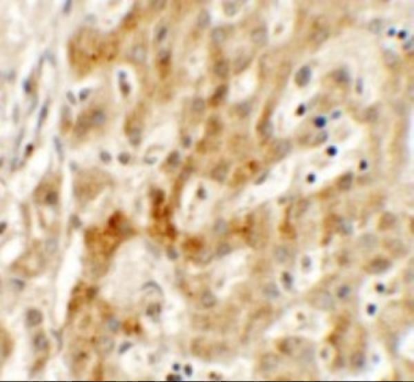
A03541 is listed for human IHC-P and IF (catalog: applications, reactivity). Its TMEM106B IHC image shows human liver at 2.5 μg/mL (IHC image caption).
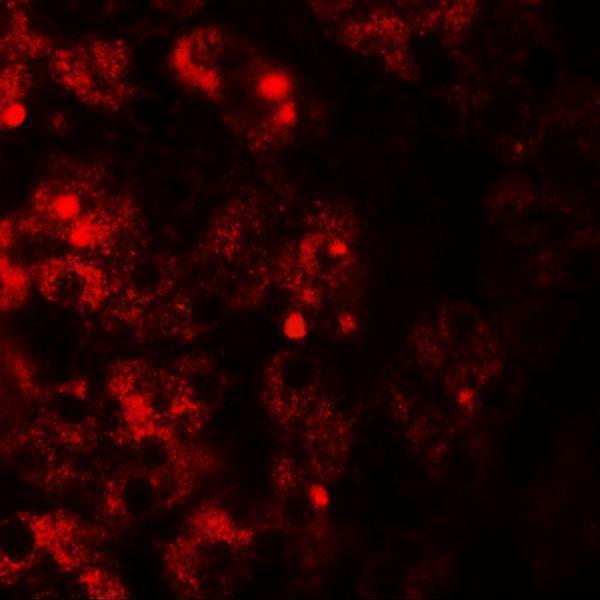
A03541 is listed for human IHC-P and IF (catalog: applications, reactivity). Its IHC image shows TMEM106B staining in human liver (IHC image caption); the IF caption instead names TMEM106A, so that image does not establish TMEM106B IF validation (IF image caption).
Which to pick: For tissue IHC, choose A03541 for human paraffin sections (catalog: Human, IHC-P); its own IHC image shows human liver at 2.5 μg/mL, but the fixative is unreported (IHC image caption). For IF/ICC, A03541 is IF-listed for human samples (catalog: applications, reactivity), although its IF caption names TMEM106A and clonality is unreported (IF image caption; catalog: clone). No listed SKU has reported nonhuman reactivity for cross-species work (catalog: Human).