TMEM59L / Transmembrane protein 59-like · IHC design guide

Design Immunohistochemistry for TMEM59L

Plan chromogenic TMEM59L IHC in paraffin sections using caudate, cerebellum, cerebral cortex or hippocampus as positive tissue controls (HPA tissue IHC). Start the catalog antibody at 2.5 μg/mL (datasheet), and interpret granular neuronal staining with the reported off-target binding in mind (HPA tissue IHC).

Evidence assembled Oct 2026 · For research use; verify linked source records and product datasheet before use
Immunohistochemistry protocol sheet for TMEM59L (IHC for TMEM59L): expected localisation Granular neuronal cytoplasm in CNS tissue (HPA tissue IHC), antibody A15285, validated IHC image, and IHC protocol steps
Printable TMEM59L IHC protocol sheet — expected localisation Granular neuronal cytoplasm in CNS tissue (HPA tissue IHC), antibody A15285, controls and protocol steps. Open the full TMEM59L IHC guide →

TMEM59L Immunohistochemistry Experimental Design Guide

Expected localisation, validated protocols, controls and antibodies — the at-a-glance facts below, then the full design guide.

Must know before staining
Expected localisation Granular neuronal cytoplasm in CNS tissue (HPA tissue IHC)
Staining pattern Granular cytoplasmic staining in CNS neurons (HPA tissue IHC)
Antigen retrieval Citrate pH 6.0 HIER, 95–98 °C, 20 min (rule: cytoplasmic / membrane antigen)
Positive control ⓘ Caudate+3 more · see all
Negative control ⓘ Adipose tissue+4 more · see all
Important caveats
Reasons your staining may differ from the expected pattern.
Fixation Keep paraffin-section fixation consistent (standard IHC practice; not target-specific); Matched tissue-IHC evidence does not establish the fixation claim. Validate the specimen-specific method before use. (selected-SKU IHC image A15285)
Caveat Presumed off-target binding may mimic signal (HPA tissue IHC)
Regulation Preferentially high expression in brain (UniProt)
Isoform / epitope No annotated isoforms; signal peptide 1–24 removed; epitope side unknown (UniProt)
Section 1

Recommended TMEM59L IHC & IF Protocols

The catalog antibody IHC-P protocol is accompanied by published TMEM59L chromogenic IHC methods from three articles (PMC13579383; PMC12894934; PMC12809716).

Recommended immunohistochemistry (IHC-P) protocol parameters
SampleTissue sections; selected-image fixative not specified (standard IHC workflow)
FixationImage fixative and duration unreported (datasheet A15285); verify before use.
Sectioning4–5 µm sections on charged slides (standard)
DeparaffinisationXylene, graded ethanol series to water (standard)
Antigen retrievalHeat-induced epitope retrieval in citrate buffer, pH 6.0, 20 min at 95–98 °C (standard rule: cytoplasmic / membrane antigen)
Peroxidase block3% H2O2, 10 min, room temperature (standard)
Blocking10% normal serum of the secondary host, 30 min, room temperature (standard)
Primary antibodyRabbit anti-TMEM59L, 2.5 μg/mL (datasheet A15285)
Primary incubationOvernight at 4 °C (standard)
DetectionHRP-polymer secondary, DAB chromogen 5–10 min (standard)
CounterstainHematoxylin, blue, dehydrate and mount (standard)
Expected resultTMEM59L-positive staining in neuronal cells of caudate (HPA tissue IHC: Medium). HPA tissue profile: Cytoplasmic expression with a granular pattern in CNS. No signal in the no-primary control.
💡Decision noteStart with citrate pH 6.0 HIER at 95–98 °C for 20 min (page antigen-retrieval rule); the colon study used pressure-cooker citrate pH 6 (PMC12894934).
Section 2

What Is the Expected TMEM59L Staining Pattern?

TMEM59L is a Golgi membrane protein with a transmembrane segment at residues 268–290 (UniProt Q9UK28). In paraffin section IHC, expect granular cytoplasmic staining in CNS neurons, including cerebellar Purkinje cells (HPA: tissue IHC). Interpret the pattern cautiously: HPA rates its IHC staining Approved but reports low consistency with RNA expression and presumed off target binding that was disregarded (HPA: IHC reliability).

What am I looking at on my slide?
Granular cytoplasmic staining in CNS neurons.This matches the reported tissue pattern; neuronal staining is Medium in caudate, cortex and hippocampus, and Purkinje cell staining is Medium in cerebellum (HPA: tissue IHC).
Predominantly nuclear staining in an otherwise positive section.A nuclear dominant pattern conflicts with the reported granular cytoplasmic IHC pattern and Golgi membrane location; assess it as possible artefact before scoring it as TMEM59L (HPA: tissue IHC; UniProt Q9UK28).
Strong staining in adipocytes or other reported negative cells.Adipocytes in adipose tissue and breast are Not detected (HPA: tissue IHC). Consider nonspecific antibody binding or endogenous chromogenic activity; staining alone cannot distinguish those causes (general IHC practice).
Diffuse colour across cells and surrounding tissue.This obscures the reported granular neuronal pattern (HPA: tissue IHC). Check background controls, blocking, washes and chromogen development before interpreting cell specific signal (general IHC practice).
No staining in a CNS section expected to be positive.Medium neuronal staining is reported in caudate, cortex and hippocampus, and Medium Purkinje cell staining in cerebellum (HPA: tissue IHC). Check tissue preservation and run controls before treating absence as biological (general IHC practice).
💡Expected TMEM59L appearanceCall a section positive when neuronal cytoplasm shows discernible granular staining, typically Medium in the listed CNS regions; nuclear dominant or strong staining in reported negative cells warrants investigation (HPA: tissue IHC; UniProt Q9UK28).
How each factor affects the staining
Cell and tissue selectionBrain enriched RNA and preferentially high brain expression support using a listed CNS region for a positive control (HPA: tissue RNA and IHC; UniProt Q9UK28). HPA IHC identifies the stained neuronal populations more precisely than the RNA summary.
Compartment and topologyA signal peptide at residues 1–24 and a transmembrane segment at 268–290 accompany a Golgi membrane assignment (UniProt Q9UK28). Use the reported granular cytoplasmic IHC pattern to judge a section; topology alone cannot locate individual chromogenic granules (HPA: tissue IHC).
Protein processingThe annotated chain spans residues 25–342, with one glycosylation site at residue 97 (UniProt Q9UK28). These annotations do not identify this antibody’s epitope or establish a retrieval condition; evaluate staining empirically with controls (general IHC practice).
Confidence in antibody stainingHPA lists antibody HPA010661 as IHC Approved, while describing low staining to RNA consistency and presumed off target binding that was disregarded (HPA: antibody and tissue IHC). Treat unexpected cell or compartment staining as a validation question.
IF/ICC Q&A: should its pattern match IHC?There are no cell line ICC-IF images and no main subcellular location available; HPA’s subcellular summary says Membrane (HPA: subcellular). UniProt assigns the Golgi membrane, but the supplied ICC-IF record cannot establish an IF staining pattern (UniProt Q9UK28).
Why is my staining missing, weak or wrong?
SituationLikely causeNext action
A listed CNS positive control has no signal.The assay may have failed, or antigen detection may be inadequate; HPA reports Medium staining in the listed neuronal populations (HPA: tissue IHC).Confirm tissue identity, include a detection control, and review retrieval and antibody incubation as general IHC workflow checks; no TMEM59L specific retrieval condition is supplied (general IHC practice).
Staining is predominantly nuclear.This conflicts with granular cytoplasmic IHC and Golgi membrane localisation (HPA: tissue IHC; UniProt Q9UK28).Inspect a known positive CNS section alongside negative controls, then reassess staining specificity before assigning a nuclear TMEM59L signal (general IHC practice).
Staining appears broadly at the cell surface.A broad surface pattern differs from the reported granular cytoplasmic IHC pattern; the UniProt Golgi assignment does not establish a surface IHC pattern (HPA: tissue IHC; UniProt Q9UK28).Compare neuronal cytoplasm with adjacent structures on the same section and check control sections before scoring the surface signal (general IHC practice).
Reported negative cells stain strongly.Nonspecific binding or endogenous detection activity is possible; HPA lists adipocytes in adipose tissue and breast as Not detected (HPA: tissue IHC; general IHC practice).Compare with a no primary control and review endogenous activity blocking; do not infer TMEM59L expression from this staining alone (general IHC practice).
Background hides cytoplasmic granules.Diffuse signal can result from inadequate blocking, washing or chromogen control (general IHC practice); it prevents assessment of the reported granular pattern (HPA: tissue IHC).Review background controls, wash steps and development time, then assess whether granules remain confined to the expected neuronal cells (general IHC practice; HPA: tissue IHC).
A weak or mixed CNS pattern is hard to score.HPA reports Medium staining in selected neuronal cells but also low consistency with RNA and presumed off target binding (HPA: tissue IHC reliability).Record compartment, cell type and intensity separately; compare the same run with a listed CNS positive and a reported negative tissue before interpreting the difference (HPA: tissue IHC; general IHC practice).

Sample controls for TMEM59L IHC & IF

🧪Run cerebellum first: Purkinje cells should stain at a medium level (HPA: Cerebellum, Purkinje cells, Medium). Use adipose tissue adipocytes as the negative tissue (HPA: Adipose tissue, adipocytes, Not detected); on the cerebellum slide, assess non-Purkinje cells as putative internal negatives and expect little specific signal, while recognizing that their TMEM59L status is not established by the supplied HPA row (HPA: Cerebellum, Purkinje cells, Medium).
Positive control tissue: Caudate (Neuronal cells, HPA Medium)
Negative control tissue: Adipose tissue (HPA Not detected)
ICC-IF cell lines (HPA subcellular resource): HPA carries no ICC-IF cell line for TMEM59L; derive a cell-line control from the positive tissue's cell type (Neuronal cells) and confirm it by RNA or western blot first.
Technical controls: Include a no-primary, secondary-only control and an irrelevant immunoglobulin control matched to the primary antibody’s host species and, for a monoclonal primary, its isotype; use knockout tissue or a peptide-block control if available (standard IHC practice). For chromogenic brain IHC, quench endogenous peroxidase and check endogenous biotin if using avidin-based detection; for IF, assess tissue autofluorescence, including lipofuscin (standard IHC/IF practice).
⚠️Feasibility: A target-specific fixation window and fixation effect are unreported in the supplied evidence, and the selected A15285 mouse-brain IHC caption does not state a fixative (A15285 tissue-IHC caption). Retrieval dependency is unreported; compare standard heat-induced retrieval with an unretrieved section during paraffin IHC optimization (standard IHC practice). The supplied evidence does not establish whether frozen sections or IF are easier for TMEM59L; brain lipofuscin can complicate IF interpretation (standard IF practice).

HPA tissue IHC evidence for TMEM59L

Comprehensive Human Protein Atlas IHC scoring per tissue (reliability: Approved — Low consistency between antibody staining and RNA expression data. Presumed off target binding observed and disregarded.). Rows are taken directly from the HPA tissue chart — click any row's HPA link to view the source.

Positive expression · recommended positive controls

TissueCell typeLevelEvidenceSource
Caudate Neuronal cells Medium Protein (IHC) HPA →
Cerebellum Purkinje cells Medium Protein (IHC) HPA →
Cerebral cortex Neuronal cells Medium Protein (IHC) HPA →
Hippocampus Neuronal cells Medium Protein (IHC) HPA →

Undetected expression · recommended negative controls

TissueCell typeLevelEvidenceSource
Adipose tissue Adipocytes Not detected Protein (IHC) HPA →
Adrenal gland Glandular cells Not detected Protein (IHC) HPA →
Appendix Glandular cells Not detected Protein (IHC) HPA →
Bone marrow Hematopoietic cells Not detected Protein (IHC) HPA →
Breast Adipocytes Not detected Protein (IHC) HPA →
Section 3

Advanced TMEM59L IHC Tips

Troubleshoot TMEM59L staining in paraffin sections by checking retrieval, cellular pattern, controls and scoring against the reported brain IHC evidence.

How should I retrieve TMEM59L in paraffin sections when staining is weak?
Start with citrate buffer at pH 6.0 for heat-induced retrieval at 95–98 °C for 20 minutes (page retrieval setting). Allow sections to cool in buffer before washing, then compare a retrieval-treated section with an otherwise matched control (standard IHC practice). The catalog antibody has a mouse brain tissue IHC image at 2.5 µg/mL, but its caption does not report fixation or retrieval (A15285 caption). If staining remains weak, test a longer citrate exposure on adjacent sections while watching for tissue damage and diffuse background (standard IHC practice). Score granular neuronal cytoplasmic signal, rather than overall brown intensity, when choosing conditions (HPA: CNS staining profile).
Could fixation explain loss of TMEM59L staining in my brain sections?
Target-specific fixation sensitivity is unknown: the selected mouse brain IHC caption does not state its fixative (A15285 caption). Record fixation method and duration for every specimen, and compare sections processed together before attributing signal loss to TMEM59L chemistry (standard IHC practice). Run the page’s citrate pH 6.0, 95–98 °C, 20-minute retrieval consistently across the comparison (page retrieval setting). Include a brain section with interpretable neuronal staining and a no-primary control in the same run (HPA: neuronal staining in brain; standard IHC practice). If signal varies with processing, report that association without assigning a target-specific fixation mechanism (A15285 caption: fixative unreported).
Which TMEM59L staining pattern should count as anatomically plausible?
Expect a granular cytoplasmic pattern in CNS cells on tissue IHC (HPA: CNS staining profile). TMEM59L is annotated at the Golgi membrane, with one transmembrane segment at residues 268–290 (UniProt Q9UK28: localisation and topology). Compare neuronal cells in caudate, cerebral cortex and hippocampus, and Purkinje cells in cerebellum, where medium staining is reported (HPA: tissue IHC). A continuous nuclear rim, isolated nuclear staining or uniform staining of every tissue element warrants review against a no-primary section (UniProt Q9UK28: Golgi localisation; standard IHC practice). Use cellular morphology and the counterstain to distinguish perinuclear granules from pigment or precipitate (standard IHC practice).
How can epitope location change TMEM59L staining after retrieval?
The record lists a cleaved signal sequence at residues 1–24, a mature chain at 25–342 and one membrane span at 268–290 (UniProt Q9UK28: processing and topology). It also lists glycosylation at residue 97 and no annotated isoforms (UniProt Q9UK28: glycosylation and isoforms). The antibody epitope is not supplied here, so neither accessibility nor a glycosylation effect can be assigned to this antibody (provided antibody evidence: epitope unspecified). If retrieval changes intensity, compare adjacent sections using the same antibody concentration and detection time (standard IHC practice). Interpret persistent granular neuronal staining alongside controls before invoking an epitope-specific explanation (HPA: CNS staining profile; standard IHC practice).
How should I adapt these IHC observations for multiplex TMEM59L IF?
Treat the catalog antibody’s mouse brain image as tissue IHC evidence, not IF validation; its caption reports 2.5 µg/mL and leaves the fixative unstated (A15285 caption). Pair TMEM59L with a validated neuronal or Purkinje cell marker appropriate to the sampled brain region, and check each channel separately before assessing overlap (HPA: brain cell staining; standard IF practice). Choose a far-red fluorophore when tissue autofluorescence obscures shorter wavelengths, and include single-label and no-primary controls (standard IF practice). Because the antibody epitope and its membrane-facing side are unspecified, compare mild and stronger permeabilisation on matched sections rather than assuming access (provided antibody evidence: epitope unspecified; UniProt Q9UK28: membrane span).
How can I reduce diffuse brown signal without losing TMEM59L staining?
First compare the stained section with a no-primary control and examine whether the brown signal follows tissue edges, vessels or deposits (standard IHC practice). Include an endogenous peroxidase block before chromogenic detection, and shorten DAB development if control sections darken (standard chromogenic IHC practice). Titrate the catalog antibody around the caption’s 2.5 µg/mL concentration on matched mouse brain sections, documenting signal and background together (A15285 caption; standard IHC practice). Prioritise granular neuronal cytoplasm when judging the result (HPA: CNS staining profile). Broad staining outside that pattern deserves extra scrutiny because presumed off-target binding was observed in the HPA assessment (HPA: reliability description).
What should I measure when comparing TMEM59L staining between sections? ⚠ ANSWER MARKED FOR VERIFICATION
Define the brain region and eligible cell type before scoring, then apply the same boundaries to every section (HPA: reported neuronal and Purkinje cell staining; standard IHC practice). An H-score can combine the percentage of cells at intensity 0–3 into a 0–300 scale, while percentage-positive cells measures prevalence (standard IHC scoring practice). For sparse populations, report positive cells per mm² of viable sampled tissue alongside total eligible cells (standard histological quantification practice). Normalise comparisons to eligible cell count or viable tissue area, and keep illumination, threshold and DAB development consistent (standard IHC practice). Exclude edge artefact and necrosis using rules set before scoring (standard IHC practice).
When is a TMEM59L-positive IHC result convincing rather than artefactual?
A credible result shows granular cytoplasmic staining in the expected brain cell population, such as neurons or cerebellar Purkinje cells (HPA: tissue IHC profile). Its intracellular pattern should be compatible with Golgi localisation, though chromogenic IHC alone cannot prove organelle identity (UniProt Q9UK28: Golgi membrane; standard IHC interpretation). Question isolated nuclear signal, staining confined to cut edges or necrotic areas, and brown deposits also present in the no-primary control (standard IHC practice). Check peroxidase blocking and DAB development when widespread brown signal masks cellular detail (standard chromogenic IHC practice). Interpret unexpected staining cautiously because HPA reports low staining–RNA consistency and presumed off-target binding (HPA: reliability description).
Boster reagents

Best TMEM59L / Transmembrane protein 59-like IHC Antibodies

The catalog lists one anti-TMEM59L antibody for IHC-P and IF in human, mouse, and rat (catalog: applications and reactivity), with pictured IHC and IF data from mouse brain tissue (image captions).

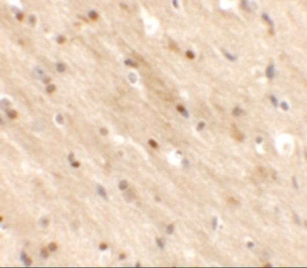
Real IHC data Immunohistochemistry of TMEM59L in mouse brain tissue with TMEM59L antibody at 2.5 μg/mL.
Anti-Transmembrane protein 59-like TMEM59L Antibody
Cat # A15285

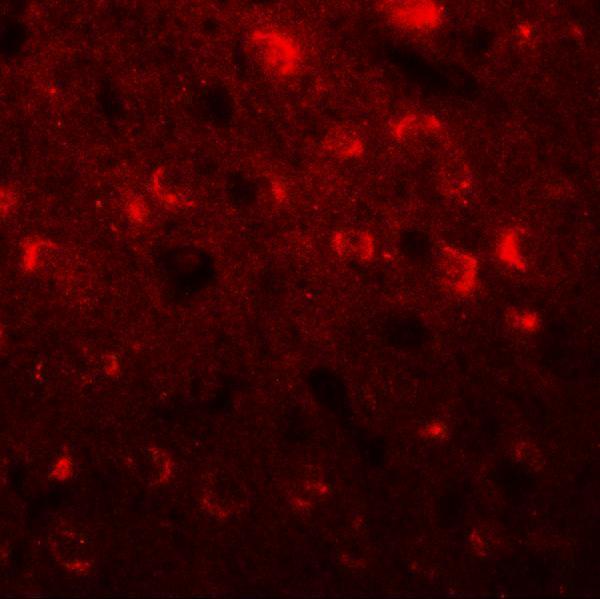
A15285 is listed for IHC-P and IF, with human, mouse, and rat reactivity (catalog: applications and reactivity). Its IHC and IF images show mouse brain tissue at 2.5 μg/mL and 20 μg/mL, respectively (IHC and IF image captions).

Which to pick: For tissue IHC, choose A15285 for paraffin sections (catalog: IHC-P); its mouse brain IHC caption reports 2.5 μg/mL and does not report the fixative (IHC image caption). For IF, A15285 has a mouse brain example at 20 μg/mL (IF image caption); ICC is not separately listed (catalog: applications). For cross-species experiments, A15285 lists human, mouse, and rat reactivity, while the pictured IHC and IF examples are from mouse brain tissue (catalog: reactivity; IHC and IF image captions).

Each figure is that product's own IHC / IF validation image from its datasheet.

References

  1. UniProt Consortium. UniProt entry Q9UK28 (TM59L_HUMAN, Transmembrane protein 59-like).
  2. Human Protein Atlas. TMEM59L tissue IHC expression (reliability: Approved).
  3. Human Protein Atlas. TMEM59L subcellular location (ICC-IF): Membrane.
  4. Human Protein Atlas. TMEM59L antibody validation summary (1 antibodies).
  5. TMEM59L promotes neuroendocrine differentiation and enzalutamide resistance in prostate cancer by regulating MUC1/β-catenin/MYC axis. Clinical and translational medicine 2026 — PMC13579383.
  6. Identification of TMEM59L as a potential diagnosis, prognosis and immunotherapy biomarker for colon adenocarcinoma. Scientific reports 2026 — PMC12894934.
  7. Transmembrane Protein TMEM59L Modulates 5-FU Resistance via PTPRN-Mediated DNA Damage Repair in Colorectal Cancer. Cancer reports (Hoboken, N.J.) 2026 — PMC12809716.
  8. Prognostic value of TMEM59L and its genomic and immunological characteristics in cancer. Frontiers in immunology 2022 — PMC9816415.
  9. PubMed PMID:10527841 — UniProt-cited evidence.
  10. PubMed PMID:15489334 — UniProt-cited evidence.
  11. PubMed PMID:20427278 — UniProt-cited evidence.