This website uses cookies to ensure you get the best experience on our website.


- Table of Contents
Plan chromogenic TRAK2 IHC in paraffin sections using cytoplasmic staining as the tissue benchmark (HPA tissue IHC). The IHC-validated antibody has a 1:100–1:300 dilution range (datasheet A07746); interpret staining cautiously because its consistency with RNA expression is low (HPA tissue IHC).

Expected localisation, validated protocols, controls and antibodies — the at-a-glance facts below, then the full design guide.
| Expected localisation | Cytoplasmic staining in most tissues (HPA tissue IHC) | |
| Staining pattern | Cytoplasmic staining across many tissue cell types (HPA tissue IHC) | |
| Antigen retrieval | Tris-EDTA pH 9.0 HIER, heat-mediated (datasheet A07746) | |
| Positive control | Appendix+4 more · see all | |
| Negative control | Hippocampus+2 more · see all |
| Fixation | Keep fixation consistent across paraffin sections (standard IHC practice; not target-specific) | |
| Caveat | Staining has low consistency with RNA expression (HPA tissue IHC) | |
| Regulation | Staining intensity varies by tissue (HPA tissue IHC) | |
| Isoform / epitope | Two isoforms; check antibody epitope coverage (UniProt) |
The catalog antibody’s IHC-P protocol (datasheet A07746) is accompanied by one published TRAK2 IHC protocol for ESCC and corresponding para-cancer tissues (PMC12125856).
| Sample | Paraffin-embedded human tonsil tissue; fixative not specified (datasheet A07746) |
| Fixation | Image fixative and duration unreported (datasheet A07746); verify before use. |
| Sectioning | 4–5 µm sections on charged slides (standard) |
| Deparaffinisation | Xylene, graded ethanol series to water (standard) |
| Antigen retrieval | Heat retrieval: Tris-EDTA pH 9.0 (datasheet A07746); 20 min, 95–100 °C (standard) |
| Peroxidase block | 3% H2O2, 10 min, room temperature (standard) |
| Blocking | 10% normal serum of the secondary host, 30 min, room temperature (standard) |
| Primary antibody | Rabbit anti-TRAK2, 1:100-1:300 (datasheet A07746) |
| Primary incubation | Overnight at 4 °C (standard) |
| Detection | HRP-polymer secondary, DAB chromogen 5–10 min (standard) |
| Counterstain | Hematoxylin, blue, dehydrate and mount (standard) |
| Expected result | TRAK2-positive staining in glandular cells of appendix (HPA tissue IHC: High). HPA tissue profile: Cytoplasmic expression in most tissues. No signal in the no-primary control. |
TRAK2 IHC is reported mainly as cytoplasmic staining across tissues, including strong staining in several glandular and epithelial cell populations (HPA tissue IHC). Cytoplasmic, early endosomal and mitochondrial localization is annotated, and TRAK2 has no transmembrane segment (UniProt O60296). Interpret a positive slide cautiously: the HPA tissue IHC profile is rated Uncertain because antibody staining and RNA expression have low consistency (HPA tissue IHC).
| Strong cytoplasmic staining in appendix glandular cells (HPA: High in appendix glandular cells). | This fits a reported positive IHC pattern. Assess the cell compartment and tissue structure together; staining intensity alone cannot establish antibody specificity because tissue IHC reliability is Uncertain (HPA tissue IHC). |
| Chromogen chiefly outlines cell membranes or fills extracellular space, with little cytoplasmic signal. | That distribution does not fit the reported cytoplasmic tissue pattern or the absence of a transmembrane segment (HPA tissue IHC; UniProt O60296 topology). Consider nonspecific staining or a slide artefact and check a control section. |
| Strong staining in smooth muscle cells, while expected positive epithelial cells remain faint. | Smooth muscle cells are reported as Not detected, so this pattern raises concern for cross-reactivity or endogenous detection activity (HPA tissue IHC). Compare matched controls before assigning the signal to TRAK2. |
| Diffuse chromogen obscures cell boundaries in both the specimen and its negative control. | Background prevents compartment-based interpretation. Check blocking, washing and the detection control as general IHC practice; the HPA tissue profile cannot establish the cause of diffuse background (HPA tissue IHC). |
| Appendix glandular cells show no convincing cytoplasmic signal (HPA: High in appendix glandular cells). | A failed positive control, an unsuitable antibody or an ineffective IHC workflow is possible. Review the catalog antibody's validated conditions and repeat with controls; one negative slide does not overturn an Uncertain tissue profile (HPA tissue IHC). |
| Tissue and cell selection | Several glandular and epithelial populations stain High, whereas smooth muscle cells are Not detected (HPA tissue IHC). Choose controls by named cell type, since adjacent cells need not share the same reported level. |
| Strength of IHC evidence | The tissue profile is Uncertain, and antibody HPA015827 has an Uncertain IHC designation (HPA tissue IHC; HPA antibodies). Treat agreement with the map as supportive, not definitive verification of specificity. |
| Compartment evidence across applications | Tissue IHC reports mainly cytoplasmic signal, while ICC-IF reports approved nucleoplasm and nuclear bodies, plus vesicles (HPA tissue IHC; HPA subcellular). Record the assay used when interpreting a compartment mismatch. |
| Topology and processing | TRAK2 has no transmembrane segment or annotated signal peptide and is listed as a single chain spanning residues 1–914 (UniProt O60296). This supports assessing intracellular staining; it does not establish an antibody epitope. |
| Isoforms and epitope coverage | Two isoforms are listed, but antibody epitope coverage is not supplied (UniProt O60296; HPA antibodies). Do not attribute a staining difference to one isoform without independent evidence. |
| Detection chemistry | Endogenous enzyme activity or nonspecific detector binding can mimic chromogen signal (general IHC practice). Use appropriate detection controls; neither HPA staining levels nor UniProt annotations identify the cause on a particular slide. |
| Situation | Likely cause | Next action |
|---|---|---|
| No cytoplasmic signal in appendix glandular cells (HPA: High in appendix glandular cells). | The positive control or staining workflow may have failed; tissue IHC reliability is Uncertain (HPA tissue IHC). | Check section quality, retrieval and the catalog antibody's IHC-P conditions; repeat alongside detection controls (general IHC practice). |
| Smooth muscle cells stain strongly (HPA: Not detected in smooth muscle cells). | Cross-reactivity or endogenous detection activity is possible; a single discordant stain does not identify which (HPA tissue IHC; general IHC practice). | Inspect morphology and a control omitting primary antibody; compare with a reported positive cell type in the same run (HPA tissue IHC; general IHC practice). |
| Background is widespread, including areas without intact cells. | Nonspecific reagent binding, insufficient washing or endogenous detection activity may contribute (general IHC practice). | Review blocking, wash steps and detector-only controls before scoring cellular intensity (general IHC practice). |
| Chromogen is chiefly extracellular or sharply membrane outlining. | This conflicts with the mainly cytoplasmic tissue IHC profile and no transmembrane segment is annotated (HPA tissue IHC; UniProt O60296). | Recheck tissue morphology and detection controls, then compare intracellular staining in a reported positive cell type (HPA tissue IHC; general IHC practice). |
| IHC appears mainly nuclear rather than cytoplasmic. | Tissue IHC reports cytoplasm, but ICC-IF reports approved nuclear locations; the assays give different compartment evidence (HPA tissue IHC; HPA subcellular). | Document the discordance and verify IHC staining with controls; do not equate an ICC-IF localization with validated paraffin-section IHC (HPA tissue IHC; HPA subcellular). |
| What should nuclear or vesicular TRAK2 signal in ICC-IF mean? | ICC-IF reports nucleoplasm and nuclear bodies as main locations and vesicles as an additional location (HPA subcellular). | Interpret that result within the separate IF/ICC workflow; antibody HPA062163 is ICC Approved, while the tissue IHC profile is Uncertain (HPA antibodies; HPA tissue IHC). |


![HPA ICC-IF BJ [Human fibroblast] — TRAK2 (HPA062163)](https://images.proteinatlas.org/62163/1368_F2_6_blue_red_green.jpg)
![HPA ICC-IF BJ [Human fibroblast] — TRAK2 (HPA062163)](https://images.proteinatlas.org/62163/1368_F2_7_blue_red_green.jpg)
Comprehensive Human Protein Atlas IHC scoring per tissue (reliability: Uncertain — Low consistency between antibody staining and RNA expression data. Pending external verification.). Rows are taken directly from the HPA tissue chart — click any row's HPA link to view the source.
| Tissue | Cell type | Level | Evidence | Source |
|---|---|---|---|---|
| Appendix | Glandular cells | High | Protein (IHC) | HPA → |
| Bronchus | Respiratory epithelial cells | High | Protein (IHC) | HPA → |
| Cervix | Glandular cells | High | Protein (IHC) | HPA → |
| Colon | Endothelial cells | High | Protein (IHC) | HPA → |
| Duodenum | Glandular cells | High | Protein (IHC) | HPA → |
Use the documented paraffin section conditions as a starting point, then assess staining by cell type and compartment (datasheet A07746; HPA tissue IHC).
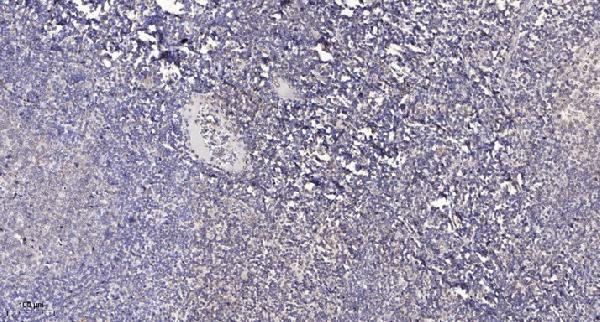
A07746 is listed for human and rat IHC and IF (catalog: applications and reactivity); its IHC figure shows paraffin-embedded human tonsil (IHC image caption). No IF figure is supplied (catalog: IF image alts).
A07746 will render with an IHC figure of paraffin-embedded human tonsil (IHC image caption). A07746 also lists IF and human and rat reactivity, but supplies no IF figure or rat tissue image (catalog: applications, reactivity, IF image alts, IHC image caption).
Which to pick: Choose A07746 for paraffin-section IHC: its human tonsil figure used 1:200 primary antibody and Tris-EDTA retrieval at pH 9.0; the fixative is unreported (IHC image caption). For IF, A07746 lists a 1:50 dilution, but supplies no IF figure or ICC application (catalog: IF dilution, IF image alts, applications). For human and rat work, A07746 lists both species; rat staining is not shown and clonality is unreported (catalog: reactivity, IHC image caption, clone).