This website uses cookies to ensure you get the best experience on our website.


- Table of Contents
Plan paraffin-section TSHZ2 IHC with catalog antibody A08863 at 1:100–1:300 and HPA-supported positive tissue controls (datasheet; HPA tissue IHC). Score nuclear and cytoplasmic staining while accounting for the uncertain tissue-IHC reliability (HPA tissue IHC).

Expected localisation, validated protocols, controls and antibodies — the at-a-glance facts below, then the full design guide.
| Expected localisation | Nuclear (UniProt); nuclear and cytoplasmic in tissue (HPA tissue IHC) | |
| Staining pattern | Widespread nuclear and cytoplasmic tissue staining (HPA tissue IHC) | |
| Antigen retrieval | Tris-EDTA pH 9.0 HIER, 95–98 °C, 20 min (rule: nuclear antigen) | |
| Positive control | Adrenal gland+4 more · see all | |
| Negative control | Liver |
| Fixation | Keep fixation consistent across paraffin sections (standard IHC practice; not target-specific) | |
| Caveat | Tissue staining has low concordance with RNA expression (HPA tissue IHC) | |
| Regulation | Reduced in elderly Alzheimer brains (UniProt) | |
| Isoform / epitope | 2 isoforms; epitope coverage is unreported (UniProt) |
Compare the catalog antibody IHC-P protocol with the published lung adenocarcinoma tissue protocol (PMC8040402).
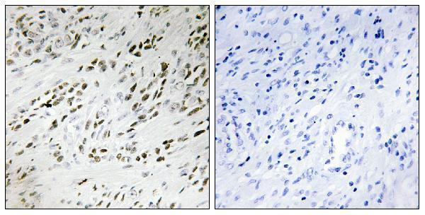
| Sample | Paraffin-embedded human prostate carcinoma tissue; fixative not specified (datasheet A08863) |
| Fixation | Image fixative and duration unreported (datasheet A08863); verify before use. |
| Sectioning | 4–5 µm sections on charged slides (standard) |
| Deparaffinisation | Xylene, graded ethanol series to water (standard) |
| Antigen retrieval | Heat-induced epitope retrieval in Tris-EDTA buffer, pH 9.0, 20 min at 95–98 °C (standard rule: nuclear antigen) |
| Peroxidase block | 3% H2O2, 10 min, room temperature (standard) |
| Blocking | 10% normal serum of the secondary host, 30 min, room temperature (standard) |
| Primary antibody | Rabbit anti-TSHZ2, 1:100 - 1:300 (datasheet A08863) |
| Primary incubation | Overnight at 4 °C (standard) |
| Detection | HRP-polymer secondary, DAB chromogen 5–10 min (standard) |
| Counterstain | Hematoxylin, blue, dehydrate and mount (standard) |
| Expected result | TSHZ2-positive staining in glandular cells of adrenal gland (HPA tissue IHC: High). HPA tissue profile: Ubiquitous nuclear and cytoplasmic expression in all tissues. No signal in the no-primary control. |
TSHZ2 is annotated in the nucleus and has no transmembrane segment (UniProt Q9NRE2). In paraffin tissue sections, HPA reports nuclear and cytoplasmic staining across tissues, including High staining in cerebral cortex neurons, adrenal glandular cells and placental decidual cells (HPA tissue IHC). Treat that broad pattern cautiously: HPA rates its tissue IHC reliability Uncertain because antibody staining and RNA expression have low consistency (HPA tissue IHC).
| Neuronal nuclei stain in cerebral cortex; some cytoplasmic signal is also present (HPA tissue IHC). | This fits the reported High neuronal staining and broad nuclear and cytoplasmic tissue pattern (HPA tissue IHC). Nuclear signal also agrees with the UniProt localization (UniProt Q9NRE2). Record nuclear and cytoplasmic scores separately; HPA's Uncertain reliability limits how confidently either signal can be assigned to TSHZ2 (HPA tissue IHC). |
| Signal is confined to cell borders, lumens or extracellular material, with no convincing cellular staining. | This does not match UniProt's nuclear localization or HPA's reported nuclear and cytoplasmic tissue pattern (UniProt Q9NRE2; HPA tissue IHC). Inspect morphology and the negative detection control before calling it TSHZ2; misplaced chromogen or detection activity is a plausible artefact (general IHC practice). |
| Cholangiocytes stain strongly while expected positive cells on the same run stain weakly or not at all. | HPA reports TSHZ2 as Not detected in liver cholangiocytes, although its tissue IHC reliability is Uncertain (HPA tissue IHC). Check for cross-reactivity or endogenous detection activity using appropriate controls (general IHC practice). A cholangiocyte signal alone cannot establish a new TSHZ2 expression pattern. |
| Brown signal spreads across stroma, section edges or blank areas and obscures cell boundaries. | Diffuse deposition is difficult to score as cellular TSHZ2; HPA's reported pattern is nuclear and cytoplasmic (HPA tissue IHC). Compare the negative detection control, then review blocking, washing and chromogen development (general IHC practice). Do not interpret background intensity as a tissue staining level. |
| No staining appears in cerebral cortex neurons or adrenal glandular cells. | Both populations are reported High by HPA tissue IHC, making them useful run controls, though HPA rates the tissue result Uncertain (HPA tissue IHC). Check section integrity, antigen retrieval, antibody dilution and detection controls before treating a blank section as biological absence (general IHC practice). |
| Tissue IHC reliability | HPA calls the pattern Uncertain because staining and RNA expression have low consistency (HPA tissue IHC). Use reported levels as comparisons, not definitive identity tests. |
| Compartment evidence differs by assay | UniProt places TSHZ2 in the nucleus; HPA tissue IHC reports nuclear and cytoplasmic staining, while HPA ICC-IF approves Golgi and nucleoplasmic localization (UniProt Q9NRE2; HPA tissue IHC; HPA subcellular). The ICC-IF result is context for interpretation, not a paraffin IHC protocol. |
| Antibody validation | HPA038123 is Uncertain for IHC and Approved for ICC (HPA antibodies). Its ICC status does not resolve uncertainty in a paraffin section; compare IHC controls and morphology (HPA antibodies; general IHC practice). |
| Isoforms and topology | UniProt lists 2 isoforms, no signal peptide and no transmembrane segment (UniProt Q9NRE2). The supplied record gives no antibody epitope, so isoform coverage cannot be inferred. |
| Fixation sensitivity | Target-specific fixation sensitivity is unreported in the supplied UniProt and HPA records. Document fixation and retrieval conditions for comparison between IHC runs (general IHC practice). |
| Situation | Likely cause | Next action |
|---|---|---|
| No signal in the expected positive control. | A failed staining step or unsuitable run conditions are possible (general IHC practice); HPA reports High staining in cerebral cortex neurons (HPA tissue IHC). | Verify tissue identity and section quality, then review retrieval, the IHC-validated antibody dilution and detection reagents (general IHC practice). |
| Strong signal appears in liver cholangiocytes. | HPA lists cholangiocytes as Not detected, but the tissue IHC pattern is Uncertain (HPA tissue IHC). Cross-reactivity or endogenous detection activity is possible (general IHC practice). | Compare an appropriate negative detection control and a positive tissue in the same run; report the discordance rather than assigning specificity from intensity alone (general IHC practice). |
| Only sharp cell-border staining is visible. | That distribution conflicts with the nuclear UniProt annotation and HPA's nuclear and cytoplasmic tissue pattern (UniProt Q9NRE2; HPA tissue IHC). | Check whether signal follows tissue edges or deposits, and compare the negative detection control before scoring cells (general IHC practice). |
| Diffuse brown background obscures nuclei. | Nonspecific deposition or excess detection signal can reduce contrast (general IHC practice). | Review blocking, washing, antibody dilution and chromogen development; score only identifiable cellular signal (general IHC practice). |
| Cytoplasmic signal exceeds nuclear signal. | HPA reports cytoplasmic tissue staining, while UniProt annotates a nuclear location and HPA rates tissue IHC Uncertain (HPA tissue IHC; UniProt Q9NRE2). | Score compartments separately and compare positive and negative controls. Do not equate cytoplasmic intensity alone with confirmed TSHZ2 localization. |
| The ICC-IF Golgi pattern is absent from a paraffin IHC section. | HPA's approved Golgi localization comes from ICC-IF; its tissue IHC summary describes nuclear and cytoplasmic staining (HPA subcellular; HPA tissue IHC). | Interpret the paraffin section against the tissue IHC pattern and controls. Use the separate IF/ICC guide for fluorescence-specific decisions. |


Comprehensive Human Protein Atlas IHC scoring per tissue (reliability: Uncertain — Low consistency between antibody staining and RNA expression data.). Rows are taken directly from the HPA tissue chart — click any row's HPA link to view the source.
| Tissue | Cell type | Level | Evidence | Source |
|---|---|---|---|---|
| Adrenal gland | Glandular cells | High | Protein (IHC) | HPA → |
| Cerebral cortex | Neuronal cells | High | Protein (IHC) | HPA → |
| Placenta | Decidual cells | High | Protein (IHC) | HPA → |
| Adipose tissue | Adipocytes | Medium | Protein (IHC) | HPA → |
| Appendix | Glandular cells | Medium | Protein (IHC) | HPA → |
| Tissue | Cell type | Level | Evidence | Source |
|---|---|---|---|---|
| Liver | Cholangiocytes | Not detected | Protein (IHC) | HPA → |
Use compartment-aware controls when optimizing chromogenic TSHZ2 IHC: UniProt and HPA report different localisation patterns, and HPA rates tissue staining reliability as uncertain.
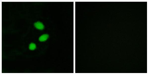
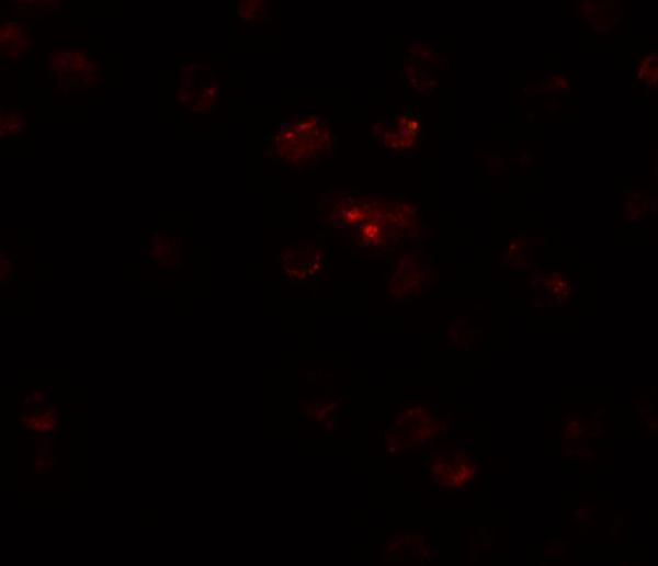
The catalog includes TSHZ2 antibodies with images from paraffin-section IHC and cell IF (catalog image captions); listed reactivity covers human and mouse for both SKUs, plus rat for A08863-1 (catalog reactivity).
A08863 is listed for IHC and IF, with images of paraffin-embedded human prostate carcinoma tissue and MCF7 cells (catalog applications and image captions). A08863-1 is listed for IF, with an image of A20 cells at 20 μg/mL (catalog applications and IF image caption).
Which to pick: Choose the polyclonal A08863 for tissue IHC: its own image shows a paraffin-embedded human prostate carcinoma section, and its listed IHC dilution is 1:100–1:300 (A08863 catalog dilution and IHC image caption); the fixative is unreported (A08863 IHC image caption). For IF/ICC, A08863 lists both applications and shows MCF7 cell IF, while A08863-1 lists IF and shows A20 cell IF at 20 μg/mL but does not list ICC (catalog applications and respective IF image captions). For rat samples, A08863-1 is the SKU with listed rat reactivity, although its pictured IF sample is A20 cells; A08863 lists human and mouse reactivity (catalog reactivity and A08863-1 IF image caption).