This website uses cookies to ensure you get the best experience on our website.


- Table of Contents
Plan chromogenic TSPAN12 IHC around widespread cytoplasmic staining with membrane staining in a few tissues (HPA tissue IHC). The annotated protein location is the cell membrane (UniProt); start the IHC-validated antibody at 2–5 μg/ml (datasheet A05472-1).

Expected localisation, validated protocols, controls and antibodies — the at-a-glance facts below, then the full design guide.
| Expected localisation | Predominantly cytoplasmic; membranous in a few tissues (HPA tissue IHC) | |
| Staining pattern | Widespread cytoplasmic staining; occasional membrane staining (HPA tissue IHC) | |
| Antigen retrieval | EDTA pH 8.0 HIER, heat-mediated (datasheet A05472-1) | |
| Positive control | Lung+4 more · see all | |
| Negative control | Lymph node+4 more · see all |
| Fixation | Keep fixation consistent across paraffin sections. (standard IHC practice; not target-specific) | |
| Caveat | Tissue staining reliability is uncertain (HPA tissue IHC) | |
| Regulation | Expression regulation is not annotated (UniProt) | |
| Isoform / epitope | 2 isoforms; check extracellular versus cytoplasmic epitope (UniProt) |
The catalog antibody’s IHC-P protocol (datasheet A05472-1) is accompanied by published TSPAN12 protocols for nude mouse tumors (PMC9308533) and human NSCLC tissues (PMC5840276).
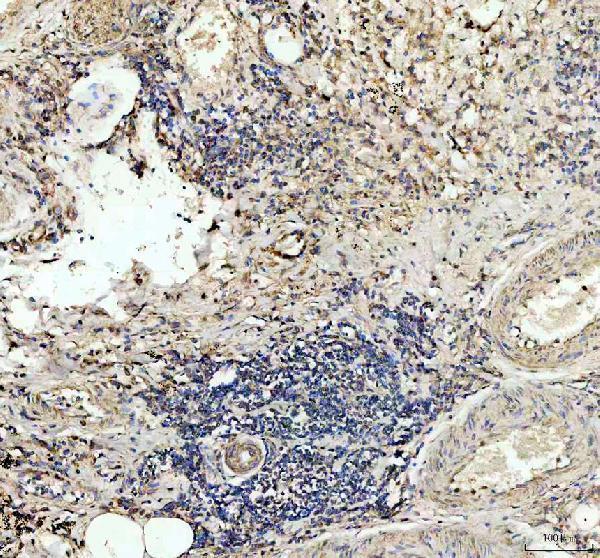
| Sample | Paraffin-embedded human lung cancer tissue; fixative not specified (datasheet A05472-1) |
| Fixation | Image fixative and duration unreported (datasheet A05472-1); verify before use. |
| Sectioning | 4–5 µm sections on charged slides (standard) |
| Deparaffinisation | Xylene, graded ethanol series to water (standard) |
| Antigen retrieval | Heat retrieval: EDTA pH 8.0 (datasheet A05472-1); 20 min, 95–100 °C (standard) |
| Peroxidase block | 3% H2O2, 10 min, room temperature (standard) |
| Blocking | 10% goat serum (datasheet A05472-1) |
| Primary antibody | Rabbit anti-TSPAN12, 2-5μg/ml (datasheet A05472-1) |
| Primary incubation | Overnight at 4 °C (datasheet A05472-1) |
| Detection | HRP-conjugated secondary, DAB chromogen (datasheet A05472-1) |
| Counterstain | Hematoxylin, blue, dehydrate and mount (standard) |
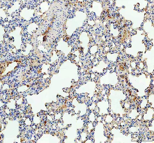
| Expected result | TSPAN12-positive staining in macrophages of lung (HPA tissue IHC: High). HPA tissue profile: Ubiquitous cytoplasmic expression and a few tissues with membranous expression. No signal in the no-primary control. |
TSPAN12 is a four-pass cell-membrane protein with extracellular and cytoplasmic regions (UniProt O95859 topology). In paraffin-section IHC, expect cytoplasmic staining across many tissues and membranous staining in some (HPA: tissue IHC profile). Lung macrophages and placental decidual cells show high staining (HPA: High in each cell type). Interpret intensity cautiously: HPA rates the tissue IHC pattern Uncertain, with medium agreement between antibody staining and RNA expression (HPA: reliability).
| Clear cytoplasmic staining, sometimes with a membrane outline, in the expected cells; strongest examples include lung macrophages and placental decidual cells (HPA: tissue IHC). | This fits the reported paraffin-section pattern: widespread cytoplasmic staining with membranous staining in a few tissues (HPA: tissue IHC profile). A membrane outline also fits the four-pass cell-membrane topology (UniProt O95859 topology). Judge the named cells within their tissue, rather than treating every cell as an expected high-intensity positive (HPA: tissue IHC). |
| Predominantly nuclear staining with little convincing cytoplasmic or membrane signal (HPA: tissue IHC profile; UniProt O95859 subcellular location). | A nuclear-only pattern conflicts with the reported IHC profile and cell-membrane annotation (HPA: tissue IHC profile; UniProt O95859 subcellular location). Treat it as a possible artefact; check the counterstain, detection controls and antibody specificity before assigning a TSPAN12-positive score (standard IHC practice). |
| Strong signal in germinal-center cells of lymph node or tonsil, alongside weak signal in the expected positive cells (HPA: Not detected in those germinal-center cells). | That cell distribution does not match the HPA observations (HPA: tissue IHC). Cross-reactivity or endogenous chromogenic activity is possible; a no-primary control can assess detection-related signal, while comparison with expected positive cells can test whether the antibody pattern is plausible (standard IHC practice). |
| A diffuse, similar-intensity deposit covers cells, stroma and slide background without a discernible cell outline (standard IHC practice). | This distribution cannot be scored confidently as cellular TSPAN12 staining (standard IHC practice). Review no-primary and substrate controls, blocking, washes and detection conditions; the reported HPA pattern describes staining in cells, with compartment and cell-type differences (HPA: tissue IHC profile). |
| No convincing signal in lung macrophages or placental decidual cells (HPA: High in those cell types). | A missing signal in these HPA-positive cells raises a technical or specimen-level question; it alone does not establish absent TSPAN12 protein (HPA: tissue IHC; standard IHC practice). Check tissue identity and preservation, then compare retrieval and detection conditions with a run control. HPA's overall IHC reliability remains Uncertain (HPA: reliability). |
| Membrane topology and antibody epitope (UniProt O95859 topology). | TSPAN12 has four transmembrane spans, extracellular regions at residues 34–59 and 111–224, and cytoplasmic regions at 1–12, 81–89 and 246–305 (UniProt O95859 topology). The supplied sources give no epitope for the IHC antibody, so topology alone cannot predict which staining conditions will expose it. |
| Antigen retrieval for paraffin sections (standard IHC practice). | If a run has weak signal, compare validated retrieval conditions while keeping controls and detection comparable (standard IHC practice). No supplied UniProt or HPA finding establishes a TSPAN12-specific retrieval requirement or fixation sensitivity; a change in signal needs empirical interpretation. |
| Tissue and cell-type variation (HPA: tissue IHC). | HPA reports high staining in lung macrophages and placental decidual cells, medium staining in several other listed cell types, and no detection in lymph-node and tonsil germinal-center cells (HPA: tissue IHC). These are cell-specific observations; avoid assigning one expected intensity to an entire section. |
| RNA evidence versus protein pattern (HPA: tissue IHC). | Kidney is tissue-enhanced at the RNA level, while the supplied HPA IHC summary rates agreement between antibody staining and RNA as medium (HPA: RNA specificity; reliability). RNA enrichment alone does not supply a kidney IHC cell pattern or an intensity threshold for scoring. |
| Antibody validation and isoforms (HPA: antibodies; UniProt O95859 isoforms). | The listed IHC antibody HPA058244 has Uncertain IHC validation, and UniProt lists two isoforms (HPA: antibodies; UniProt O95859 isoforms). The payload does not identify the antibody epitope or isoform coverage. Resolve disputed staining with controls and independent evidence before assigning specificity (standard IHC practice). |
| IF/ICC Q: should it mirror paraffin-section IHC (HPA: subcellular ICC-IF; tissue IHC)? | HPA reports approved microtubule and additional vesicle localisation by ICC-IF, while its tissue IHC profile is largely cytoplasmic with some membranous staining (HPA: subcellular ICC-IF; tissue IHC). They are different preparations and observations; do not use the ICC-IF pattern as an IHC compartment requirement (standard IHC practice). |
| Situation | Likely cause | Next action |
|---|---|---|
| No signal in a section expected to contain lung macrophages or placental decidual cells (HPA: High in those cells). | A failed staining run, unsuitable retrieval or detection, or a specimen issue may explain the absence (standard IHC practice). HPA's Uncertain reliability also limits the certainty of any single expected result (HPA: reliability). | Confirm that the named cells are present, examine the run control, and compare retrieval and detection settings under matched conditions (standard IHC practice). Record any lack of signal as an observation until the controls support interpretation. |
| Only faint staining where HPA reports high signal (HPA: lung macrophages; placental decidual cells). | Signal may vary with the section and IHC workflow (standard IHC practice); the supplied sources specify no TSPAN12-specific fixation effect or required antibody dilution. | Check tissue identity, section quality, control performance, reagent preparation and incubation consistency (standard IHC practice). Optimize one IHC condition at a time against an expected positive cell population; do not infer a numeric cutoff from HPA's qualitative level. |
| Cytoplasmic signal is present, but no membrane outline is visible (HPA: tissue IHC profile). | This can still fit the observed profile, which describes ubiquitous cytoplasmic staining and membranous expression in only a few tissues (HPA: tissue IHC profile). | Score the observed compartment and cell type as seen, then compare with an appropriate HPA tissue example if membrane localisation matters to the study (HPA: tissue IHC). Avoid calling the run failed solely because a membrane rim is absent. |
| Germinal-center cells stain strongly in lymph node or tonsil (HPA: Not detected in those cells). | Off-target binding or endogenous detection activity may account for unexpected chromogen (standard IHC practice); HPA's negative call applies to the named cells, not every cell in either tissue (HPA: tissue IHC). | Check morphology and run a no-primary control; review blocking and endogenous enzyme quenching when the detection chemistry calls for it (standard IHC practice). Compare the suspect cells with HPA-positive cell types before interpreting the deposit. |
| Brown deposit is widespread across cell-free areas or obscures cell boundaries (standard IHC practice). | Nonspecific detection, inadequate washing or excessive chromogen development can produce diffuse background (standard IHC practice). That appearance cannot establish the cellular pattern reported by HPA (HPA: tissue IHC profile). | Inspect no-primary controls, wash and blocking steps, and chromogen development; adjust the contributing step and repeat with a comparable positive section (standard IHC practice). Score only interpretable cells after background is controlled. |
| Different IHC runs give different compartments or cell rankings (HPA: tissue IHC profile). | Run conditions or interpretation may differ (standard IHC practice), while the available antibody validation is Uncertain and does not resolve every discordant pattern (HPA: HPA058244 IHC status; reliability). | Compare matched sections, controls, cell identification and scoring criteria across runs (standard IHC practice). Record cytoplasmic and membranous staining separately, and treat persistent discordance as unresolved rather than forcing a positive call (HPA: tissue IHC profile). |


Comprehensive Human Protein Atlas IHC scoring per tissue (reliability: Uncertain — Medium consistency between antibody staining and RNA expression data.). Rows are taken directly from the HPA tissue chart — click any row's HPA link to view the source.
| Tissue | Cell type | Level | Evidence | Source |
|---|---|---|---|---|
| Lung | Macrophages | High | Protein (IHC) | HPA → |
| Placenta | Decidual cells | High | Protein (IHC) | HPA → |
| Adipose tissue | Adipocytes | Medium | Protein (IHC) | HPA → |
| Adrenal gland | Glandular cells | Medium | Protein (IHC) | HPA → |
| Appendix | Glandular cells | Medium | Protein (IHC) | HPA → |
| Tissue | Cell type | Level | Evidence | Source |
|---|---|---|---|---|
| Lymph node | Germinal center cells | Not detected | Protein (IHC) | HPA → |
| Parathyroid gland | Glandular cells | Not detected | Protein (IHC) | HPA → |
| Spleen | Cells in red pulp | Not detected | Protein (IHC) | HPA → |
| Tonsil | Germinal center cells | Not detected | Protein (IHC) | HPA → |
| Vagina | Squamous epithelial cells | Not detected | Protein (IHC) | HPA → |
Troubleshoot TSPAN12 chromogenic IHC by checking retrieval, compartment, cell identity, controls, and scoring before interpreting staining.
A05472-1 has real paraffin-section IHC images from human lung cancer and rat lung tissue (catalog IHC captions). No IF/ICC images are provided (catalog IF image alts).
A05472-1 is listed for IHC and has images of human lung cancer and rat lung paraffin sections (catalog applications; catalog IHC captions). Its listed reactivity is human, mouse, and rat, but no mouse IHC image is provided (catalog reactivity; catalog IHC image alts).
Which to pick: Choose A05472-1 for paraffin-section tissue IHC based on its human lung cancer and rat lung images; the fixative is unreported (catalog IHC captions). No supplied SKU has IF/ICC validation, so there is no supported IF/ICC pick (catalog applications; catalog IF image alts). For cross-species planning, A05472-1 lists human, mouse, and rat reactivity, although pictured IHC covers only human and rat; its host is rabbit and clonality is unreported (catalog reactivity; catalog IHC captions; catalog host/clone).