TSPAN8 / Tetraspanin-8 · IHC design guide

Design Immunohistochemistry for TSPAN8

Plan paraffin-section TSPAN8 IHC around glandular membrane staining and renal tubular luminal staining (HPA tissue IHC). The IHC-validated antibody A06997 has a recommended dilution of 1:100–1:300 (datasheet A06997); compare staining with matched negative controls (standard IHC practice).

Evidence assembled Oct 2026 · For research use; verify linked source records and product datasheet before use
Immunohistochemistry protocol sheet for TSPAN8 (IHC for TSPAN8): expected localisation Membranous in glands; luminal in renal tubules (HPA tissue IHC), antibody A06997, validated IHC image, and IHC protocol steps
Printable TSPAN8 IHC protocol sheet — expected localisation Membranous in glands; luminal in renal tubules (HPA tissue IHC), antibody A06997, controls and protocol steps. Open the full TSPAN8 IHC guide →

TSPAN8 Immunohistochemistry Experimental Design Guide

Expected localisation, validated protocols, controls and antibodies — the at-a-glance facts below, then the full design guide.

Must know before staining
Expected localisation Membranous in glands; luminal in renal tubules (HPA tissue IHC)
Staining pattern GI, gallbladder and pancreatic glands: membranous; kidney: luminal (HPA tissue IHC)
Antigen retrieval Tris-EDTA pH 9.0 HIER, heat-mediated (datasheet A06997)
Positive control ⓘ Appendix+4 more · see all
Negative control ⓘ Adipose tissue+4 more · see all
Important caveats
Reasons your staining may differ from the expected pattern.
Fixation Keep fixation consistent across sections (standard IHC practice; not target-specific)
Caveat GI endocrine cells can also stain strongly (HPA tissue IHC)
Regulation Regulatory drivers not specified (UniProt)
Isoform / epitope No isoforms annotated; extracellular vs cytoplasmic epitope matters (UniProt)
Section 1

Recommended TSPAN8 IHC & IF Protocols

The catalog antibody has a datasheet IHC-P protocol (datasheet A06997). Two published TSPAN8 IHC methods provide paraffin tissue and tissue microarray examples (PMC8553440; PMC6599078).

Recommended immunohistochemistry (IHC-P) protocol parameters
SampleParaffin-embedded human colon cancer tissue; fixative not specified (datasheet A06997)
FixationImage fixative and duration unreported (datasheet A06997); verify before use.
Sectioning4–5 µm sections on charged slides (standard)
DeparaffinisationXylene, graded ethanol series to water (standard)
Antigen retrievalHeat retrieval: Tris-EDTA pH 9.0 (datasheet A06997); 20 min, 95–100 °C (standard)
Peroxidase block3% H2O2, 10 min, room temperature (standard)
Blocking10% normal serum of the secondary host, 30 min, room temperature (standard)
Primary antibodyRabbit anti-TSPAN8, 1:100-1:300 (datasheet A06997)
Primary incubationOvernight at 4 °C (standard)
DetectionHRP-polymer secondary, DAB chromogen 5–10 min (standard)
CounterstainHematoxylin, blue, dehydrate and mount (standard)
Expected resultTSPAN8-positive staining in endocrine cells of appendix (HPA tissue IHC: High). HPA tissue profile: Membranous expression in glandular cells of the gastrointestinal tract, gall bladder, exocrine pancreas with luminal expression in renal tubules. No signal in the no-primary control.
💡Decision noteStart with Tris-EDTA pH 9.0 heat retrieval for the catalog antibody (datasheet A06997). Optimize published primary dilutions separately for their respective antibodies (PMC8553440; PMC6599078).
Section 2

What Is the Expected TSPAN8 Staining Pattern?

TSPAN8 is a four-pass cell-membrane protein (UniProt P19075 topology and subcellular location). In paraffin-section IHC, expect membranous staining in gastrointestinal and gallbladder glandular cells, exocrine pancreas, and the luminal surface of renal tubules (HPA tissue IHC profile). HPA rates the tissue staining pattern Enhanced, citing consistency between antibody staining and RNA expression (HPA tissue IHC reliability).

What am I looking at on my slide?
Crisp staining outlines glandular cells or faces a glandular lumen.This fits the reported membranous glandular pattern (HPA tissue IHC profile) and four-pass membrane topology (UniProt P19075 topology). Judge the stained cell and membrane surface together: HPA also reports luminal renal-tubule staining (HPA tissue IHC profile).
A known-positive section shows predominantly nuclear or diffuse cytoplasmic stain, with little membrane definition.That distribution does not match the expected IHC pattern (HPA tissue IHC profile; UniProt P19075 subcellular location). Treat it as suspect and compare controls and detection conditions before assigning it to TSPAN8 (standard IHC practice).
Strong staining appears in a cell population reported as undetected, such as adipocytes in adipose tissue.HPA reports Not detected in those adipocytes (HPA adipose tissue IHC). Investigate cross-reactivity or endogenous detection activity; an unexpected positive alone does not establish either cause (standard IHC practice).
Color spreads across tissue or the slide without clear cell borders.Diffuse background cannot be scored as the reported membrane pattern (HPA tissue IHC profile). Review blocking, washes, primary-antibody concentration and chromogen development using appropriate controls (standard chromogenic IHC practice).
Expected positive cells remain unstained in stomach or gallbladder.HPA reports High staining in stomach and gallbladder glandular cells (HPA tissue IHC). Confirm that the relevant cells are present, then review the assay controls and staining workflow (standard IHC practice). Absence in one section does not establish absent TSPAN8 expression.
💡Expected TSPAN8 appearanceCall a result positive when distinct membrane-associated staining marks the relevant glandular or luminal cells, with High signal reported in stomach and gallbladder glandular cells and renal proximal-tubule microvilli (HPA tissue IHC); broad background or dominant nuclear color is suspect (HPA tissue IHC profile; UniProt P19075 subcellular location; standard IHC practice).
How each factor affects the staining
Membrane topology and epitope locationTSPAN8 has 4 transmembrane segments and extracellular and cytoplasmic regions (UniProt P19075 topology). Interpret staining by its cellular distribution; the payload does not identify the catalog antibody’s epitope, so its accessibility cannot be inferred from topology alone.
GlycosylationUniProt annotates one glycosylation site at residue 118 in an extracellular region (UniProt P19075 glycosylation and topology). Its effect on recognition by the catalog antibody is unreported in the supplied sources.
Antibody validationHPA lists HPA044337 as IHC Enhanced and CAB026009 as IHC Supported; HPA022273 is ICC Approved with no IHC status listed (HPA antibody validation). Apply each status only to its stated assay and antibody.
Tissue and cell selectionHPA reports High staining in gastrointestinal endocrine cells, stomach and gallbladder glandular cells, and renal proximal-tubule microvilli (HPA tissue IHC). Use the stated cell population when interpreting a control; a tissue label alone is insufficient.
IF/ICC: what location should I expect?HPA reports an approved vesicular ICC-IF location and images in OE19, PC-3 and U2OS (HPA subcellular ICC-IF). This is an IF/ICC observation; the paraffin-section IHC expectation remains the HPA tissue membranous pattern (HPA tissue IHC profile).
Why is my staining missing, weak or wrong?
SituationLikely causeNext action
A stomach or gallbladder positive control is blank.The target cell population may be absent from the section, or an IHC workflow step may have failed (HPA tissue IHC; standard IHC practice).Locate glandular cells first; then check the positive control, primary-antibody application, antigen-retrieval run and detection reagents (standard IHC practice). The supplied sources do not establish TSPAN8-specific retrieval or fixation sensitivity.
Only nuclear or widespread cytoplasmic color is visible.The distribution conflicts with the reported membrane pattern (HPA tissue IHC profile; UniProt P19075 subcellular location). Background or nonspecific detection is possible (standard IHC practice).Compare a primary-omission control and a known-positive section, and assess whether membrane borders resolve (standard IHC practice). Avoid scoring compartment-mismatched color as specific.
Unexpected cells stain strongly.HPA reports some cell populations as Not detected, including adipose adipocytes and bronchial respiratory epithelium (HPA tissue IHC). Cross-reactivity or endogenous activity is possible (standard IHC practice).Verify cell identity and compare primary-omission and detection controls; interpret any residual signal against the HPA cell-specific pattern (standard IHC practice; HPA tissue IHC).
Background obscures cell borders.Excess primary signal, inadequate blocking or washing, or excessive chromogen development can raise background (standard chromogenic IHC practice).Review blocking and wash steps; adjust primary concentration and development time against positive and omission controls (standard chromogenic IHC practice). No target-specific dilution is supplied.
Renal staining seems limited to the lumen.Luminal proximal-tubule microvilli are a reported High site (HPA kidney tissue IHC).Check that color follows the luminal surface of proximal tubules before calling it background; compare tissue morphology and controls (HPA kidney tissue IHC; standard IHC practice).
IF/ICC vesicles appear inconsistent with the IHC slide.HPA reports vesicles for ICC-IF and a membranous tissue IHC pattern (HPA subcellular ICC-IF; HPA tissue IHC profile).Interpret each result within its assay and specimen context (HPA subcellular ICC-IF; HPA tissue IHC profile). Use the separate IF/ICC guide for that assay; no IF/ICC protocol is specified here.

Sample controls for TSPAN8 IHC & IF

🧪Run colon first and score its endocrine cells, which show High TSPAN8 staining (HPA: colon endocrine cells, High). Use bronchus respiratory epithelium as the negative tissue (HPA: bronchus respiratory epithelial cells, Not detected); on the colon slide, use nonendocrine cells that lack specific signal as internal negatives, showing counterstain without target-specific chromogen.
Positive control tissue: Appendix (Endocrine cells, HPA High)
Negative control tissue: Adipose tissue (HPA Not detected)
ICC-IF cell lines (HPA subcellular resource): HPA ICC-IF images show TSPAN8 in OE19, PC-3, U2OS, with annotated localisation: Vesicles (approved) (HPA subcellular).
Technical controls: Include a no-primary (secondary-only) slide, an isotype control matched to the primary antibody’s host species and immunoglobulin class at the same concentration, and a TSPAN8-knockout specimen as a biological negative. For colon IHC, block endogenous peroxidase before chromogenic detection and assess any residual pigment against the control slides.
⚠️Feasibility: Paraffin-section IHC is supported by the selected A06997 colon cancer caption, which reports Tris-EDTA retrieval at pH 9.0 and primary antibody at 1:200 overnight at 4°C (selected A06997 tissue-IHC caption). The fixative and any target-specific fixation window or fixation effect are unreported (selected A06997 tissue-IHC caption); the caption supports testing that retrieval condition, but does not establish whether retrieval is essential. The supplied evidence does not establish that frozen sections or IF are easier; in colon, distinguish staining in endocrine cells from background in neighboring cells (HPA: colon endocrine cells, High).

HPA tissue IHC evidence for TSPAN8

Comprehensive Human Protein Atlas IHC scoring per tissue (reliability: Enhanced — High consistency between antibody staining and RNA expression data.). Rows are taken directly from the HPA tissue chart — click any row's HPA link to view the source.

Positive expression · recommended positive controls

TissueCell typeLevelEvidenceSource
Appendix Endocrine cells High Protein (IHC) HPA →
Colon Endocrine cells High Protein (IHC) HPA →
Duodenum Endocrine cells High Protein (IHC) HPA →
Gallbladder Glandular cells High Protein (IHC) HPA →
Kidney Proximal tubules (microvilli) High Protein (IHC) HPA →

Undetected expression · recommended negative controls

TissueCell typeLevelEvidenceSource
Adipose tissue Adipocytes Not detected Protein (IHC) HPA →
Adrenal gland Glandular cells Not detected Protein (IHC) HPA →
Bone marrow Hematopoietic cells Not detected Protein (IHC) HPA →
Breast Adipocytes Not detected Protein (IHC) HPA →
Bronchus Respiratory epithelial cells Not detected Protein (IHC) HPA →
Section 3

Advanced TSPAN8 IHC Tips

Troubleshoot TSPAN8 staining in paraffin sections by checking retrieval, controls, cell type and subcellular pattern before scoring chromogenic signal.

What retrieval should I start with for TSPAN8 paraffin sections?
Start with heat-mediated Tris-EDTA retrieval at pH 9.0 (datasheet A06997). The catalog antibody stained paraffin-embedded human colon cancer after this retrieval, with primary antibody at 1:200 overnight at 4°C (datasheet A06997). Keep retrieval conditions consistent across comparison slides, and include a section with expected gastrointestinal glandular staining to detect a failed run (HPA tissue IHC profile; general IHC practice). If staining is weak, optimize heating and cooling on adjacent sections before considering another buffer; the caption supplies no validated alternative (datasheet A06997). Assess membrane detail as well as overall intensity, since TSPAN8 is a cell-membrane protein (UniProt P19075 localization).
How should I assess whether fixation is affecting TSPAN8 staining?
The catalog image identifies a paraffin-embedded colon cancer section but does not report its fixative, so target-specific fixation sensitivity is unknown (datasheet A06997 caption). Record fixative, fixation duration and processing history for each specimen, and compare similarly processed sections with the same retrieval and detection run (general IHC practice). Use the documented Tris-EDTA pH 9.0 retrieval and 1:200 primary dilution as a starting point when checking a suspect block (datasheet A06997). If staining differs between blocks, examine morphology and an expected-positive control before attributing the difference to TSPAN8 biology (HPA tissue IHC profile; general IHC practice).
Where should convincing TSPAN8 chromogenic staining appear?
Prioritize staining along cell borders or luminal surfaces: TSPAN8 is annotated at the cell membrane (UniProt P19075 localization), and tissue IHC shows membranous gastrointestinal glandular staining and luminal renal tubular staining (HPA tissue IHC profile). Its 4 transmembrane segments support a membrane-centered interpretation, but topology alone does not validate an individual stained structure (UniProt P19075 topology; general IHC practice). HPA also reports vesicles in ICC/IF images, which can inform investigation of intracellular signal without establishing that pattern for this chromogenic assay (HPA subcellular). Compare adjacent epithelium, tissue architecture and a no-primary control before accepting diffuse cytoplasmic deposits (general IHC practice).
Could epitope location explain inconsistent TSPAN8 staining?
Check the catalog antibody’s immunogen or epitope documentation before assigning an epitope side; none is specified in the supplied image caption (datasheet A06997 caption). TSPAN8 has extracellular segments at residues 34–57 and 110–205, cytoplasmic segments at 1–9, 73–83 and 231–237, and one annotated glycosylation site at 118 (UniProt P19075 topology and glycosylation). Accessibility may vary with processing and retrieval, so compare serial sections under controlled conditions rather than assigning a cause from intensity alone (general IHC practice). No isoforms are listed in the supplied record; do not explain discordant staining by an unreported isoform (UniProt P19075 isoforms).
How can IF help resolve an ambiguous chromogenic TSPAN8 pattern?
On a matched section, multiplex TSPAN8 with a marker that identifies the glandular or endocrine cell population being evaluated; those populations are reported in the tissue IHC profile (HPA tissue IHC). Choose spectrally separated fluorophores and inspect single-stain and unstained controls because tissue autofluorescence can mimic weak IF signal (general IF practice). Set permeabilisation after confirming the antibody epitope: an extracellular epitope may be accessible without it, whereas a cytoplasmic epitope requires access across the membrane (UniProt P19075 topology; general IF practice). HPA reports vesicular ICC/IF localization, so compare membrane and punctate signals without treating either IF pattern as direct validation of the chromogenic assay (HPA subcellular).
What should I check when TSPAN8 DAB staining is widespread?
First inspect a no-primary section and the expected-positive control under identical detection conditions; widespread brown signal in the no-primary section implicates the workflow (general IHC practice). Apply an appropriate endogenous peroxidase block before DAB detection, and check whether tissue edges or damaged areas stain disproportionately (general chromogenic IHC practice). The catalog image used primary antibody at 1:200 overnight at 4°C and secondary at 1:200 for 45 minutes at room temperature; treat those values as the documented starting conditions (datasheet A06997). Recheck washing and blocking while judging specificity against the reported membranous gastrointestinal pattern (general IHC practice; HPA tissue IHC profile).
How should I score TSPAN8 across paraffin tissue sections? ⚠ ANSWER MARKED FOR VERIFICATION
Define the cell population and compartment before scoring: HPA reports gastrointestinal glandular membrane staining, high signal in several endocrine cell populations, and luminal renal tubular staining (HPA tissue IHC profile). For a consistent epithelial population, record percent positive cells and membrane intensity, then calculate an H-score from 0–300 if intensity categories are reproducible (general IHC scoring practice). Normalize counts or stained area to viable cells or viable tissue area, and report compartment-specific results separately (general IHC scoring practice). Exclude folds, edges and necrosis using the same rules across slides; retain a common retrieval and detection run when comparing groups (general IHC practice).
How do I distinguish genuine TSPAN8 signal from tissue artefact?
A credible result follows the sampled cell population and a plausible membrane or luminal pattern: TSPAN8 is membrane annotated, and HPA tissue IHC reports gastrointestinal glandular membranes and renal tubular luminal staining (UniProt P19075 localization; HPA tissue IHC profile). Compare expected-positive tissue with a no-primary control, and inspect whether staining instead tracks cut edges, necrosis or endogenous enzyme activity (HPA tissue IHC profile; general IHC practice). Strong nuclear-only deposits or uniform signal across unrelated cells warrant investigation before calling the section positive (UniProt P19075 localization; general IHC practice). Interpret carcinoma staining against adjacent morphology, since the supplied record reports TSPAN8 in gastric, colon, rectal and pancreatic carcinomas without making staining alone diagnostic (UniProt P19075 tissue specificity).
Boster reagents

Best TSPAN8 / Tetraspanin-8 IHC Antibodies

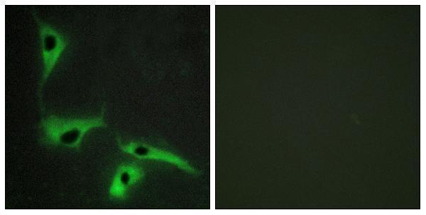
Two anti-TSPAN8 antibodies have paraffin-section IHC images (catalog IHC captions); one also has an IF image in HeLa cells (A06997 IF caption). Catalog reactivity covers human, mouse, and rat (catalog reactivity lists).

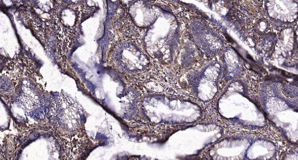
Real IHC data Immunohistochemical analysis of paraffin-embedded human colon cancer. 1, Tris-EDTA, pH9.0 was used for antigen retrieval. 2 Antibody was diluted at 1:200 (4° overnight.3, Secondary antibody was diluted at 1:200 (room temperature, 45min).
Anti-Tetraspanin-8 TSPAN8 Antibody
Cat # A06997
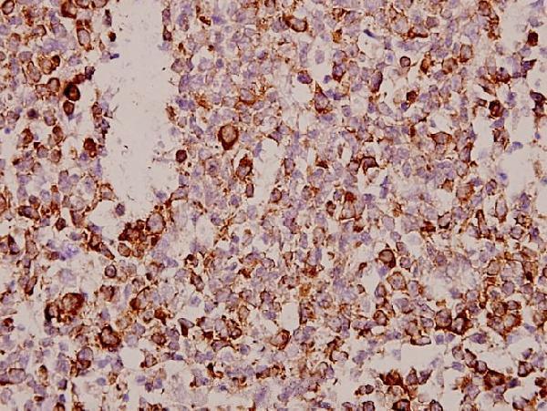
Real IHC data Immunohistochemistry (IHC) analyzes of Tetraspanin-8 (S129) pAb in paraffin-embedded human tonsil carcinoma tissue at 1:50.
Anti-Tetraspanin-8 (S129) TSPAN8 Antibody
Cat # A06997S129

A06997 has an IHC image of paraffin-embedded human colon cancer and an IF image of HeLa cells; its listed reactivity is human and mouse (A06997 image captions; catalog applications and reactivity). A06997S129 has an IHC image of paraffin-embedded human tonsil carcinoma, with human, mouse, and rat listed as reactive species (A06997S129 IHC caption; catalog reactivity).

Which to pick: For paraffin-section IHC, choose either rabbit polyclonal antibody according to the relevant tissue example: A06997 for human colon cancer or A06997S129 for human tonsil carcinoma (catalog host and clonality; each SKU’s IHC caption). For IF/ICC, choose A06997 because IF is listed and its IF image uses HeLa cells; A06997S129 has no listed IF application (catalog applications; A06997 IF caption). For rat samples, A06997S129 is the listed option, although its IHC image shows human tissue; neither IHC caption reports the fixative (catalog reactivity; IHC captions).

Each figure is that product's own IHC / IF validation image from its datasheet.

References

  1. UniProt Consortium. UniProt entry P19075 (TSN8_HUMAN, Tetraspanin-8).
  2. Human Protein Atlas. TSPAN8 tissue IHC expression (reliability: Enhanced).
  3. Human Protein Atlas. TSPAN8 subcellular location (ICC-IF): Localized to vesicles..
  4. Human Protein Atlas. TSPAN8 antibody validation summary (3 antibodies).
  5. Expression of Tspan8 in Patients with Intrahepatic Cholangiocarcinoma and Its Relationship with Clinicopathological Features and Prognosis. Evidence-based complementary and alternative medicine : eCAM 2021 — PMC8553440.
  6. Chronic hepatitis B and perioperative chemotherapy efficacy in colorectal liver metastases: an exploratory analysis of clinical and mechanistic evidence. Frontiers in immunology 2026 — PMC13585926.
  7. Tetraspanin 8 is a novel regulator of ILK-driven β1 integrin adhesion and signaling in invasive melanoma cells. Oncotarget 2017 — PMC5370029.
  8. TSPAN8 promotes cancer cell stemness via activation of sonic Hedgehog signaling. Nature communications 2019 — PMC6599078.
  9. PubMed PMID:2395876 — UniProt-cited evidence.
  10. PubMed PMID:14702039 — UniProt-cited evidence.
  11. PubMed PMID:15489334 — UniProt-cited evidence.