TTN / Titin · IHC design guide

Design Immunohistochemistry for TTN

Plan TTN paraffin IHC using heart and skeletal muscle as positive tissues (HPA tissue IHC) and the catalog antibody at 1:500 (datasheet M00540). Assess cytoplasmic staining in cardiomyocytes and skeletal myocytes (HPA tissue IHC), while considering tissue-specific isoform expression (UniProt).

Evidence assembled Oct 2026 · For research use; verify linked source records and product datasheet before use
Immunohistochemistry protocol sheet for TTN (IHC for TTN): expected localisation Cytoplasmic in heart and skeletal muscle (HPA tissue IHC), antibody M00540, validated IHC image, and IHC protocol steps
Printable TTN IHC protocol sheet — expected localisation Cytoplasmic in heart and skeletal muscle (HPA tissue IHC), antibody M00540, controls and protocol steps. Open the full TTN IHC guide →

TTN Immunohistochemistry Experimental Design Guide

Expected localisation, validated protocols, controls and antibodies — the at-a-glance facts below, then the full design guide.

Must know before staining
Expected localisation Cytoplasmic in heart and skeletal muscle (HPA tissue IHC)
Staining pattern High cardiomyocyte and medium myocyte cytoplasmic staining (HPA tissue IHC)
Antigen retrieval EDTA pH 9 HIER, heat-mediated (datasheet M00540)
Positive control ⓘ Heart muscle+1 more · see all
Negative control ⓘ Adipose tissue+4 more · see all
Important caveats
Reasons your staining may differ from the expected pattern.
Fixation Formaldehyde-fixed paraffin sections (datasheet M00540 IHC-P) (selected-SKU IHC image M00540)
Caveat Presumed off-target tissue staining was observed (HPA tissue IHC)
Regulation Muscle-specific isoform expression (UniProt)
Isoform / epitope 13 isoforms; verify epitope retention in the target isoform (UniProt)
Section 1

Recommended TTN IHC & IF Protocols

The catalog antibody’s IHC-P protocol (datasheet: M00540) is accompanied by one published TTN IHC protocol in paraffin sections (PMC10538261).

Recommended immunohistochemistry (IHC-P) protocol parameters
SampleFormaldehyde-fixed, paraffin-embedded Human skeletal muscle tissue (datasheet M00540)
FixationImage formalin-fixed; duration unreported (datasheet M00540); verify before use.
Sectioning4–5 µm sections on charged slides (standard)
DeparaffinisationXylene, graded ethanol series to water (standard)
Antigen retrievalHeat retrieval: EDTA pH 9 (datasheet M00540); 20 min, 95–100 °C (standard)
Peroxidase block3% H2O2, 10 min, room temperature (standard)
Blocking10% normal serum of the secondary host, 30 min, room temperature (standard)
Primary antibodyRabbit anti-TTN, 1:500 (datasheet M00540)
Primary incubation1 hours at room temperature (datasheet M00540)
DetectionHRP-conjugated secondary, DAB chromogen (datasheet M00540)
CounterstainHematoxylin, blue, dehydrate and mount (standard)
Expected resultTTN-positive staining in cardiomyocytes of heart muscle (HPA tissue IHC: High). HPA tissue profile: Cytoplasmic expression in skeletal muscle and heart muscle. No signal in the no-primary control.
💡Decision noteStart with heat-mediated EDTA pH 9 retrieval (datasheet: M00540); the published protocol used citrate or Tris/EDTA buffer (PMC10538261).
Section 2

What Is the Expected TTN Staining Pattern?

In paraffin-section IHC, TTN is expected in the cytoplasm of heart cardiomyocytes and skeletal-muscle myocytes (HPA: tissue IHC, Enhanced reliability). Heart staining is High and skeletal-muscle staining is Medium (HPA: tissue IHC). TTN is a cytoplasmic protein with no transmembrane segment; UniProt also lists a nuclear location, which does not change the observed tissue-IHC pattern (UniProt Q8WZ42: location and topology; HPA: tissue IHC).

What am I looking at on my slide?
Strong cytoplasmic staining in cardiomyocytes; weaker staining in skeletal-muscle myocytes.This matches the reported High heart-muscle and Medium skeletal-muscle IHC levels (HPA: tissue IHC). Compare signal within the relevant cells; the two tissues need not have equal intensity (HPA: tissue IHC).
Predominantly nuclear staining in muscle cells, with little cytoplasmic signal.This differs from the reported tissue-IHC pattern (HPA: cytoplasmic expression). UniProt lists a nuclear location, so nuclear signal alone cannot prove an artefact; review the control and antibody result before assigning it to TTN (UniProt Q8WZ42: location; general IHC practice).
Strong staining in adipocytes or other listed negative cells.Adipocytes are reported Not detected in adipose tissue and breast (HPA: tissue IHC). Treat strong signal there as suspect; cross-reactivity or endogenous detection activity are possible explanations requiring control slides (general IHC practice).
Broad, diffuse colour across muscle and surrounding tissue.This is less convincing than cytoplasmic staining confined to the expected muscle cells (HPA: tissue IHC). Check the no-primary control for detection background, then assess blocking, antibody concentration and washes (general IHC practice).
No staining in heart muscle or skeletal muscle.A blank known-positive tissue conflicts with High cardiomyocyte and Medium myocyte staining (HPA: tissue IHC). Check tissue identity and positive-control performance before calling the sample TTN negative (general IHC practice).
💡Expected TTN appearanceCall positive when muscle-cell cytoplasm stains, typically High in cardiomyocytes and Medium in skeletal-muscle myocytes; strong staining in listed negative cells is suspect (HPA: tissue IHC).
How each factor affects the staining
Tissue and cell choiceHeart cardiomyocytes provide a High reference; skeletal-muscle myocytes provide a Medium reference (HPA: tissue IHC). Listed Not detected cells can help reveal nonspecific staining (HPA: tissue IHC).
Isoform coverageUniProt lists 13 isoforms and assigns several to cardiac or skeletal muscle (UniProt Q8WZ42: isoforms and tissue specificity). An antibody's epitope is needed to predict which isoforms it detects; that information is absent here.
Antibody validationThree listed antibodies have Enhanced IHC status (HPA: antibodies). The tissue profile has Enhanced reliability, while presumed off-target binding was observed and disregarded (HPA: tissue IHC); assess unexpected staining against controls.
Compartment and IF evidenceTTN has no transmembrane segment (UniProt Q8WZ42: topology). HPA's ICC-IF summary says Membrane, but gives no main location or cell-line images (HPA: subcellular); it does not establish a membrane pattern for paraffin IHC.
Antigen retrievalNo TTN-specific retrieval condition or fixation sensitivity is supplied. Select and optimise retrieval using the IHC-validated antibody's instructions and tissue controls (general IHC practice); do not infer a TTN-specific effect from HPA staining levels.
Why is my staining missing, weak or wrong?
SituationLikely causeNext action
Known-positive heart muscle is blank.The run may have failed, or the sampled area may lack evaluable cardiomyocytes (general IHC practice). Heart cardiomyocytes are reported High (HPA: tissue IHC).Confirm tissue identity and cell preservation; review the concurrent positive control, then check antibody incubation, detection reagents and retrieval settings against the antibody instructions (general IHC practice).
Heart stains, but skeletal muscle is faint.This can fit the reported High versus Medium tissue levels (HPA: tissue IHC). It may also reflect differing assay conditions (general IHC practice).Judge each tissue against its expected level and same-run controls before increasing detection sensitivity (HPA: tissue IHC; general IHC practice).
Nuclei dominate the muscle-cell signal.UniProt permits nuclear localisation, but HPA reports cytoplasmic tissue-IHC staining (UniProt Q8WZ42: location; HPA: tissue IHC). The signal is therefore unresolved.Inspect the no-primary control and compare cytoplasmic staining in known-positive muscle; avoid scoring nuclear-only staining as the expected tissue pattern (general IHC practice; HPA: tissue IHC).
Listed negative cells stain strongly.Such staining conflicts with HPA Not detected calls; cross-reactivity or endogenous detection activity may contribute (HPA: tissue IHC; general IHC practice).Compare no-primary and other assay controls, then review blocking and detection chemistry; record the cell type rather than treating all coloured tissue as TTN positive (general IHC practice).
Diffuse background obscures muscle cells.Nonspecific antibody binding, insufficient washes or detection background may reduce contrast (general IHC practice).Review the no-primary control; optimise blocking, antibody concentration and washes while retaining the known-positive muscle control (general IHC practice; HPA: tissue IHC).
IF appears membranous despite cytoplasmic paraffin IHC.HPA's ICC-IF entry says Membrane but provides no main location or cell-line images; its tissue-IHC profile is cytoplasmic (HPA: subcellular; HPA: tissue IHC).Interpret the paraffin slide against the tissue-IHC pattern. Assess IF with controls on its separate guide page; the supplied ICC-IF record cannot validate an IF protocol (HPA: tissue IHC and subcellular; general IF practice).

Sample controls for TTN IHC & IF

🧪Run heart muscle first: cardiomyocytes should show TTN staining (HPA: High in cardiomyocytes). Use adipose tissue as the negative comparator (HPA: Not detected in adipocytes); on the heart slide, assess non-cardiomyocyte areas for background rather than assuming every cell is TTN-negative.
Positive control tissue: Heart muscle (Cardiomyocytes, HPA High)
Negative control tissue: Adipose tissue (HPA Not detected)
ICC-IF cell lines (HPA subcellular resource): HPA carries no ICC-IF cell line for TTN; derive a cell-line control from the positive tissue's cell type (Cardiomyocytes) and confirm it by RNA or western blot first.
Technical controls: Include no-primary (secondary-only) and host- and isotype-matched antibody controls, plus TTN-knockout tissue or a peptide-block control if the immunizing peptide is available. For chromogenic heart IHC, quench endogenous peroxidase and check a no-primary section for residual signal (standard IHC practice).
⚠️Feasibility: A target-specific fixation window and fixation effect are unreported in the supplied evidence; the selected M00540 tissue-IHC caption does not report a fixative. That skeletal-muscle IHC-P example uses heat-mediated EDTA retrieval at pH 9.0 and primary antibody at 1:500 (M00540 caption), but it does not establish that those settings work for heart tissue. The evidence does not establish whether frozen sections or IF are easier; in heart sections, distinguish cardiomyocyte staining from endogenous peroxidase background with the no-primary control (standard IHC practice).

HPA tissue IHC evidence for TTN

Comprehensive Human Protein Atlas IHC scoring per tissue (reliability: Enhanced — High consistency between antibody staining and RNA expression data. Presumed off target binding observed and disregarded.). Rows are taken directly from the HPA tissue chart — click any row's HPA link to view the source.

Positive expression · recommended positive controls

TissueCell typeLevelEvidenceSource
Heart muscle Cardiomyocytes High Protein (IHC) HPA →
Skeletal muscle Myocytes Medium Protein (IHC) HPA →

Undetected expression · recommended negative controls

TissueCell typeLevelEvidenceSource
Adipose tissue Adipocytes Not detected Protein (IHC) HPA →
Adrenal gland Glandular cells Not detected Protein (IHC) HPA →
Appendix Glandular cells Not detected Protein (IHC) HPA →
Bone marrow Hematopoietic cells Not detected Protein (IHC) HPA →
Breast Adipocytes Not detected Protein (IHC) HPA →
Section 3

Advanced TTN IHC Tips

Troubleshoot TTN staining in paraffin sections by checking retrieval, tissue morphology, epitope coverage and the cellular pattern before scoring signal.

What retrieval should I try when TTN staining is weak in paraffin sections?
Start with heat-mediated EDTA retrieval at pH 9 (datasheet M00540). The selected skeletal-muscle IHC image used that retrieval, followed by a 1:500 primary-antibody dilution for 1 hour at room temperature (datasheet M00540); use its staining pattern as a reference while keeping retrieval time consistent across slides. If signal remains weak, compare a small series of heating times and inspect morphology after each condition (standard IHC practice). Because TTN has many repeated Ig-like and fibronectin type-III domains (UniProt Q8WZ42 domains), retrieval performance for one antibody should not be assumed for another epitope.
How can I troubleshoot weak TTN staining after fixation?
TTN-specific sensitivity to fixation is unknown from the supplied evidence, so evaluate fixation as a processing variable without assigning a target-specific effect. Compare sections with documented, consistent fixation and processing histories, then run the same EDTA pH 9 retrieval and detection conditions on each (datasheet M00540; standard IHC practice). Check whether muscle architecture and nuclear counterstaining remain interpretable before increasing antibody concentration or retrieval time (standard IHC practice). Include a skeletal-muscle positive control in each run, since myocytes show TTN staining in tissue IHC (HPA: skeletal-muscle myocytes, Medium), and record any difference between runs before interpreting a negative specimen.
Which cellular pattern should support a TTN-positive IHC result?
Expect staining in muscle cells, with a predominantly cytoplasmic tissue pattern: HPA reports expression in skeletal muscle and heart muscle (HPA: tissue IHC profile). Cardiomyocytes show High staining and skeletal-muscle myocytes Medium staining in the supplied tissue data (HPA: positive tissues); compare the candidate signal with these cell populations on matched control sections. UniProt also lists cytoplasm and nucleus and describes a possible nuclear role outside muscle (UniProt Q8WZ42 subcellular location and function), so localisation alone does not establish specificity. If staining instead concentrates in unrelated tissue compartments, check the negative control, section edges and morphology before calling it TTN (standard IHC practice).
Could TTN isoforms explain different staining across muscle samples?
Yes: TTN has 13 listed isoforms, with isoforms 3, 7 and 8 reported in cardiac muscle and isoforms 4 and 6 in skeletal muscle (UniProt Q8WZ42 isoforms and tissue specificity). Ask for the antibody’s mapped epitope and check whether that sequence is retained in the isoforms relevant to the specimen; the supplied caption does not map the epitope (datasheet M00540 caption). TTN’s extensive Ig-like and fibronectin type-III repeats make a domain name alone insufficient to establish isoform coverage (UniProt Q8WZ42 domains). Compare staining within the same muscle type and processing batch before attributing differences to expression (standard IHC practice).
How should I adapt the TTN question to a multiplex IF experiment?
For IF, multiplex TTN with a marker identifying the expected muscle cell population, then assess whether the signals occupy the same cells (HPA: cardiomyocytes High; skeletal-muscle myocytes Medium). Choose a fluorophore channel with low tissue autofluorescence and examine an unstained section in each channel before interpreting weak signal (standard IF practice). TTN has no transmembrane segment and is listed in cytoplasm and nucleus (UniProt Q8WZ42 topology and subcellular location); select permeabilisation for the antibody’s mapped intracellular epitope and preserve cell morphology (standard IF practice). The selected 1:500 dilution and EDTA pH 9 retrieval come from IHC tissue evidence (datasheet M00540), so optimise IF conditions independently.
What should I check when TTN IHC shows diffuse brown background?
First compare the stained section with a no-primary control and inspect whether color follows tissue edges, damaged regions or endogenous pigment rather than muscle-cell structure (standard IHC practice). Confirm adequate peroxidase quenching, protein blocking and washing in the chromogenic workflow, then shorten chromogen development if background persists (standard IHC practice). The selected IHC image used a 1:500 primary dilution for 1 hour at room temperature (datasheet M00540); use those conditions as a documented starting point when troubleshooting this antibody. HPA reports cytoplasmic expression in skeletal and heart muscle (HPA: tissue IHC profile), so broad unrelated staining warrants a specificity check.
How should I score TTN staining without conflating signal and muscle content? ⚠ ANSWER MARKED FOR VERIFICATION
Define a muscle-cell region of interest before scoring and exclude folds, torn edges and nonviable areas from the analysis (standard IHC practice). Record the percentage of positive myocytes and intensity on a 0–3 scale, or calculate an H-score from intensity-weighted percentages on a 0–300 scale (standard IHC practice). Normalise the result to the number or area of evaluable myocytes, rather than total section area, because the measured tissue may contain differing proportions of muscle cells (standard IHC practice). Report cardiac and skeletal samples separately: their listed staining levels differ (HPA: cardiomyocytes High; skeletal-muscle myocytes Medium).
When is an apparent TTN-positive IHC signal likely to be artefactual?
A convincing result follows intact muscle cells and the expected cytoplasmic tissue pattern, with heart and skeletal muscle providing relevant positive contexts (HPA: tissue IHC profile and positive tissues). Treat staining restricted to section edges, necrotic areas or unrelated cells as suspect, and compare it with a no-primary control and an adjacent stained section (standard IHC practice). Check peroxidase-related color in the control before attributing diffuse brown signal to TTN (standard IHC practice). UniProt lists a possible nuclear role outside muscle (UniProt Q8WZ42 function and subcellular location), but an isolated nuclear IHC pattern still needs independent antibody and morphology checks before it supports that interpretation.
Boster reagents

Best TTN / Titin IHC Antibodies

Anti-TTN antibodies have IHC images from human heart and skeletal muscle (catalog: IHC captions); one also lists IF and mouse and rat reactivity (catalog: A00540 applications, reactivity).

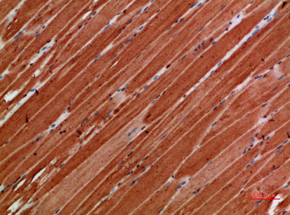
Real IHC data Immunohistochemical analysis of TITIN in Human skeletal muscle tissue sections (IHC-P -paraformaldehyde-fixed, paraffin-embedded sections). Tissue was fixed with formaldehyde at room temperature; antigen retrieval was by heat mediation with a EDTA buffer (pH9. 0). Samples were incubated with primary antibody (1:500) for 1 hours at room temperature. A undiluted biotinylated CRF Anti-Polyvalent HRP Polymer antibody was used as the secondary antibody.
Anti-TTN Antibody (N-term)
Cat # M00540
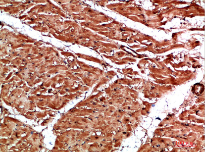
Real IHC data Immunohistochemical analysis of paraffin-embedded human-heart, antibody was diluted at 1:200
Anti-TTN/Titin Antibody
Cat # A00540

M00540 is listed for human IHC-P and has images from human skeletal muscle and heart sections (catalog: M00540 applications, reactivity, IHC captions). A00540 lists IHC and IF, with IHC images from paraffin-embedded human heart and skeletal muscle (catalog: A00540 applications, IHC captions).

Which to pick: For human tissue IHC, choose M00540 when its documented conditions fit: formaldehyde-fixed paraffin sections, heat retrieval in EDTA at pH 9.0, and primary antibody at 1:500 (catalog: M00540 IHC captions); it is listed as polyclonal (catalog: M00540 dilution notes). For IF/ICC, A00540 is the IF-listed option at 1:50, but ICC-specific validation is unreported (catalog: A00540 applications, IF dilution); its IHC captions show paraffin sections and do not report a fixative (catalog: A00540 IHC captions). For mouse or rat samples, A00540 lists reactivity with both species, while its supplied IHC images show human tissue (catalog: A00540 reactivity, IHC captions).

Each figure is that product's own IHC / IF validation image from its datasheet.

References

  1. UniProt Consortium. UniProt entry Q8WZ42 (TITIN_HUMAN, Titin).
  2. Human Protein Atlas. TTN tissue IHC expression (reliability: Enhanced).
  3. Human Protein Atlas. TTN subcellular location (ICC-IF): Membrane.
  4. Human Protein Atlas. TTN antibody validation summary (3 antibodies).
  5. Long noncoding RNA TTN-AS1 facilitates tumorigenesis and metastasis by maintaining TTN expression in skin cutaneous melanoma. Cell death & disease 2020 — PMC7441063.
  6. Deciphering Prognostic Value of TTN and Its Correlation With Immune Infiltration in Lung Adenocarcinoma. Frontiers in oncology 2022 — PMC9304871.
  7. Titin as a potential novel therapeutic target in colorectal cancer. Journal of cellular and molecular medicine 2023 — PMC10538261.
  8. Titin founder mutation is a common cause of myofibrillar myopathy with early respiratory failure. Journal of neurology, neurosurgery, and psychiatry 2014 — PMC6558248.
  9. PubMed PMID:7569978 — UniProt-cited evidence.
  10. PubMed PMID:10850961 — UniProt-cited evidence.
  11. PubMed PMID:11717165 — UniProt-cited evidence.