This website uses cookies to ensure you get the best experience on our website.


- Table of Contents
Plan TUBA1A paraffin-section IHC around its general cytoplasmic staining pattern (HPA tissue IHC). Compare high staining in cortical neurons with staining reported as undetected in skeletal myocytes, while accounting for possible recognition of proteins from more than one gene (HPA tissue IHC).

Expected localisation, validated protocols, controls and antibodies — the at-a-glance facts below, then the full design guide.
| Expected localisation | General cytoplasmic tissue staining (HPA tissue IHC) | |
| Staining pattern | High cytoplasmic staining in cortical and caudate neurons (HPA tissue IHC) | |
| Antigen retrieval | Citrate pH 6.0 HIER, 95–98 °C, 20 min (rule: cytoplasmic / membrane antigen) | |
| Positive control | Adrenal gland+4 more · see all | |
| Negative control | Skeletal muscle |
| Fixation | Keep fixation consistent across paraffin sections (standard IHC practice; not target-specific) | |
| Caveat | Staining may reflect protein from more than one gene (HPA tissue IHC) | |
| Regulation | High expression in fetal brain (UniProt) | |
| Isoform / epitope | 2 isoforms; terminal processing may alter epitopes (UniProt) |
Compare the catalog antibody’s IHC-P protocol with published tissue-microarray and confocal tissue-section IHC methods (PMC11975906; PMC9818799).
| Sample | Tissue sections; selected-image fixative not specified (standard IHC workflow) |
| Fixation | Image fixative and duration unreported (datasheet A03989); verify before use. |
| Sectioning | 4–5 µm sections on charged slides (standard) |
| Deparaffinisation | Xylene, graded ethanol series to water (standard) |
| Antigen retrieval | Heat-induced epitope retrieval in citrate buffer, pH 6.0, 20 min at 95–98 °C (standard rule: cytoplasmic / membrane antigen) |
| Peroxidase block | 3% H2O2, 10 min, room temperature (standard) |
| Blocking | 10% normal serum of the secondary host, 30 min, room temperature (standard) |
| Primary antibody | Rabbit anti-TUBA1A, 2.5 μg/mL (datasheet A03989) |
| Primary incubation | Overnight at 4 °C (standard) |
| Detection | HRP-polymer secondary, DAB chromogen 5–10 min (standard) |
| Counterstain | Hematoxylin, blue, dehydrate and mount (standard) |
| Expected result | TUBA1A-positive staining in glandular cells of adrenal gland (HPA tissue IHC: High). HPA tissue profile: General cytoplasmic expression. No signal in the no-primary control. |
TUBA1A is a cytoplasmic microtubule protein with no transmembrane segment (UniProt Q71U36). In paraffin-section IHC, expect predominantly cytoplasmic staining in HPA-positive cells, including cortical and caudate neurons and several glandular cell populations (HPA: tissue IHC). HPA rates its tissue IHC reliability Enhanced, while reporting medium agreement with RNA data and warning that the staining may represent proteins from more than one gene (HPA: tissue IHC).
| Cytoplasmic staining in cortical or caudate neurons, with little staining in skeletal myocytes. | This fits the reported high neuronal staining and undetected myocyte staining (HPA: tissue IHC). Interpret the result by cell type and compartment; HPA cautions that its tissue pattern may include protein from more than one gene (HPA: tissue IHC). |
| Staining is predominantly nuclear or confined to cell membranes. | That distribution does not fit the reported cytoplasmic IHC profile or cytoskeletal localization (HPA: tissue IHC; UniProt Q71U36). Review morphology and controls before scoring it as target staining (general IHC practice). |
| Strong staining appears in skeletal myocytes while the section's expected positive cells stain weakly. | HPA reports TUBA1A staining as undetected in skeletal myocytes (HPA: tissue IHC). Consider cross-reactivity or endogenous detection activity; this pattern alone cannot identify which is responsible (general IHC practice). |
| Color spreads across tissue, stroma, and blank areas without a clear cell pattern. | Diffuse deposition is difficult to score as cytoplasmic staining (general IHC practice). Compare with a no-primary control to assess detection background, then review washing, blocking, and chromogen development (general IHC practice). |
| No signal appears in cortical neurons or another documented high-staining population. | The result conflicts with HPA's reported high staining in those cells (HPA: tissue IHC). Check a documented positive section and the detection run before concluding that the specimen lacks TUBA1A (general IHC practice). |
| Tissue and cell type | HPA reports high staining in cortical and caudate neurons, glandular cells at several listed sites, and marrow hematopoietic cells; heart cardiomyocytes are low and skeletal myocytes undetected (HPA: tissue IHC). |
| Attribution to TUBA1A | The tissue profile is rated Enhanced, but HPA notes medium staining–RNA consistency and possible detection of protein from more than one gene (HPA: tissue IHC). A positive slide therefore supports a pattern, not gene-specific identity. |
| Protein forms and epitope | UniProt lists two isoforms, a full-length chain, a detyrosinated chain, and modified residues (UniProt Q71U36). The supplied sources do not locate this antibody's epitope, so they do not establish which forms it detects. |
| Detection background | Endogenous enzyme activity can contribute color in chromogenic IHC; a no-primary control and an appropriate blocking step help assess it (general IHC practice). This is workflow guidance, not a reported TUBA1A-specific effect. |
| IF/ICC Q: What pattern should I expect? | A: HPA reports microtubules as the main location and the primary cilium as an additional location (HPA: subcellular ICC-IF). The separate IF/ICC guide covers its workflow. |
| Situation | Likely cause | Next action |
|---|---|---|
| A documented high-staining tissue shows no signal. | The run or primary-antibody detection may have failed; the slide alone cannot distinguish a technical failure from specimen variation (general IHC practice). | Check a concurrently processed positive section with reported high-staining cells, such as cortical neurons (HPA: tissue IHC), then review the IHC detection steps (general IHC practice). |
| Skeletal myocytes show strong color. | This conflicts with HPA's undetected skeletal-myocyte result and may reflect nonspecific binding or endogenous detection activity (HPA: tissue IHC; general IHC practice). | Compare a no-primary control and the expected positive cells on the run; reassess blocking and detection conditions if the control develops color (general IHC practice). |
| Staining is mainly nuclear or outlines membranes. | The distribution conflicts with the cytoplasmic IHC profile and cytoskeletal annotation (HPA: tissue IHC; UniProt Q71U36). | Confirm cell boundaries and counterstain, compare the no-primary control, and score only a reproducible compartment-consistent pattern (general IHC practice). |
| Most structures have diffuse brown background. | Nonspecific deposition or overdevelopment can obscure cell-level interpretation (general IHC practice). | Use a no-primary control to localize background; review washing, blocking, and chromogen development against the IHC workflow (general IHC practice). |
| A low-staining heart section seems negative. | HPA reports low staining in cardiomyocytes, so absence of a clear signal there is less informative than in a high-staining population (HPA: tissue IHC). | Assess a concurrently processed high-staining tissue before interpreting the heart result, and compare like cell types when scoring (HPA: tissue IHC; general IHC practice). |
| Strong cytoplasmic staining is taken as proof of TUBA1A alone. | HPA explicitly cautions that its tissue staining may detect protein from more than one gene (HPA: tissue IHC). | Report the observed cell pattern and antibody-based result without claiming gene-specific identity; use independent specificity evidence if that distinction is required (HPA: tissue IHC; general IHC practice). |
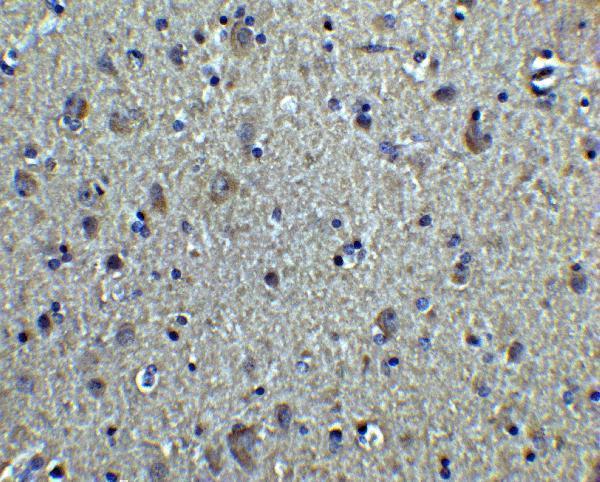
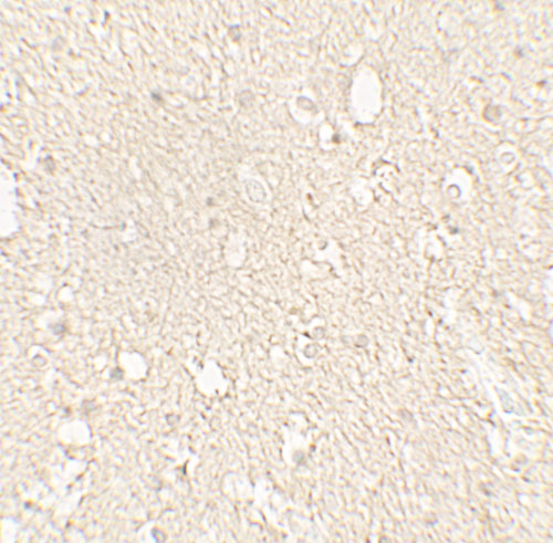
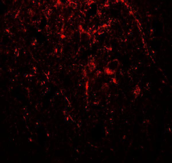


Comprehensive Human Protein Atlas IHC scoring per tissue (reliability: Enhanced — Medium consistency between antibody staining and RNA expression data. Caution, targets protein from more than one gene.). Rows are taken directly from the HPA tissue chart — click any row's HPA link to view the source.
| Tissue | Cell type | Level | Evidence | Source |
|---|---|---|---|---|
| Adrenal gland | Glandular cells | High | Protein (IHC) | HPA → |
| Appendix | Glandular cells | High | Protein (IHC) | HPA → |
| Bone marrow | Hematopoietic cells | High | Protein (IHC) | HPA → |
| Breast | Glandular cells | High | Protein (IHC) | HPA → |
| Bronchus | Basal cells | High | Protein (IHC) | HPA → |
| Tissue | Cell type | Level | Evidence | Source |
|---|---|---|---|---|
| Skeletal muscle | Myocytes | Not detected | Protein (IHC) | HPA → |
Troubleshoot chromogenic TUBA1A staining in paraffin sections by checking retrieval, staining pattern, antibody specificity and cell-level controls.
The IHC-validated antibody A03989 has images of human brain tissue by IHC and IF (catalog image captions); its listed reactivity covers human, mouse, and rat (catalog reactivity).
A03989 is the SKU with a rendered card and an IHC image of human brain tissue at 2.5 μg/mL (catalog image caption). Its IF image shows human brain tissue at 20 μg/mL, and its application list includes IHC-P and IF (catalog image caption; catalog applications).
Which to pick: For paraffin-section IHC, choose A03989 (catalog applications: IHC-P); its IHC caption shows human brain tissue but does not report the fixative (catalog image caption). For IF, choose A03989 (catalog applications: IF); ICC validation is unreported (catalog applications). For work requiring chicken reactivity, consider mouse monoclonal M03989-5, which lists chicken alongside human, mouse, and rat and an IHC paraffin-section dilution of 1–2 μg/mL (catalog host, clone, reactivity, and dilution); no IHC image caption or fixative is reported for that SKU (catalog image alts).