This website uses cookies to ensure you get the best experience on our website.


- Table of Contents
Plan chromogenic TUBB2A IHC in paraffin sections around the cytoplasmic staining reported in CNS and peripheral nerves (HPA tissue IHC). Compare cell populations with reported staining levels, and interpret positive signal in light of HPA's warning that the antibody targets products from more than one gene (HPA tissue IHC).

Expected localisation, validated protocols, controls and antibodies — the at-a-glance facts below, then the full design guide.
| Expected localisation | Cytoplasmic tissue staining (HPA tissue IHC) | |
| Staining pattern | General cytoplasmic staining, strongest in CNS and peripheral nerves (HPA tissue IHC) | |
| Antigen retrieval | Citrate pH 6.0 HIER, heat-mediated (datasheet M06868) | |
| Positive control | Placenta+3 more · see all | |
| Negative control | Adipose tissue+4 more · see all |
| Fixation | Keep fixation consistent across sections (standard IHC practice; not target-specific). Selected-image fixative and duration unreported (datasheet M06868); verify before use. | |
| Caveat | Staining may include products from other genes (HPA tissue IHC) | |
| Regulation | Brain enriched expression (UniProt) | |
| Isoform / epitope | No annotated isoforms; one 1–445 chain (UniProt) |
The catalog antibody’s IHC-P protocol is accompanied by published protocols for hepatocellular carcinoma tissue (PMC13033567) and mouse xenograft tumors (PMC5390261).
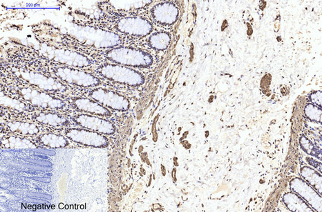
| Sample | Paraffin-embedded Human colon tissue; fixative not specified (datasheet M06868) |
| Fixation | Image fixative and duration unreported (datasheet M06868); verify before use. |
| Sectioning | 4–5 µm sections on charged slides (standard) |
| Deparaffinisation | Xylene, graded ethanol series to water (standard) |
| Antigen retrieval | Heat retrieval: Citrate pH 6.0, 20min (datasheet M06868) |
| Peroxidase block | 3% H2O2, 10 min, room temperature (standard) |
| Blocking | 10% normal serum of the secondary host, 30 min, room temperature (standard) |
| Primary antibody | Mouse monoclonal (clone Mix) anti-TUBB2A, 1:50 recommended; image 1:200 (datasheet M06868) |
| Primary incubation | Overnight at 4 °C (standard) |
| Detection | HRP-polymer secondary, DAB chromogen 5–10 min (standard) |
| Counterstain | Hematoxylin, blue, dehydrate and mount (standard) |
| Expected result | TUBB2A-positive staining in endothelial cells of placenta (HPA tissue IHC: High). HPA tissue profile: General cytoplasmic expression, most abundant in CNS and peripheral nerves. No signal in the no-primary control. |
TUBB2A is a cytoplasmic microtubule protein with no transmembrane segment (UniProt Q13885). In IHC, expect cytoplasmic staining, including signal in peripheral nerves and CNS neuropil, with prominent staining reported in placental endothelial cells, skin granular layer cells and pachytene spermatocytes (HPA tissue IHC). Interpret positive staining cautiously: HPA rates the tissue pattern Supported, but notes that its antibodies can recognize proteins from more than one gene and that presumed off-target staining was disregarded (HPA tissue IHC).
| Cytoplasmic staining in placental endothelial cells, skin granular layer cells or pachytene spermatocytes. | This matches HPA's High IHC observations in those cell populations (HPA tissue IHC). Check the identity of stained cells and the surrounding architecture; a strong color reaction alone cannot establish TUBB2A specificity because HPA reports recognition of proteins from more than one gene (HPA tissue IHC). |
| Medium signal in cerebral cortex neuropil, or weak staining in hippocampal neuronal cells. | These intensities fit the respective Medium and Low HPA observations (HPA tissue IHC). Do not require every brain cell to stain strongly: HPA reports no detection in caudate glial cells or cerebellar granular layer cells, despite describing CNS enrichment overall (HPA tissue IHC). |
| Predominantly nuclear, membranous or extracellular staining, with little cytoplasmic signal. | The compartment disagrees with UniProt's cytoplasm and cytoskeleton assignment and its lack of a transmembrane segment (UniProt Q13885). Review slide morphology and detection controls before scoring it as specific; a compartment mismatch alone does not identify the artifact's cause (standard IHC practice). |
| Strong color in an HPA-negative population, or widespread color outside recognizable cells. | Adipocytes and adrenal glandular cells are among HPA's Not detected populations (HPA tissue IHC). Staining there warrants a specificity or endogenous detection check; widespread color may be background from the staining workflow (standard IHC practice). Neither appearance proves a particular cause. |
| No cytoplasmic signal in a well-preserved, expected-positive control section. | Compare the control with HPA's High placental endothelial, skin granular layer or pachytene spermatocyte observations (HPA tissue IHC). If it is also blank, check the primary antibody, retrieval, detection and counterstain workflow before interpreting a test section as negative (standard IHC practice). |
| Cell population and intensity | HPA reports High staining in three specified populations, Medium in cerebral cortex neuropil, Low in hippocampal neuronal cells and Not detected in several other populations (HPA tissue IHC). Score the identified cell population rather than assigning one intensity to an entire tissue. |
| Antibody specificity | The HPA tissue profile is Supported, and three listed antibodies have Supported IHC status (HPA antibodies; HPA tissue IHC). HPA also cautions that antibodies may recognize proteins from more than one gene and that presumed off-target staining was disregarded; morphology and controls therefore matter. |
| Localization and processing | UniProt places TUBB2A in the cytoplasm and cytoskeleton, with no transmembrane segment, signal peptide or propeptide; the annotated chain spans residues 1–445 (UniProt Q13885). These annotations support a cytoplasmic interpretation, but do not predict antigen retrieval requirements or fixation sensitivity. |
| Antigen retrieval in paraffin IHC | Use a documented, working retrieval condition and compare positive controls when optimizing staining (standard IHC practice). The supplied UniProt and HPA records provide no TUBB2A-specific fixation or retrieval sensitivity, so a weak result cannot be attributed to epitope masking from these sources. |
| IF/ICC Q&A: where should fluorescence appear? | Primarily along microtubules; HPA also reports the mitotic spindle, cytokinetic bridge and specified ciliary or sperm structures (HPA subcellular ICC-IF). This is an IF/ICC localization reference, while the tissue intensities above come from IHC (HPA tissue IHC). |
| Situation | Likely cause | Next action |
|---|---|---|
| Expected-positive cells are blank. | A failed staining run or unsuitable working conditions are possible; the supplied sources establish expected cell populations but no TUBB2A-specific fixation effect (HPA tissue IHC; standard IHC practice). | Confirm morphology, primary antibody application, retrieval and detection with a positive control containing an HPA High population; assess the test section only after that control works (HPA tissue IHC; standard IHC practice). |
| Nuclear color dominates the section. | That pattern conflicts with the cytoplasmic and cytoskeletal annotation (UniProt Q13885); the image alone cannot distinguish nonspecific binding from a workflow artifact (standard IHC practice). | Check compartment boundaries against the counterstain and compare a no-primary control. Score cytoplasmic signal separately from nuclear color (standard IHC practice). |
| An HPA Not detected cell population stains strongly. | Possible cross-reactivity merits attention because HPA cautions that its antibodies may recognize proteins from more than one gene (HPA tissue IHC). Endogenous chromogenic activity is another general IHC possibility (standard IHC practice). | Verify cell identity, compare an expected-positive population and run a no-primary detection control; investigate persistent cell-associated color with another validated antibody if available (HPA tissue IHC; standard IHC practice). |
| Color is diffuse across tissue and empty spaces. | This distribution is inconsistent with the expected cell-associated cytoplasmic pattern (UniProt Q13885; HPA tissue IHC). Background from blocking, washing or detection can produce diffuse color (standard IHC practice). | Review a no-primary control, washing and blocking, then reassess the cytoplasmic pattern in identifiable positive cells before assigning a score (standard IHC practice). |
| All brain cells appear equally intense. | HPA describes abundant CNS expression overall, yet reports Medium cortex neuropil, Low hippocampal neuronal cells and no detection in listed caudate glial and cerebellar granular layer cells (HPA tissue IHC). | Score each identified cell population and compartment separately; verify that strong, uniform color is absent from the no-primary control before calling it specific (HPA tissue IHC; standard IHC practice). |
| Two IHC antibodies give different patterns. | HPA lists three antibodies with Supported IHC status but cautions about recognition of proteins from more than one gene (HPA antibodies; HPA tissue IHC). Discordance therefore needs review rather than an automatic positive call. | Compare localization and expected cell populations on matched sections, check detection controls, and report the disagreement when assigning confidence to the result (UniProt Q13885; HPA tissue IHC; standard IHC practice). |


Comprehensive Human Protein Atlas IHC scoring per tissue (reliability: Supported — Caution, targets protein from more than one gene. Presumed off target binding observed and disregarded. External characterization data supports antibody staining.). Rows are taken directly from the HPA tissue chart — click any row's HPA link to view the source.
| Tissue | Cell type | Level | Evidence | Source |
|---|---|---|---|---|
| Placenta | Endothelial cells | High | Protein (IHC) | HPA → |
| Skin | Cells in granular layer | High | Protein (IHC) | HPA → |
| Testis | Pachytene spermatocytes | High | Protein (IHC) | HPA → |
| Cerebral cortex | Neuropil | Medium | Protein (IHC) | HPA → |
| Tissue | Cell type | Level | Evidence | Source |
|---|---|---|---|---|
| Adipose tissue | Adipocytes | Not detected | Protein (IHC) | HPA → |
| Adrenal gland | Glandular cells | Not detected | Protein (IHC) | HPA → |
| Appendix | Glandular cells | Not detected | Protein (IHC) | HPA → |
| Bone marrow | Hematopoietic cells | Not detected | Protein (IHC) | HPA → |
| Caudate | Glial cells | Not detected | Protein (IHC) | HPA → |
Use the IHC-validated antibody’s paraffin-section conditions as a starting point, then judge staining by cellular location, tissue context and controls.
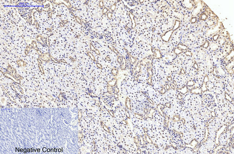
M06868 has IHC images from paraffin-embedded human colon, rat kidney and mouse testis (catalog image captions); IF is listed as an application, with no IF image supplied (catalog).
M06868 has IHC images from paraffin-embedded human colon, rat kidney and mouse testis (catalog image captions). M06868 lists IF among its applications and human, mouse and rat reactivity, but supplies no IF image (catalog).
Which to pick: Choose M06868 for tissue IHC: its own captions document staining of paraffin-embedded human colon, rat kidney and mouse testis at 1:200 (catalog image captions). For IF/ICC, M06868 lists IF and a 1:50 dilution, but supplies no IF image or ICC validation (catalog); its listed human, mouse and rat reactivity makes it the cross-species option (catalog). The IHC captions do not report the fixative (catalog image captions).