This website uses cookies to ensure you get the best experience on our website.


- Table of Contents
For paraffin-section IHC, start with the catalog antibody at 1:100–1:300 (datasheet A05834). Assess cytoplasmic and membranous staining against the high staining reported in cervical or esophageal squamous cells (HPA tissue IHC).

Expected localisation, validated protocols, controls and antibodies — the at-a-glance facts below, then the full design guide.
| Expected localisation | Cytoplasmic and membranous tissue staining (HPA tissue IHC) | |
| Staining pattern | High staining in squamous epithelia; cytoplasmic and membranous (HPA tissue IHC) | |
| Antigen retrieval | Tris-EDTA pH 9.0 HIER, heat-mediated (datasheet A05834) | |
| Positive control | Cervix+4 more · see all | |
| Negative control | Adipose tissue+4 more · see all |
| Fixation | Keep fixation conditions consistent across sections (standard IHC practice; not target-specific); target-specific effects are unknown. | |
| Caveat | The antibody may detect protein from more than one gene (HPA tissue IHC) | |
| Regulation | Broad non-lymphoid expression (UniProt) | |
| Isoform / epitope | 2 isoforms; map epitopes to ER-lumenal or cytoplasmic regions (UniProt) |
The catalog antibody’s IHC-P protocol is accompanied by four published TUSC3 staining protocols for paraffin tissue sections (PMC4752499; PMC5825997; PMC5581085; PMC6507427).
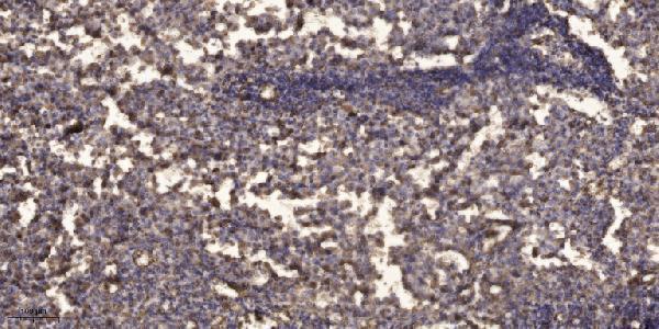
| Sample | Paraffin-embedded human Squamous cell carcinoma of lung tissue; fixative not specified (datasheet A05834) |
| Fixation | Image fixative and duration unreported (datasheet A05834); verify before use. |
| Sectioning | 4–5 µm sections on charged slides (standard) |
| Deparaffinisation | Xylene, graded ethanol series to water (standard) |
| Antigen retrieval | Heat retrieval: Tris-EDTA pH 9.0 (datasheet A05834); 20 min, 95–100 °C (standard) |
| Peroxidase block | 3% H2O2, 10 min, room temperature (standard) |
| Blocking | 10% normal serum of the secondary host, 30 min, room temperature (standard) |
| Primary antibody | Rabbit anti-TUSC3, 1:100-1:300 (datasheet A05834) |
| Primary incubation | Overnight at 4 °C (standard) |
| Detection | HRP-polymer secondary, DAB chromogen 5–10 min (standard) |
| Counterstain | Hematoxylin, blue, dehydrate and mount (standard) |
| Expected result | TUSC3-positive staining in squamous epithelial cells of cervix (HPA tissue IHC: High). HPA tissue profile: Cytoplasmic and membranous expression in most tissues. No signal in the no-primary control. |
TUSC3 is an endoplasmic reticulum membrane protein with 4 transmembrane segments (UniProt Q13454 topology). In paraffin sections, expect cytoplasmic and membranous staining, particularly in squamous epithelial cells and selected glandular cells (HPA: tissue IHC). HPA rates the tissue staining “Approved,” with medium consistency against RNA data, and cautions that its antibodies target protein from more than one gene (HPA: reliability description).
| Cytoplasmic and membranous staining is strong in esophageal squamous epithelial cells or small intestinal glandular cells (HPA: High in both cell populations). | This fits the reported tissue pattern and an endoplasmic reticulum membrane location (HPA: tissue IHC; UniProt Q13454 subcellular location). Record the stained cell population as well as intensity; a positive area alone does not establish which cells express TUSC3 (standard IHC practice). |
| Staining is confined to nuclei, with no convincing cytoplasmic or membranous signal. | A nucleus-only result conflicts with the reported compartment pattern and membrane topology (HPA: tissue IHC; UniProt Q13454 topology). Treat it as suspect; compare the same run with a known-positive tissue and review counterstain, detection and background before assigning TUSC3 localisation (standard IHC practice). |
| Strong staining appears in adipocytes or bone marrow hematopoietic cells (HPA: Not detected in these cell populations). | That distribution conflicts with the reported tissue observations (HPA: tissue IHC). Cross-reactivity or endogenous detection activity is possible; HPA also cautions that its antibodies target protein from more than one gene (HPA: reliability description). Check control slides before calling these cells positive (standard IHC practice). |
| Diffuse colour covers tissue and empty spaces, obscuring cell boundaries. | The distribution cannot be scored reliably as cytoplasmic or membranous staining (HPA: tissue IHC pattern; standard IHC practice). Uneven blocking, residual detection activity or excessive chromogen development are general IHC possibilities; this appearance does not identify a TUSC3-specific cause (standard IHC practice). |
| No staining is seen in esophageal squamous epithelium processed alongside the study sections (HPA: High in squamous epithelial cells). | The expected positive control has failed, so negative study sections are inconclusive (HPA: tissue IHC; standard IHC practice). Review tissue preservation, retrieval, primary antibody and detection steps as general workflow checks; no target-specific fixation sensitivity is established by the supplied record. |
| Cell population and tissue | HPA reports High staining in cervical and esophageal squamous epithelial cells, skin keratinocytes and small intestinal glandular cells, but Not detected in adipocytes (HPA: tissue IHC). Compare like cell populations when choosing controls; whole-organ labels can hide these differences (standard IHC practice). |
| Membrane topology and epitope position | TUSC3 has 4 transmembrane segments and lumenal and cytoplasmic regions (UniProt Q13454 topology). The supplied record does not map either antibody's epitope, so topology alone cannot determine an antibody-specific retrieval or permeabilisation setting. |
| Processing and isoforms | UniProt lists a signal sequence at residues 1–41, a chain at 42–348 and 2 isoforms (UniProt Q13454 processing and isoforms). Without an epitope map or isoform-specific validation, staining cannot identify which isoform was detected or whether an antibody recognises the processed chain. |
| Strength of IHC evidence | Both listed antibodies, HPA049851 and HPA049974, are IHC “Approved”; neither is reported here as IHC “Enhanced” (HPA: antibody validation). The tissue profile has medium consistency with RNA data and a multiple-gene target caution, so its levels are useful reference observations rather than proof of antibody specificity (HPA: reliability description). |
| IF/ICC: should the IHC pattern be carried over? | HPA's ICC-IF summary says “Membrane,” but gives no main location or cell-line images; ICC status is unlisted for both supplied antibodies (HPA: subcellular record and antibody validation). Treat that as limited IF evidence. This section provides no IF/ICC protocol or prediction of a validated cell-line pattern. |
| Situation | Likely cause | Next action |
|---|---|---|
| The known-positive esophageal squamous epithelium is blank (HPA: High in squamous epithelial cells). | A run or reagent problem is possible; the supplied sources do not establish TUSC3-specific fixation sensitivity (standard IHC practice). | Repeat with a positive control and verify retrieval, primary antibody, detection reagent and chromogen steps against the chosen IHC protocol (standard IHC practice). |
| The slide shows only nuclear colour. | This conflicts with the cytoplasmic and membranous pattern and endoplasmic reticulum membrane location (HPA: tissue IHC; UniProt Q13454). | Check a positive-control section and a no-primary control; reassess counterstain and background before scoring the nuclei (standard IHC practice). |
| Adipocytes stain strongly (HPA: Not detected in adipocytes). | Cross-reactivity or endogenous detection activity is possible; HPA warns of protein targeting from more than one gene (HPA: reliability description; standard IHC practice). | Compare a no-primary control and the expected positive tissue in the same run; record the unexpected cell-specific pattern (standard IHC practice). |
| Colour is widespread and cell borders cannot be resolved. | General IHC background or excessive chromogen development can obscure the reported compartment pattern (standard IHC practice; HPA: tissue IHC). | Review blocking, washes and chromogen development; score only after cell boundaries and compartments are distinguishable (standard IHC practice). |
| A low-staining glandular population is called negative. | HPA reports Low staining in thyroid glandular cells and pancreatic exocrine glandular cells; faint signal may be missed (HPA: tissue IHC). | Compare the named cells with a high-staining control under the same scoring conditions, and report weak and absent signal separately (HPA: tissue IHC; standard IHC practice). |
| An ICC-IF result is used to settle an ambiguous paraffin-section result. | HPA supplies a “Membrane” ICC-IF summary without cell-line images or ICC validation for the listed antibodies (HPA: subcellular record and antibody validation). | Interpret the paraffin section against its IHC controls and named tissue pattern; describe any IF observation separately (HPA: tissue IHC; standard IHC practice). |
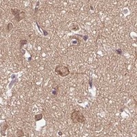


Comprehensive Human Protein Atlas IHC scoring per tissue (reliability: Approved — Medium consistency between antibody staining and RNA expression data. Caution, targets protein from more than one gene.). Rows are taken directly from the HPA tissue chart — click any row's HPA link to view the source.
| Tissue | Cell type | Level | Evidence | Source |
|---|---|---|---|---|
| Cervix | Squamous epithelial cells | High | Protein (IHC) | HPA → |
| Esophagus | Squamous epithelial cells | High | Protein (IHC) | HPA → |
| Fallopian tube | Ciliated cells (ciliary rootlets) | High | Protein (IHC) | HPA → |
| Nasopharynx | Basal cells | High | Protein (IHC) | HPA → |
| Oral mucosa | Squamous epithelial cells | High | Protein (IHC) | HPA → |
| Tissue | Cell type | Level | Evidence | Source |
|---|---|---|---|---|
| Adipose tissue | Adipocytes | Not detected | Protein (IHC) | HPA → |
| Bone marrow | Hematopoietic cells | Not detected | Protein (IHC) | HPA → |
| Liver | Cholangiocytes | Not detected | Protein (IHC) | HPA → |
| Lung | Alveolar cells | Not detected | Protein (IHC) | HPA → |
| Parathyroid gland | Glandular cells | Not detected | Protein (IHC) | HPA → |
Use the catalog antibody’s paraffin-section image as a starting point, then assess staining by cell type, compartment, and controls (caption A05834; HPA tissue IHC; UniProt Q13454 localisation).
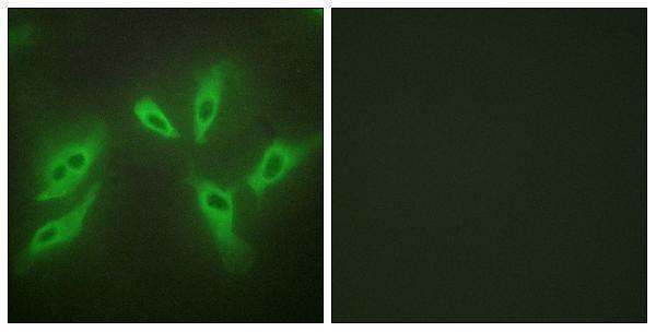
Two anti-TUSC3 antibodies have human paraffin-section IHC images; A05834 also has a HeLa-cell IF image, and both list Human, Mouse and Rat reactivity (IHC/IF image captions; catalog reactivity).
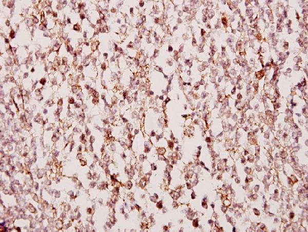
A05834 will render with an IHC image of paraffin-embedded human lung squamous cell carcinoma and has an IF image in HeLa cells (A05834 image captions). A05834-1 will render with an IHC image of paraffin-embedded human tonsil carcinoma; its listed applications do not include IF (A05834-1 IHC image caption; catalog applications).
Which to pick: For tissue IHC, A05834 has a documented paraffin-section example using Tris-EDTA retrieval at pH 9.0; A05834-1 has a paraffin-section example, and neither caption reports the fixative (IHC image captions). For IF/ICC, choose A05834 because IF is listed and illustrated in HeLa cells (catalog applications; A05834 IF image caption). For cross-species work, both are rabbit polyclonals listing Human, Mouse and Rat reactivity, though their shown IHC examples are human tissue (catalog host, dilution_raw and reactivity; IHC image captions).